Manyata Tech Park ( Call Girls ) Bangalore ✔ 6297143586 ✔ Hot Model With Sexy...
112 118
1. Adaptive Medicine 3(2): 112-118, 2011
DOI: 10.4247/AM.2011.ABB015
IGF-1 Partially Reproduces Beneficial Effect of Exercise Training on
Glucose Tolerance in Normal Rats
Ching-Yu Tzeng1, 2, Yu-Chiang Lai1, Chien-Wen Hou1, Chung-Yu Chen1, Shin-Da Lee3, 6,
Chih-Yang Huang4, Chiu-Chou Chen 1, Te-Chih Liu 1, Yuh-Feng Liou 1, Chung-Lan Kao 5,
and Chia-Hua Kuo 1, 3
1
Laboratory of Exercise Biochemistry, Taipei Physical Education College, Taipei, Taiwan, ROC
2
Department of Physical Education, Fu Jen Catholic University, Taipei, Taiwan, ROC
3
Department of Physical Therapy, Graduate Institute of Rehabilitation Science, China Medical University, Taichung, Taiwan, ROC
4
Graduate Institute of Basic Medical Science, China Medical University, Taichung, Taiwan, ROC
5
Department of Physical Medicine and Rehabilitation, Taipei Veterans General Hospital and National Yang Ming University, Taipei,
Taiwan, ROC
6
Department of Healthcare Administration, Asia University, Taichung, Taiwan, Republic of China
Exercise transiently elevates the IGF-1 (insulin-like growth Introduction
factor 1) level, but whether exogenous IGF-1 adminis-
tration can reproduce exercise training benefit in glycemic Under postprandial conditions, skeletal muscle be-
control is currently unknown. This study compared the comes the main site for glucose disposal. Thus, this
effect of IGF-1 administration and exercise training on tissue plays a pivotal role in regulating whole body
glycogen storage, glucose tolerance, and muscle glucose
glucose homeostasis (3, 9). Glucose transporter 4
transporter 4 (GLUT4) protein expression in normal rats.
(GLUT4) is the main glucose transporter isoform
Forty rats were weight matched and evenly assigned to
the following 4 groups: control (C), exercise trained (E),
expressed in skeletal muscle, which can be rapidly
IGF-1 treated (I), and exercise-trained + IGF-1 (EI). Same recruited to the plasma membrane upon insulin
volume of saline or IGF-1 (2 µg/kg BW) was injected daily stimulation. This protein translocation increases the
to the rats. Exercise training consisted of daily 90 min of membrane permeability to circulating glucose and
swimming for the first week and gradually increased to thus increases glucose disposal in skeletal muscle.
180 min, twice for the third week. Oral glucose tolerance Several early reports found that the amount of GLUT4
test (OGTT) was performed in all rats under fasted protein is strongly correlated with maximal insulin-
condition. Muscle tissues were removed at the end of stimulated glycogen storage in skeletal muscle (13,
the 3-week treatments (3 days after OGTT). The levels of 17). Therefore, interventions that enhance muscle
GLUT4 protein and mRNA were determined in red and GLUT4 protein expression might be a possible method
white portions of the quadriceps muscle (RQ and WQ). for treating patients with insulin resistance and type
Both exercise training and chronic IGF-1 administration 2 diabetes.
increased GLUT4 expression and improved glucose
The beneficial consequence of regular exercise
tolerance without an observed additive effect. Exercise
training on glucose tolerance, insulin sensitivity, and
training increased glycogen level in RQ and WQ above
control level. Despites chronic IGF-1 administration
muscle glycogen storage has been previously reported
increased muscle GLUT4 expression above control level, in humans and animals (6, 7, 12). This improvement
glycogen increase was not observed. Our data suggest is thought, to some extent, associated with the in-
that IGF-1 can partially reproduce exercise training effect creased GLUT4 protein expression in exercised muscle
on improving glycemic control. (6, 17, 27). The underlying mechanism for the ex-
ercise-induced increase in GLUT4 expression has
Key Words: insulin-like growth factor, insulin resistance, not been fully understood. Available data suggest
diabetes, glycogen, aging that exercise can transiently elevate the IGF-1 (insulin-
Corresponding author: Chia-Hua Kuo, Ph.D., Laboratory of Exercise Biochemistry, Taipei Physical Education College, 101, Sec. 2, Jhongcheng
Rd., Shihlin District, Taipei City 111, Taiwan, Republic of China. Fax: +886-2-28753383, E-mail: kuochiahua@gmail.com
*Chung-Lan Kao and Chia-Hua Kuo equally contributed to this work.
Received: July 29, 2011; Revised: August 22, 2011; Accepted: August 29, 2011.
2011 by The Society of Adaptive Science in Taiwan and Airiti Press Inc. ISSN : 2076-944X. http://www.sast.org.tw
112
2. Exercise and Muscle Fiber Conversion 113
like growth factor 1) level (5, 24, 28), and this signal ercise, according to Hou et al. (11). This recovery pe-
has been suggested to be associated with exercise riod included a 12-h fast prior to the glucose intubation
training-dependent improvement in muscle prop- for OGTT. Blood samples were withdrawn from the
erties (5, 19). In this study, we compared the effect tail at 0 (fasted sample), 15, and 45 min after the oral
of IGF-1 administration and exercise training on glucose load (1 g/kg BW) for blood glucose and se-
GLUT4 protein expression in rat muscles, as well as rum insulin measurements, according to the procedure
its association to glycogen storage and whole-body given in Cortez et al. (8). A glucose analyzer (Lifescan,
glucose tolerance. Milpitas, CA, USA) was used for glucose concentra-
tion determination; the glucose oxidase method was
Materials and Methods used. The consistency of the analyzer was tested
using real blood samples twice before use. Serum
Animal Care and Experimental Design insulin levels were measured using enzyme-linked
immunosorbent assay (ELISA) with anti-insulin
Forty male Sprague-Dawley rats from the National monoclonal antibody.
Animal Laboratory of the NSC (National Science
Council, Taipei, Taiwan, ROC) weighing 200 g each GLUT4 Protein
were housed 3 per cage and were provided normal rat
chow (PMI Nutrition International, Brentwood, MO, Three days after the OGTT, and 18 h after the last ex-
USA) and water ad libitum. The temperature of the ercise bout with an immediate glucose intubation (1
animal room was maintained at 23°C, with a 12-h g/kg BW), muscles were surgically removed for
light-dark cycle. After 1 week of familiarization, the analysis of glycogen, and GLUT4 protein levels, and
rats were weight-matched and divided into 4 groups: citrate synthase activity assay. Muscle samples for
control (C, n = 10), exercise (E, n = 10), IGF-1 (I, n = GLUT4 protein were homogenized in ice-cold HES
10), and exercise + IGF-1 (EI, n = 10). Exercise train- (20 mM N-2-hydroxyethylpiperazine-N’-2-ethane-
ing protocol consisted of 90 min of swimming for sulfonic acid, 1 mM EDTA, and 250 mM sucrose, pH
the first week and gradually increased to 180 × 2 min 7.4) buffer (1: 20) with a Polytron homogenizer
(1-h rest in between) for the third week. Swimming (Kinematica, Littau, Switzerland). Sample homo-
training started at 9 am every morning with a break on genates and standards were diluted 1: 1 with Laemmli
Sunday. The temperature of the water, 25 cm in depth sample buffer (125 mM Tris, 20% glycerol, 2% SDS,
in a plastic barrel, was maintained at 34° ± 1°C; 3 rats and 0.008% bromophenol blue, pH 6.8). The Western
were placed in each barrel at the same time. For IGF- blotting procedure for GLUT4 analysis was followed
1 administration, recombinant human IGF-1 was pro- the previously described method (11). Muscle homo-
duced from eukaryotic cells (Leinco Technologies, genates containing 75 µg (red gastrocnemius and
St. Louis, MO, USA). IGF1 was dissolved in saline plantaris muscles) of protein were subjected to SDS-
and was injected (ip) 2 µg/kg 1 h after exercise polyacrylamide gel electrophoresis (PAGE) and
training (15). At the end of the third week, rats were electrophoretically transferred to a PVDF membrane.
anesthetized at a time when they had recovered 18 h Two heart homogenates containing 15 and 30 µg of
after the cessation of the last exercise bout, and red protein were loaded in parallel with the muscle sam-
and white portion of the quadriceps muscles were ex- ples. GLUT4 antiserum (Chemicon, Temecula, CA,
cised and examined for GLUT4 protein level, glycogen USA) was used for immunoblotting (directly against
content, and citrate synthase activity. Oral glucose the carboxyl-terminus of the GLUT4 protein) in a di-
tolerance test (OGTT) was performed 3 days before lution of 1: 5000. GLUT4 protein was visualized us-
muscle sample excision; and the muscle tissue analysis ing an ECL Western blot detection kit (Amersham,
for glycogen, GLUT4 protein, and citrate synthase Arlington Heights, IL, USA) on x-ray film according
was performed 3 days after the OGTT to avoid the to the manufacturer’s instructions.
potential interference effect of OGTT procedure on
the muscle glycogen result (IGF-1 was continuously GLUT4 mRNA
given during the next 3 days after OGTT). The same
volume of saline was daily injected as a placebo into For RNA extraction, muscle tissues were homogenized
control animals. In the EI group, IGF-1 was dissolved in guanidium isothiocyanate-beta-mercaptoethanol
in saline and injected (2 µg/kg) 1 h after exercise buffer with a Polytron. Total RNA was isolated from
training. frozen tissue samples. For Northern blotting analysis,
equal amounts of total RNA (20 µg) were denatured
Oral Glucose Tolerance Test (OGTT) by heating at 60°C for 10 min and separated on 1%
agarose-formaldehyde gels. Ethidium bromide stain-
OGTT was performed 18 h after the last bout of ex- ing of the formaldehyde gel and the transferred blots
3. 114 Tzeng, Lai, Hou, Chen, Lee, Huang, Chen, Liu, Liou, Kao and Kuo
were used for determining the quality of the RNA A
sample. Treatment groups were always analyzed in 2500
parallel. GLUT-4 mRNA level was determined by
hybridization with DIG-labeled anti-sense GLUT-4
Area under Curve of Glucose
cRNA. GLUT4 mRNA was quantified on the blots 2000
using densitometric analysis with NIH image software. *
Both 28S ribosomal RNA and beta-actin mRNA were 1500
*
used as an internal standard on each blot. The amount *
of GLUT-4 mRNA present in each sample was deter-
mined by comparing the intensity of the treatment 1000
band with control band on each membrane.
Glycogen 500
About 50 mg of muscle sample was dissolved in 1 N 0
KOH at 70°C for 30 min. Dissolved homogenate was C E I IE
neutralized by glacial acetic acid and incubated over-
night in acetate buffer (0.3 M sodium acetate, pH 4.8) B
containing amyloglucosidase. The reaction mixture 50
was neutralized with 1 N NaOH. Samples were then 45
analyzed by measuring glucosyl units using the Trinder
40
Area under Curve of Insulin
reaction (Sigma, St. Louis, MO, USA).
35
Citrate Synthase Activity 30
25
Citrate synthase (CS) activity was determined in the
plantaris and red gastrocnemius muscles as originally 20
described by Srere (25). Briefly, samples were homo- 15
genized in HES buffer in a 1: 40 dilution. The super-
10
natant was assayed spectrophotometrically using
DTNB. Assays were performed at 37°C in a spectro- 5
photometer (Beckman, Fullerton, CA, USA) equipped 0
with a thermoelectric flow cell and a 1-cm light path. C E I IE
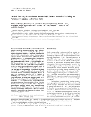
Fig. 1. Area under curve of glucose (A) and insulin (B) of oral
Statistical Analysis glucose tolerance. C: control; E: exercise-training; I:
IGF-1 administration; IE: IGF-1 administration + exer-
A two-way analysis of variance among the experi- cise training. *Significance against C group, P < 0.05.
mental groups was performed for all variables. Fisher’s
protected least significance test, which holds the val-
ue of type I errors to 0.05 for each test, was used to Data for muscle glycogen content are showed in
distinguish significant differences between pairs of Fig. 2. Exercise training significantly elevated glyco-
groups. P < 0.05 was considered statistically signifi- gen content in RQ and WQ (P < 0.05), whether IGF-
cant. All values are expressed as the means ± SE. 1 was treated or not. IGF-1 administration alone did
not cause significant difference in glycogen content
Results from control for both muscles. For both RQ and WQ,
no significant difference in glycogen content was ob-
OGTT was performed under overnight fasted con- served between the E and IE groups.
dition. Fig. 1 displays the mean value for area under All GLUT4 proteins given are relative to mean
curves (AUC) of glucose (Fig. 1A) and insulin (Fig. control level. Data for muscle GLUT4 protein is dis-
1B) during OGTT, as indicators for the whole body played in Fig. 3 (3A for RQ; 3B for WQ). Both ex-
glucose tolerance and insulin sensitivity. The AUC ercise training and IGF-1 administration elevated
of glucose for the E, I, and IE groups was significantly GLUT4 protein levels in RQ and WQ (P < 0.05).
lower than that in the C group (P < 0.05). No signifi- Muscles for the IE group also showed greater GLUT4
cant difference was found among the E, I, and IE protein level than those for the C group (P < 0.05).
groups. The AUC of insulin level was not different For both muscles, IGF-1 administration, with exercise
among all groups. or not, significantly elevated GLUT4 protein level
4. Exercise and Muscle Fiber Conversion 115
A A
60 3.0
50 * * 2.5 *# *#
RQ GLUT4 Protein (% control)
RQ Glycogen (mmol/g)
40 2.0
*
30 1.5
20 1.0
10 0.5
0
C E I IE 0.0
C E I IE
B B
45
1.8
40 *
1.6 *#
* *#
WQ GLUT4 Protein (% control)
35
1.4
WQ Glycogen (mmol/g)
*
30
1.2
25 1.0
20 0.8
15 0.6
10 0.4
5 0.2
0 0.0
C E I IE C E I IE
Fig. 2. Glycogen. RQ: red portion of quadriceps muscle; Fig. 3. GLUT4 protein. RQ: red portion of quadriceps muscle;
WQ: white portion of quadriceps muscle. *Significance WQ: white portion of quadriceps muscle. *Significance
against C group, P < 0.05. against C group, P < 0.05. #Significance against E group,
P < 0.05.
above control and exercise-trained levels (P < 0.05). Result for muscle CS activity as a mitochondria
There is no difference in GLUT4 protein level of RQ marker is shown in Fig. 5 (5A for RQ; 5B for WQ). The
and WQ between the I and IE groups. activity of CS in the E, I, and IE groups was signifi-
All GLUT4 mRNA given are relative to mean cantly greater than that in both RQ and WQ (P < 0.05).
control level. Data for muscle GLUT4 mRNA are There is no difference among the E, I, and IE groups.
displayed in Fig. 4 (4A for RQ; 4B for WQ). Both ex-
ercise training and IGF-1 administration elevated Discussion
GLUT4 mRNA levels in RQ and WQ (P < 0.05).
Muscles for the IE group also showed greater GLUT4 Previous studies on animal muscle have shown that
mRNA level than those of the C group (P < 0.05). For following weight overload there is an increase in
both muscles, the IGF-1 treated group, with exercise the expression of IGF-1 mRNA in muscle (21, 22).
or not, displays greater GLUT4 protein level than Recently, Bamman et al. (2) reported a 62% increase
those in the control and exercise-trained groups (P < in IGF-1 mRNA concentration in human muscle 48 h
0.05). For RQ and WQ, no significant difference in after a single bout of exercise. It was unknown how
GLUT4 protein level was observed between the I much, or in what aspect, the training effect is mediated
and IE groups. by exercise-induced IGF-1 production. Thus we hy-
5. 116 Tzeng, Lai, Hou, Chen, Lee, Huang, Chen, Liu, Liou, Kao and Kuo
A A
4.0 60
*# *
*
*# *
3.5
50
RQ GLUT4 mRNA (% control)
RQ CS Activity (µmol/g/min)
3.0
40
2.5 *
2.0 30
1.5
20
1.0
10
0.5
0.0 0
C E I IE C E I IE
B 3.0 B
30
2.5 *#
WQ GLUT4 mRNA (% control)
*# 25 *
WQ CS Activity (µmol/g/min)
*
*
2.0
20
*
1.5
15
1.0 10
0.5 5
0.0 0
C E I IE C E I IE
Fig. 4. GLUT4 mRNA. RQ: red portion of quadriceps muscle; Fig. 5. Citrate synthase activity. RQ: red portion of quadriceps
WQ: white portion of quadriceps muscle. *Significance muscle; WQ: white portion of quadriceps muscle. *Sig-
against C group, P < 0.05. #Significance against E group, nificance against C group, P < 0.05.
P < 0.05.
pothesized that, without exercise training, chronic GLUT4 protein expression, regardless of treatment
IGF-1 administration can simulate the exercise training of IGF-1 or exercise training. This result suggests
effect on GLUT4 protein expression, glycogen storage, that exercise training-induced GLUT4 elevation could
and thus affecting the whole-body glucose tolerance. be mediated by IGF-1 signaling pathway. Alterna-
This hypothesis was partially demonstrated by the tively, a cross-talk between exercise-derived signal
current study. If exercise and IGF-1 mediate different and IGF-1 signaling pathways to enhance GLUT4
mechanism to enhance GLUT4 protein expression, protein expression was reciprocally inhibited to each
then additive effect should be observed. Here we other (14).
found IGF-1 significantly elevated GLUT4 protein Although the increased insulin AUC in the IGF-
and mRNA level in both IGF-1 treated and exercise- 1 injected rats did not reach statistical significance,
trained muscle, and no additive effect was found. we could not rule out the possibility that reduced glu-
Both white and red skeletal muscles had similar cose AUC was affected by greater insulin secretion.
training adaptation in GLUT4 increases, suggesting It has been reported that IGF-1 administration can
that both muscle groups were recruited by our exercise stimulate beta-cell proliferation (16). To further
training protocol. Cortez has previously reported that clarify whether IGF-1 effect is completely mediated
exercise training-induced GLUT-4 gene and protein by GLUT4 protein expression requires data concerned
expression occurred only in the recruited muscle (8). insulin-stimulated glucose uptake and GLUT-4 protein
It is noteworthy that the improvement in the whole- translocation in skeletal muscle. IGF-1 can also in-
body glucose tolerance is associated with greater crease the angiogenesis in skeletal muscle which may
6. Exercise and Muscle Fiber Conversion 117
also play a role for the observed improvement in an improvement in insulin sensitivity. This result
glucose tolerance (18). suggests that the elevation of muscle oxidative
The generally observed post-exercise glycogen capacity with both treatments may also take part in
supercompensation (greater than pre-exercise glyco- improving glucose tolerance and insulin sensitivity.
gen storage) is partly associated with increased GLUT4 In addition, this result also implies that the generally
protein expression. The causal relationship was dem- observed exercise training effect can be partly simu-
onstrated in the muscle with greater GLUT4 protein lated by IGF-1 treatment. The fact that no additive
expression by germ-line manipulation that leads to a effect by exercise-training and IGF-1 also suggests
greater insulin-stimulated glycogen storage (26). The that both stimulations share common signaling
present study that increased glycogen storage by ex- mechanism for inducing mitochondria biogenesis.
ercise training was occurred in parallel with an in- Previous study has shown that the beneficial ef-
crease in GLUT4 protein following a 16-h recovery fect of exercise training on insulin sensitivity is at-
also supports this idea. However, in the IGF-1 treated tenuated in elderly and high body mass index
group glycogen level was not elevated even though individuals (20), the population generally displays
we saw a greater GLUT4 protein level. This result lower IGF-1 level (1, 23). Apparently, IGF-1 signaling
implicates that, during the post-exercise recovery system is one anabolic signal that can be stimulated
phase, metabolic need for glycogen utilization was by exercise (24, 28). In this study, the evidence
also elevated by IGF-1 administration. IGF-1 is gen- implicates that IGF-1 is relevant for upregulating
erally known as an enhancer for metabolism in skeletal GLUT4 protein expression and citrate synthase
muscle due to greater energy requirement for muscle activity. Apparently, the benefit of IGF-1 adminis-
growth or protein turnover (10). Therefore, the lower tration could not simulate all the benefits of exercise
glycogen storage in IGF-1 treated muscle compared training. Greater glycogen storage was only observed
to the trained muscle could be due to greater glyco- in exercise-trained muscle, which highlights the
gen turnover for the enhanced rate of synthesis for unique effect of exercise over the chronic IGF-1
protein. administration. Glycogen is an important anaerobic
The fact that glycogen supercompensation phe- substrate relevant for the body to encounter metabolic
nomenon was only observed in the exercise-trained stress, and thus is important for survival under various
muscle, also suggests that motor unit recruitment is challenge in normal life.
essential for the training effect. It is known that the We found that IGF-1 administration can partly
glucose transport across the plasma membrane for mimic the effect of exercise training in enhancing
providing substrate for glycogen synthesis is relied GLUT4 protein expression, citrate synthase activity,
on the number of GLUT4 protein presence on plasma and glucose tolerance. However, training-induced
membrane, which can be regulated by insulin (12). glycogen supercompensation was absent with IGF-1
Brozinick et al. (4) has shown that exercise training treatment alone, suggesting the importance of muscle
can increase GLUT4 protein concentration in the contraction component in the full benefit of exercise
proximity adjacent to plasma membrane, as observed training.
48-h post exercise. Therefore, increased distribution
of GLUT4 protein to plasma membrane by exercise Acknowledgments
training could lead to a faster rate of glucose transport
for glycogen synthesis after meal, which might in turn The present work was partly sponsored by Ministry of
explain why greater glycogen level did not occur in Education.
IGF-1 treated muscle without exercise.
The ability to maintain glucose homeostasis References
after carbohydrate ingestion relies on the efficient
glucose metabolism in skeletal muscle, resulting from 1. Alderete, T.L., Byrd-Williams, C.E., Toledo-Corral, C.M., Conti,
parallel expression of the proteins controlling glucose D.V., Weigensberg, M.J. and Goran, M.I. Relationships between
uptake and disposal, including glucose transporter IGF-1 and IGFBP-1 and adiposity in obese African-American and
Latino adolescents. Obesity. 19: 933-938, 2011.
and mitochondria enzymes for glucose oxidation.
2. Bamman, M.M., Shipp, J.R., Jiang, J., Gower, B.A., Hunter, G.R.,
Therefore, alteration in the oxidative capacity of a Goodman, A., McLafferty, C.L. and Urban, R.J. Mechanical load
muscle may also contribute a change in glucose dis- increases muscle IGF-I and androgen receptor mRNA concentra-
posal property. Many studies have reported that both tions in humans. Am. J. Physiol. Endocrinol. Metab. 280: E383-
citrate synthase and GLUT4 protein are regulated in E390, 2001.
3. Baron, A.D., Brechtel, G., Wallace, P. and Edelman, S.V. Rates
parallel by exercise training (12). In the present study,
and tissue sites of non-insulin- and insulin-mediated glucose uptake
we found that exercise training and IGF-1 administra- in humans. Am. J. Physiol. Endocrinol. Metab. 255: E769-E774,
tion significantly elevated citrate synthase activity 1988.
(as a generic mitochondrial marker) concurrent with 4. Brozinick, J.T., Etgen, G.J., Yaspelkis, B.B., Kang, H.Y. and Ivy,
7. 118 Tzeng, Lai, Hou, Chen, Lee, Huang, Chen, Liu, Liou, Kao and Kuo
J.L. Effects of exercise training on muscle GLUT-4 protein content D.R. and Rhodes, C.J. Activation of IRS-2 mediated signal trans-
and translocation in obese Zucker rats. Am. J. Physiol. Endocrinol. duction by IGF-1, but not TGF-β or EGF, augments pancreatic
Metab. 265: E419-E427, 1993. β-cell proliferation. Diabetes 51: 966-976, 2002.
5. Chang, H.C., Yang, Y.R., Wang, P.S., Kuo, C.H. and Wang, R.Y. 17. MacLean, P.S., Zheng, D. and Dohm, G.L. Muscle glucose trans-
Effects of insulin-like growth factor 1 on muscle atrophy and motor porter (GLUT 4) gene expression during exercise. Exerc. Sport
function in rats with brain ischemia. Chinese J. Physiol. 53: 337- Sci. Rev. 28: 148-152, 2000.
348, 2010. 18. Mallikarjuna, K., Hou, C.W., Chen, C.Y., Lee, J.P., Sathyavelu,
6. Chen, C.Y., Tsai, Y.L., Kao, C.L., Lee, S.D., Wu, M.C., Mallikarjuna, R.K. and Kuo, C.H. Angiogenesis: Role of exercise training and
K., Liao, Y.H., Ivy, J.L. and Kuo, C.H. Effect of mild intermittent aging. Adapt. Med. 2: 29-41, 2010.
hypoxia on glucose tolerance, muscle morphology and AMPK- 19. Manetta, J., Brun, J., Maimoun, L., Callis, A., Prefaut, C. and
PGC-1α signaling. Chinese J. Physiol. 53: 62-71, 2010. Mercier, J. Effect of training on the GH/IGF-I axis during exercise
7. Chen, H.H., Chen, Y.L., Huang, C.Y., Lee, S.D., Chen, S.C. and in middle-aged men: relationship to glucose homeostasis. Am. J.
Kuo, C.H. Effects of one-year swimming training on blood pressure Physiol. Endocrinol. Metab. 283: E929-E936, 2002.
and insulin sensitivity in mild hypertensive young patients. Chinese 20. Marin, P., Krotkiewski, M., Holm, G., Gustafsson, C. and Bjorntorp,
J. Physiol. 53: 185-189, 2010. P. Effects of acute exercise on insulin and non-insulin-dependent
8. Cortez, M.Y., Torgan, C.E., Brozinick, J.T. and Ivy, J.L. Insulin glucose uptake in normal and moderately obese women. Eur. J.
resistance of obese Zucker rats exercise trained at two different Med. 2: 199-204, 1993.
intensities. Am. J. Physiol. Endocrinol. Metab. 261: E613-E619, 21. McKoy, G., Ashley, W., Mander, J., Yang, S.Y., Williams, N.,
1991. Russell, B. and Goldspink, G. Expression of insulin growth factor-
9. DeFronzo, R.A., Jacot, E., Jequier, E., Maeder, E., Wahren, J. and 1 splice variants and structural genes in rabbit skeletal muscle
Felber, J.P. The effect of insulin on the disposal of intravenous induced by stretch and stimulation. J. Physiol. 516: 583-592, 1999.
glucose. Results from indirect calorimetry and hepatic and femoral 22. Owino, V., Yang, S.Y. and Goldspink, G. Age-related loss of
venous catheterization. Diabetes 30: 1000-1007, 1981. skeletal muscle function and the inability to express the autocrine
10. Glass, D.J. Molecular mechanisms modulating muscle mass. form of insulin-like growth factor-1 (MGF) in response to mechani-
Trends Mol. Med. 9: 344-350, 2003. cal overload. FEBS Lett. 505: 259-263, 2001.
11. Hou, C.W., Chou, S.W., Ho, H.Y., Lee, W.C., Lin, C.H. and Kuo, 23. Ruiz-Torres, A. and Soares de Melo Kirzner, M. Ageing and
C.H. Effect of exercise training and growth hormone administra- longevity are related to growth hormone/insulin-like growth factor-
tion on glucose tolerance and muscle GLUT4 protein expression. 1 secretion. Gerontology 48: 401-407, 2002.
J. Biomed. Sci. 10: 689-696, 2003. 24. Schwarz, A.J., Brasel, J.A., Hintz, R.L., Mohan, S. and Cooper,
12. Ivy, J.L., Zderic, T.W. and Fogt, D.L. Prevention and treatment of D.M. Acute effect of brief low- and high-intensity exercise on
non-insulin-dependent diabetes mellitus. Exerc. Sport Sci. Rev. circulating insulin-like growth factor (IGF) I, II, and IGF-binding
27: 1-35, 1999. protein-3 and its proteolysis in young healthy men. J. Clin.
13. Kern, M., Wells, J.A., Stephens, J.M., Elton, C.W., Friedman, J.E., Endocrinol. Metab. 81: 3492-3497, 1996.
Tapscott, E.B., Pekala, P.H. and Dohm, G.L. Insulin respon- 25. Srere, P.A. Citrate synthase. Methods in Enzymol. 230: 946-950,
siveness in skeletal muscle is determined by glucose transporter 1969.
(Glut4) protein level. Biochem. J. 270: 397-400, 1990. 26. Tsao, T.S., Li, J., Chang, K.S., Stenbit, A.E., Galuska, D., Anderson,
14. Kim, J., Yoon, M.Y., Choi, S.L., Kang, I., Kim, S.S., Kim, Y.S., J.E., Zierath, J.R., McCarter, R.J. and Charron, M.J. Metabolic
Choi, Y.K. and Ha, J. Effects of stimulation of AMP-activated adaptations in skeletal muscle overexpressing GLUT4: effects on
protein kinase on insulin-like growth factor 1- and epidermal muscle and physical activity. FASEB. J. 15: 958-969, 2001.
growth factor-dependent extracellular signal-regulated kinase 27. Winder, W.W. Energy-sensing and signaling by AMP-activated
pathway. J. Biol. Chem. 276: 19102-19110, 2001 protein kinase in skeletal muscle. J. Appl. Physiol. 91: 1017-1028,
15. Korolkiewicz, R.P., Tashima, K., Fujita, A., Kato, S. and Takeuchi, 2001.
K. Exogenous insulin-like growth factor (IGF)-1 improves the 28. Yeh, J.K., Aloia, J.F., Chen, M., Ling, N., Koo, H.C. and Millard,
impaired healing of gastric mucosal lesion in diabetic rats. W.J. Effect of growth hormone administration and treadmill
Pharmacol. Res. 41: 221-229, 2000. exercise on serum and skeletal IGF-I in rats. Am. J. Physiol.
16. Lingohr, M.K., Dickson, L.M., McCuaig, J.F., Hugl, S.R., Twardzik, Endocrinol. Metab. 266: E129-E135, 1994.