SlideShare ist ein Scribd-Unternehmen logo
1 von 20
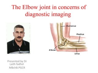
The Elbow joint in concerns of
diagnostic imaging

Presented by Dr
Laith fadhel
MBchB.PGCR
X-Ray ) bones and articulation
Ossification centers and bone age
Ryan –page 254 /Sutton –page 1847
Elbow joint movement
Anatomy and relation study
When you study the anatomy of the elbow, it is good to use the inside-out approach.
First study the bones and then continue with the ligaments and the tendons and then the
surrounding structures.
MRI technique
• Scan planes
• Just like in the shoulder you
need to be sure to get the
imaging planes correctly in a
standardized way.
Use the axis of the epicondyles
on a axial localizer to plan the
coronal scan.
The Sagittal images are scanned
perpendicular to the coronal
scan.
• In this way you get very
persistent images and you will
get used to the normal anatomy.
Imaging sequences
T1
In every joint that is studied you should
have at least one T1-sequence not only
to look at the anatomy, but also as a back
up for looking at the marrow.
Of course the T2-fatsat images will show
marrow abnormalities, but T1 can be
helpful in telling us what is really going
on.
T1 is certainly used in MR-arthrography.

T2-fatsat
T2 will show us most of the pathology,
whether it is in the bone marrow,
ligaments or muscle because of the high
water content. It can also be used to
image cartilage.
Gradient echo
With gradient echo we can use 3-D thin
sections to image the cartilage and the
ligaments.
In the MR-protocol we do T1 and T2-fatsat in all three imaging
planes.
Sometimes STIR is used.
Tendon attachments
Common flexor tendon
Attaches at the medial epicondyle
Ulnar collateral ligament or UCL
Starts at the undersurface of the medial
epicondyle and runs down to the sublime
tubercle, which is the medial side of the coronoid
process.
Common extensor tendon
Originates at the lateral epicondyle.
Lateral collateral ligament
Originates just underneath the attachment of the
common extensor tendon.
Lateral ulnar collateral ligament
This is a somewhat confusing term for a tendon
that also originates just underneath the common
extensor tendon. It swings down behind the radial
head and attaches at the area of the ulna that is
called the supinator crest - see lateral view.
Biceps tendon
Attaches on the radial tuberosity.
Brachialis tendon
Attaches on the coronoid process.
Annular ligament
Attaches on the volar side of the sigmoid notch of
the ulna and runs around the radial head and
attaches on the dorsal side of the sigmoid notch.

Attachment sites
Medical epicondyle avulsion
fracture
A fat-suppressed T2 weighted
coronal image in a 15 year old
baseball pitcher reveals an avulsion
fracture (arrow) of the medial
epicondyle apophysis,
Ulnar Collateral ligament
If you look at the medial epicondyle you will
notice the posterior bundle as a thin
structure (blue arrow).
The posterior bundle forms the floor of the
cubital tunnel.
A retinaculum covers the cubital tunnel.
Notice that the anterior bundle is much
thicker (white arrow).
As we go distally we'll see that they merge
together to attach to the sublime tubercle
It is normal to see some high signal in the
proximal part (arrow).
UCL tear
Remember that the UCL should attach very tightly on
the sublime tubercle.
In this case it doesn't, so even on these two images
you can tell that there is a complete tear.
Notice that there is some marrow edema in the
sublime tubercle.
The mechanism of injury to the UCL is usually chronic
tensile forces, which create micro tears.
This is seen in pitchers and other overhead throwingathletes.
A tear can also occur in a fall on the outstretched hand.
Most commonly there is a complete tear, but
sometimes there is a partial tear which can be very
hard to see.
That is why in these athletes MR- arthrogram is usually
performed.
This is a 18 year old baseball pitcher with medial elbow
pain.
A partial tear is seen creating a 'T-sign‘
Notice that the anterior bundle is intact and firmly
attaches to the sublime tubercle (yellow arrow).
On the next two images there is some soft tissue
edema and more abnormal signal posteriorly (red
arrow). So we suspect pathology of the posterior
bundle.
UCL tear
On the axial image we nicely see the
anterior bundle is o.k. (red arrow).
There is only some edema next to it.
However the posterior bundle is not o.k.
This is partial tearing.
We see this occasionally in throwing
athletes, where the anterior bundle is
intact and their elbow is not unstable.
They somehow have torn their posterior
bundle, which causes pain.
They do not need surgery, but it still
may keep them out of the game for
quite a while.
Now here is the last case.
This is a 38 year old male who has been
weight-lifting for 20 years.
He complains of intermittent elbow pain
and popping.
Notice that the UCL is abnormal with
some areas of very high signal indicating
a partial tear.
On the lateral side there is subchondral
edema and cartilage.
This is arthrosis secondary to chronic
instability due to the chronic partial
tearing.
Lateral Collateral Ligament
When you look for the radial collateral
ligament, first try to identify the common
extensor tendon, because right
underneath it you will find the radial
collateral ligament (yellow arrow).
As you go more posteriorly you will see
the LUCL - the lateral ulnar collateral
ligament, which sweeps behind the radial
head (white arrows).
The annular ligament is usually difficult to
differentiate from the RCL, but sometimes
it can be identified on a Sagittal MRartrogram.
Muscles tendons
The common flexor
tendon originates at the
medial epicondyle.
On a T1W-images the
tendon should have a low
signal intensity (red
arrow).

The common extensor
tendon originates at the
lateral epicondyle.
On a T1W-images the
tendon should have a low
signal intensity (yellow
arrow).
Muscles tendons
through the axial images
of the biceps tendon
from the
musculotendinous
junction to the
attachment on the radial
tuberositas.
Muscles tendons
The Brachialis originates
from the lower half of the
front of the hummers, near
the insertion of the deltoid
muscle.
It lies deeper than the
biceps brachii, and is a
synergist that assists the
biceps in flexing the elbow.
The thick tendon inserts on
the anterior surface of the
coronoid process of the
ulna.
On a Sagittal view, when you
compare the Brachialis
tendon (yellow arrows) with
the biceps tendon (red
arrows), notice that the
Brachialis is almost all
muscle.
It only has a very short
tendon distally
Nerves
Soft Tissue
Masses & bursitis

On MR a mass was seen
just above the medial
epicondyle, where the
epitrochlear lymph
nodes live.
The mass is very
heterogeneous as is the
enhancement.
Based on the MRfindings you still have to
call this mass
indeterminate.
The final diagnosis was

cat scratch disease
based on high Bartonella
henselae titers.
Soft Tissue
Masses & bursitis
Here images of a 26 year old
female who also came with a
mass in the peritrochlear
region.
It looks quite homogeneous
and cystic.
Continue with the post-Gd
image.
Notice the inhomogeneous
enhancement on the MRI and
prominent internal vascularity
on the Sagittal ultrasound
image.
So this was not a cystic mass.
Again this was diagnosed as
indeterminate.
The final diagnosis at biopsy
was Lymphoma
Soft Tissue Masses &
bursitis
Olecranon
Brachioradilus
bursitis

Weitere ähnliche Inhalte

Was ist angesagt?

Presentation1.pptx, radiological anatomy of the knee joint.
Presentation1.pptx, radiological anatomy of the knee joint.Presentation1.pptx, radiological anatomy of the knee joint.
Presentation1.pptx, radiological anatomy of the knee joint.
Abdellah Nazeer
 
Presentation1.pptx mri of elbow joint
Presentation1.pptx mri of elbow jointPresentation1.pptx mri of elbow joint
Presentation1.pptx mri of elbow joint
Abdellah Nazeer
 
Radiographic anatomy of abd and pelvis r a
Radiographic anatomy of abd and pelvis r aRadiographic anatomy of abd and pelvis r a
Radiographic anatomy of abd and pelvis r a
mr_koky
 
Presentation1.pptx, radiological imaging of salivary glands diseases.
Presentation1.pptx, radiological imaging of salivary glands diseases.Presentation1.pptx, radiological imaging of salivary glands diseases.
Presentation1.pptx, radiological imaging of salivary glands diseases.
Abdellah Nazeer
 
Presentation1.pptx, radiological anatomy of the shoulder joint.
Presentation1.pptx, radiological anatomy of the shoulder joint.Presentation1.pptx, radiological anatomy of the shoulder joint.
Presentation1.pptx, radiological anatomy of the shoulder joint.
Abdellah Nazeer
 
Presentation1.pptx, radiological imaging of metabolic bone diseases.
Presentation1.pptx, radiological imaging of metabolic bone diseases.Presentation1.pptx, radiological imaging of metabolic bone diseases.
Presentation1.pptx, radiological imaging of metabolic bone diseases.
Abdellah Nazeer
 
Presentation1.pptx, radiological anatomy of the thigh and leg.
Presentation1.pptx, radiological anatomy of the thigh and leg.Presentation1.pptx, radiological anatomy of the thigh and leg.
Presentation1.pptx, radiological anatomy of the thigh and leg.
Abdellah Nazeer
 
Presentation1.pptx, ultrasound examination of the hip joint
Presentation1.pptx, ultrasound examination of the hip jointPresentation1.pptx, ultrasound examination of the hip joint
Presentation1.pptx, ultrasound examination of the hip joint
Abdellah Nazeer
 

Was ist angesagt? (20)

Presentation1.pptx, radiological anatomy of the knee joint.
Presentation1.pptx, radiological anatomy of the knee joint.Presentation1.pptx, radiological anatomy of the knee joint.
Presentation1.pptx, radiological anatomy of the knee joint.
 
Anatomy & Radiology of pelvis
Anatomy & Radiology of pelvisAnatomy & Radiology of pelvis
Anatomy & Radiology of pelvis
 
Presentation1.pptx mri of elbow joint
Presentation1.pptx mri of elbow jointPresentation1.pptx mri of elbow joint
Presentation1.pptx mri of elbow joint
 
Mri of knee
Mri of kneeMri of knee
Mri of knee
 
Hip joint
Hip jointHip joint
Hip joint
 
Imaging in Skull base
Imaging in Skull baseImaging in Skull base
Imaging in Skull base
 
Radiographic anatomy of abd and pelvis r a
Radiographic anatomy of abd and pelvis r aRadiographic anatomy of abd and pelvis r a
Radiographic anatomy of abd and pelvis r a
 
Journal club final
Journal club finalJournal club final
Journal club final
 
MRI fistulogram
MRI fistulogramMRI fistulogram
MRI fistulogram
 
Presentation1.pptx, radiological imaging of salivary glands diseases.
Presentation1.pptx, radiological imaging of salivary glands diseases.Presentation1.pptx, radiological imaging of salivary glands diseases.
Presentation1.pptx, radiological imaging of salivary glands diseases.
 
Mri anatomy of ankle
Mri anatomy of ankleMri anatomy of ankle
Mri anatomy of ankle
 
Presentation1.pptx, radiological anatomy of the shoulder joint.
Presentation1.pptx, radiological anatomy of the shoulder joint.Presentation1.pptx, radiological anatomy of the shoulder joint.
Presentation1.pptx, radiological anatomy of the shoulder joint.
 
Presentation1.pptx, radiological imaging of metabolic bone diseases.
Presentation1.pptx, radiological imaging of metabolic bone diseases.Presentation1.pptx, radiological imaging of metabolic bone diseases.
Presentation1.pptx, radiological imaging of metabolic bone diseases.
 
Larynx anatomy ct and mri
Larynx anatomy ct and mriLarynx anatomy ct and mri
Larynx anatomy ct and mri
 
Cartilage imaging simplified
Cartilage imaging simplifiedCartilage imaging simplified
Cartilage imaging simplified
 
Presentation1.pptx, radiological anatomy of the thigh and leg.
Presentation1.pptx, radiological anatomy of the thigh and leg.Presentation1.pptx, radiological anatomy of the thigh and leg.
Presentation1.pptx, radiological anatomy of the thigh and leg.
 
Orbit imaging anatomy
Orbit imaging anatomyOrbit imaging anatomy
Orbit imaging anatomy
 
Presentation1.pptx. shoulder joint.
Presentation1.pptx. shoulder joint.Presentation1.pptx. shoulder joint.
Presentation1.pptx. shoulder joint.
 
Presentation1.pptx, ultrasound examination of the hip joint
Presentation1.pptx, ultrasound examination of the hip jointPresentation1.pptx, ultrasound examination of the hip joint
Presentation1.pptx, ultrasound examination of the hip joint
 
Skull base imaging
Skull base imagingSkull base imaging
Skull base imaging
 

Andere mochten auch

Forearm And Elbow Pathologies Dr. Mark Davies Sjsu, Spring 2008
Forearm And Elbow Pathologies   Dr. Mark Davies   Sjsu, Spring 2008Forearm And Elbow Pathologies   Dr. Mark Davies   Sjsu, Spring 2008
Forearm And Elbow Pathologies Dr. Mark Davies Sjsu, Spring 2008
JLS10
 
Anatomy and Biomechanics of the Elbow Joint
Anatomy and Biomechanics of the Elbow JointAnatomy and Biomechanics of the Elbow Joint
Anatomy and Biomechanics of the Elbow Joint
orthoprince
 
Ernia storia naturale
Ernia storia naturaleErnia storia naturale
Ernia storia naturale
nicolmed
 
Instability around elbow -1st part
Instability around elbow -1st partInstability around elbow -1st part
Instability around elbow -1st part
Vishnu Raja
 
Musculoskeletal Imaging 09
Musculoskeletal Imaging 09Musculoskeletal Imaging 09
Musculoskeletal Imaging 09
Steven Sager
 
Elbow Anatomy And Examination
Elbow Anatomy And ExaminationElbow Anatomy And Examination
Elbow Anatomy And Examination
med027972
 
Presentation2.pptx wrist joint.
Presentation2.pptx wrist joint.Presentation2.pptx wrist joint.
Presentation2.pptx wrist joint.
Abdellah Nazeer
 
Clinical examination of elbow joint
Clinical examination of elbow jointClinical examination of elbow joint
Clinical examination of elbow joint
varuntandra
 

Andere mochten auch (20)

Forearm And Elbow Pathologies Dr. Mark Davies Sjsu, Spring 2008
Forearm And Elbow Pathologies   Dr. Mark Davies   Sjsu, Spring 2008Forearm And Elbow Pathologies   Dr. Mark Davies   Sjsu, Spring 2008
Forearm And Elbow Pathologies Dr. Mark Davies Sjsu, Spring 2008
 
Radiology of the Elbow Joint. Dr. Sumit Sharma
Radiology of the Elbow Joint. Dr. Sumit SharmaRadiology of the Elbow Joint. Dr. Sumit Sharma
Radiology of the Elbow Joint. Dr. Sumit Sharma
 
Anatomy and Biomechanics of the Elbow Joint
Anatomy and Biomechanics of the Elbow JointAnatomy and Biomechanics of the Elbow Joint
Anatomy and Biomechanics of the Elbow Joint
 
Elbow joint
Elbow joint Elbow joint
Elbow joint
 
Elbow joint imaging
Elbow joint imagingElbow joint imaging
Elbow joint imaging
 
Ten Common Orthopedic Injuries - Tennis Elbow
Ten Common Orthopedic Injuries - Tennis ElbowTen Common Orthopedic Injuries - Tennis Elbow
Ten Common Orthopedic Injuries - Tennis Elbow
 
Ernia storia naturale
Ernia storia naturaleErnia storia naturale
Ernia storia naturale
 
Instability around elbow -1st part
Instability around elbow -1st partInstability around elbow -1st part
Instability around elbow -1st part
 
Cubital fossa
Cubital fossaCubital fossa
Cubital fossa
 
elbow sports injuries
elbow sports injurieselbow sports injuries
elbow sports injuries
 
Musculoskeletal Imaging 09
Musculoskeletal Imaging 09Musculoskeletal Imaging 09
Musculoskeletal Imaging 09
 
dislocations & fractures of Elbow in adults
dislocations & fractures of Elbow in adultsdislocations & fractures of Elbow in adults
dislocations & fractures of Elbow in adults
 
Elbow and forearm fractures
Elbow and forearm fracturesElbow and forearm fractures
Elbow and forearm fractures
 
Elbow Anatomy And Examination
Elbow Anatomy And ExaminationElbow Anatomy And Examination
Elbow Anatomy And Examination
 
The Elbow, Examination
The Elbow, ExaminationThe Elbow, Examination
The Elbow, Examination
 
Rotator cuff
Rotator cuffRotator cuff
Rotator cuff
 
Presentation2.pptx wrist joint.
Presentation2.pptx wrist joint.Presentation2.pptx wrist joint.
Presentation2.pptx wrist joint.
 
Anatomy of Hip joint
Anatomy of Hip joint Anatomy of Hip joint
Anatomy of Hip joint
 
Clinical examination of elbow joint
Clinical examination of elbow jointClinical examination of elbow joint
Clinical examination of elbow joint
 
Elbow examination
Elbow examinationElbow examination
Elbow examination
 

Ähnlich wie The elbow joint in concern of diagnostic imaging .pptx 1

shoulder_anatomy_166103_284_41995_v1.ppt
shoulder_anatomy_166103_284_41995_v1.pptshoulder_anatomy_166103_284_41995_v1.ppt
shoulder_anatomy_166103_284_41995_v1.ppt
pejuangskripsi6
 
06 Appendicular Skeleton Pectoral Girdle And Upper Limbs
06 Appendicular Skeleton   Pectoral Girdle And Upper Limbs06 Appendicular Skeleton   Pectoral Girdle And Upper Limbs
06 Appendicular Skeleton Pectoral Girdle And Upper Limbs
guest334add
 
09 Articulations Selected Articulations In Depth
09 Articulations   Selected Articulations In Depth09 Articulations   Selected Articulations In Depth
09 Articulations Selected Articulations In Depth
guest334add
 
Presentation1.pptx, radiological anatomy of the lower limb anatomy.
Presentation1.pptx, radiological anatomy of the lower limb anatomy.Presentation1.pptx, radiological anatomy of the lower limb anatomy.
Presentation1.pptx, radiological anatomy of the lower limb anatomy.
Abdellah Nazeer
 
Presentation1.pptx, radiological anatomy of the lower limb anatomy.
Presentation1.pptx, radiological anatomy of the lower limb anatomy.Presentation1.pptx, radiological anatomy of the lower limb anatomy.
Presentation1.pptx, radiological anatomy of the lower limb anatomy.
Abdellah Nazeer
 
Atlanto occipital and atlanto axial joint
Atlanto occipital and atlanto axial jointAtlanto occipital and atlanto axial joint
Atlanto occipital and atlanto axial joint
Shubham Singh
 
Biomechanics Assignment
Biomechanics AssignmentBiomechanics Assignment
Biomechanics Assignment
Liz Sims
 
Presentation1.pptx, radiological anatomy of the upper limb joint.
Presentation1.pptx, radiological anatomy of the upper limb joint.Presentation1.pptx, radiological anatomy of the upper limb joint.
Presentation1.pptx, radiological anatomy of the upper limb joint.
Abdellah Nazeer
 

Ähnlich wie The elbow joint in concern of diagnostic imaging .pptx 1 (20)

Elbow MRI
Elbow MRIElbow MRI
Elbow MRI
 
shoulder_anatomy_166103_284_41995_v1.ppt
shoulder_anatomy_166103_284_41995_v1.pptshoulder_anatomy_166103_284_41995_v1.ppt
shoulder_anatomy_166103_284_41995_v1.ppt
 
06 Appendicular Skeleton Pectoral Girdle And Upper Limbs
06 Appendicular Skeleton   Pectoral Girdle And Upper Limbs06 Appendicular Skeleton   Pectoral Girdle And Upper Limbs
06 Appendicular Skeleton Pectoral Girdle And Upper Limbs
 
appendicular trauma in radiology. .pptx
appendicular trauma in radiology.  .pptxappendicular trauma in radiology.  .pptx
appendicular trauma in radiology. .pptx
 
06 Appendicular Skeleton Pectoral Girdle And Upper Limbs
06 Appendicular Skeleton   Pectoral Girdle And Upper Limbs06 Appendicular Skeleton   Pectoral Girdle And Upper Limbs
06 Appendicular Skeleton Pectoral Girdle And Upper Limbs
 
06 Appendicular Skeleton Pectoral Girdle And Upper Limbs
06 Appendicular Skeleton   Pectoral Girdle And Upper Limbs06 Appendicular Skeleton   Pectoral Girdle And Upper Limbs
06 Appendicular Skeleton Pectoral Girdle And Upper Limbs
 
Craniovertebral junction
Craniovertebral junction Craniovertebral junction
Craniovertebral junction
 
Radiology of the lower extremity dr asif ali khan
Radiology of the lower extremity   dr asif ali khanRadiology of the lower extremity   dr asif ali khan
Radiology of the lower extremity dr asif ali khan
 
09 Articulations Selected Articulations In Depth
09 Articulations   Selected Articulations In Depth09 Articulations   Selected Articulations In Depth
09 Articulations Selected Articulations In Depth
 
09 Articulations Selected Articulations In Depth
09 Articulations   Selected Articulations In Depth09 Articulations   Selected Articulations In Depth
09 Articulations Selected Articulations In Depth
 
Presentation1.pptx, radiological anatomy of the lower limb anatomy.
Presentation1.pptx, radiological anatomy of the lower limb anatomy.Presentation1.pptx, radiological anatomy of the lower limb anatomy.
Presentation1.pptx, radiological anatomy of the lower limb anatomy.
 
Presentation1.pptx, radiological anatomy of the lower limb anatomy.
Presentation1.pptx, radiological anatomy of the lower limb anatomy.Presentation1.pptx, radiological anatomy of the lower limb anatomy.
Presentation1.pptx, radiological anatomy of the lower limb anatomy.
 
MRI sholdure
MRI sholdureMRI sholdure
MRI sholdure
 
MSK L017 Upper 06 Joints of upper limb anatomy.pdf
MSK L017 Upper 06 Joints of upper limb anatomy.pdfMSK L017 Upper 06 Joints of upper limb anatomy.pdf
MSK L017 Upper 06 Joints of upper limb anatomy.pdf
 
Atlanto occipital and atlanto axial joint
Atlanto occipital and atlanto axial jointAtlanto occipital and atlanto axial joint
Atlanto occipital and atlanto axial joint
 
Skeleton of the upper limb
Skeleton of the upper limbSkeleton of the upper limb
Skeleton of the upper limb
 
Anterior knee pain
Anterior knee painAnterior knee pain
Anterior knee pain
 
Biomechanics Assignment
Biomechanics AssignmentBiomechanics Assignment
Biomechanics Assignment
 
5-Cervical Spine.pdf
5-Cervical Spine.pdf5-Cervical Spine.pdf
5-Cervical Spine.pdf
 
Presentation1.pptx, radiological anatomy of the upper limb joint.
Presentation1.pptx, radiological anatomy of the upper limb joint.Presentation1.pptx, radiological anatomy of the upper limb joint.
Presentation1.pptx, radiological anatomy of the upper limb joint.
 

Mehr von DR Laith

Mehr von DR Laith (20)

Diagnostic imaging modalities
Diagnostic imaging modalities Diagnostic imaging modalities
Diagnostic imaging modalities
 
Biliary system
Biliary systemBiliary system
Biliary system
 
Spleen
SpleenSpleen
Spleen
 
Carpal tunnel syndrome ( cts )us criteria
Carpal tunnel syndrome ( cts )us criteria  Carpal tunnel syndrome ( cts )us criteria
Carpal tunnel syndrome ( cts )us criteria
 
Skeletal trauma
Skeletal traumaSkeletal trauma
Skeletal trauma
 
Ankle joint
Ankle jointAnkle joint
Ankle joint
 
Brain anatomy in concerin of diagnostic imaging
Brain anatomy in concerin of diagnostic imaging Brain anatomy in concerin of diagnostic imaging
Brain anatomy in concerin of diagnostic imaging
 
Soft tissue tumours
Soft tissue tumoursSoft tissue tumours
Soft tissue tumours
 
Wrist Joint
Wrist Joint Wrist Joint
Wrist Joint
 
Brain infection
Brain infection  Brain infection
Brain infection
 
Sinuses & neck lesions
Sinuses & neck lesions Sinuses & neck lesions
Sinuses & neck lesions
 
Salviary glands
Salviary glands Salviary glands
Salviary glands
 
Ear
EarEar
Ear
 
Spine degenerative disease
Spine  degenerative  disease     Spine  degenerative  disease
Spine degenerative disease
 
Congenital lesions and variants of renal system
Congenital lesions and variants of renal systemCongenital lesions and variants of renal system
Congenital lesions and variants of renal system
 
Von hippel –lindau disease
Von hippel –lindau diseaseVon hippel –lindau disease
Von hippel –lindau disease
 
Urethra lesions
Urethra lesions Urethra lesions
Urethra lesions
 
Mri in diagnosis of acute appendicitis during pregnancy
Mri in diagnosis of acute appendicitis  during pregnancy Mri in diagnosis of acute appendicitis  during pregnancy
Mri in diagnosis of acute appendicitis during pregnancy
 
Focal hepatic lesions in concerns of diagnostic imaging .pptx 1
Focal hepatic lesions in concerns of diagnostic imaging .pptx 1Focal hepatic lesions in concerns of diagnostic imaging .pptx 1
Focal hepatic lesions in concerns of diagnostic imaging .pptx 1
 
Skeletal manifestations of eosinophilic granuloma (eg)
Skeletal manifestations of eosinophilic granuloma (eg)Skeletal manifestations of eosinophilic granuloma (eg)
Skeletal manifestations of eosinophilic granuloma (eg)
 

Kürzlich hochgeladen

Dehradun Call Girls Service {8854095900} ❤️VVIP ROCKY Call Girl in Dehradun U...
Dehradun Call Girls Service {8854095900} ❤️VVIP ROCKY Call Girl in Dehradun U...Dehradun Call Girls Service {8854095900} ❤️VVIP ROCKY Call Girl in Dehradun U...
Dehradun Call Girls Service {8854095900} ❤️VVIP ROCKY Call Girl in Dehradun U...
Sheetaleventcompany
 
Call Girls in Gagan Vihar (delhi) call me [🔝 9953056974 🔝] escort service 24X7
Call Girls in Gagan Vihar (delhi) call me [🔝  9953056974 🔝] escort service 24X7Call Girls in Gagan Vihar (delhi) call me [🔝  9953056974 🔝] escort service 24X7
Call Girls in Gagan Vihar (delhi) call me [🔝 9953056974 🔝] escort service 24X7
9953056974 Low Rate Call Girls In Saket, Delhi NCR
 

Kürzlich hochgeladen (20)

Call Girls Mumbai Just Call 8250077686 Top Class Call Girl Service Available
Call Girls Mumbai Just Call 8250077686 Top Class Call Girl Service AvailableCall Girls Mumbai Just Call 8250077686 Top Class Call Girl Service Available
Call Girls Mumbai Just Call 8250077686 Top Class Call Girl Service Available
 
Coimbatore Call Girls in Thudiyalur : 7427069034 High Profile Model Escorts |...
Coimbatore Call Girls in Thudiyalur : 7427069034 High Profile Model Escorts |...Coimbatore Call Girls in Thudiyalur : 7427069034 High Profile Model Escorts |...
Coimbatore Call Girls in Thudiyalur : 7427069034 High Profile Model Escorts |...
 
Top Quality Call Girl Service Kalyanpur 6378878445 Available Call Girls Any Time
Top Quality Call Girl Service Kalyanpur 6378878445 Available Call Girls Any TimeTop Quality Call Girl Service Kalyanpur 6378878445 Available Call Girls Any Time
Top Quality Call Girl Service Kalyanpur 6378878445 Available Call Girls Any Time
 
Russian Call Girls Service Jaipur {8445551418} ❤️PALLAVI VIP Jaipur Call Gir...
Russian Call Girls Service  Jaipur {8445551418} ❤️PALLAVI VIP Jaipur Call Gir...Russian Call Girls Service  Jaipur {8445551418} ❤️PALLAVI VIP Jaipur Call Gir...
Russian Call Girls Service Jaipur {8445551418} ❤️PALLAVI VIP Jaipur Call Gir...
 
Call Girls Mysore Just Call 8250077686 Top Class Call Girl Service Available
Call Girls Mysore Just Call 8250077686 Top Class Call Girl Service AvailableCall Girls Mysore Just Call 8250077686 Top Class Call Girl Service Available
Call Girls Mysore Just Call 8250077686 Top Class Call Girl Service Available
 
Call Girls Ahmedabad Just Call 9630942363 Top Class Call Girl Service Available
Call Girls Ahmedabad Just Call 9630942363 Top Class Call Girl Service AvailableCall Girls Ahmedabad Just Call 9630942363 Top Class Call Girl Service Available
Call Girls Ahmedabad Just Call 9630942363 Top Class Call Girl Service Available
 
Call Girls Coimbatore Just Call 8250077686 Top Class Call Girl Service Available
Call Girls Coimbatore Just Call 8250077686 Top Class Call Girl Service AvailableCall Girls Coimbatore Just Call 8250077686 Top Class Call Girl Service Available
Call Girls Coimbatore Just Call 8250077686 Top Class Call Girl Service Available
 
Premium Call Girls In Jaipur {8445551418} ❤️VVIP SEEMA Call Girl in Jaipur Ra...
Premium Call Girls In Jaipur {8445551418} ❤️VVIP SEEMA Call Girl in Jaipur Ra...Premium Call Girls In Jaipur {8445551418} ❤️VVIP SEEMA Call Girl in Jaipur Ra...
Premium Call Girls In Jaipur {8445551418} ❤️VVIP SEEMA Call Girl in Jaipur Ra...
 
Mumbai ] (Call Girls) in Mumbai 10k @ I'm VIP Independent Escorts Girls 98333...
Mumbai ] (Call Girls) in Mumbai 10k @ I'm VIP Independent Escorts Girls 98333...Mumbai ] (Call Girls) in Mumbai 10k @ I'm VIP Independent Escorts Girls 98333...
Mumbai ] (Call Girls) in Mumbai 10k @ I'm VIP Independent Escorts Girls 98333...
 
Andheri East ^ (Genuine) Escort Service Mumbai ₹7.5k Pick Up & Drop With Cash...
Andheri East ^ (Genuine) Escort Service Mumbai ₹7.5k Pick Up & Drop With Cash...Andheri East ^ (Genuine) Escort Service Mumbai ₹7.5k Pick Up & Drop With Cash...
Andheri East ^ (Genuine) Escort Service Mumbai ₹7.5k Pick Up & Drop With Cash...
 
Independent Call Girls In Jaipur { 8445551418 } ✔ ANIKA MEHTA ✔ Get High Prof...
Independent Call Girls In Jaipur { 8445551418 } ✔ ANIKA MEHTA ✔ Get High Prof...Independent Call Girls In Jaipur { 8445551418 } ✔ ANIKA MEHTA ✔ Get High Prof...
Independent Call Girls In Jaipur { 8445551418 } ✔ ANIKA MEHTA ✔ Get High Prof...
 
Dehradun Call Girls Service {8854095900} ❤️VVIP ROCKY Call Girl in Dehradun U...
Dehradun Call Girls Service {8854095900} ❤️VVIP ROCKY Call Girl in Dehradun U...Dehradun Call Girls Service {8854095900} ❤️VVIP ROCKY Call Girl in Dehradun U...
Dehradun Call Girls Service {8854095900} ❤️VVIP ROCKY Call Girl in Dehradun U...
 
9630942363 Genuine Call Girls In Ahmedabad Gujarat Call Girls Service
9630942363 Genuine Call Girls In Ahmedabad Gujarat Call Girls Service9630942363 Genuine Call Girls In Ahmedabad Gujarat Call Girls Service
9630942363 Genuine Call Girls In Ahmedabad Gujarat Call Girls Service
 
Premium Bangalore Call Girls Jigani Dail 6378878445 Escort Service For Hot Ma...
Premium Bangalore Call Girls Jigani Dail 6378878445 Escort Service For Hot Ma...Premium Bangalore Call Girls Jigani Dail 6378878445 Escort Service For Hot Ma...
Premium Bangalore Call Girls Jigani Dail 6378878445 Escort Service For Hot Ma...
 
Low Rate Call Girls Bangalore {7304373326} ❤️VVIP NISHA Call Girls in Bangalo...
Low Rate Call Girls Bangalore {7304373326} ❤️VVIP NISHA Call Girls in Bangalo...Low Rate Call Girls Bangalore {7304373326} ❤️VVIP NISHA Call Girls in Bangalo...
Low Rate Call Girls Bangalore {7304373326} ❤️VVIP NISHA Call Girls in Bangalo...
 
Andheri East ) Call Girls in Mumbai Phone No 9004268417 Elite Escort Service ...
Andheri East ) Call Girls in Mumbai Phone No 9004268417 Elite Escort Service ...Andheri East ) Call Girls in Mumbai Phone No 9004268417 Elite Escort Service ...
Andheri East ) Call Girls in Mumbai Phone No 9004268417 Elite Escort Service ...
 
Call Girls in Gagan Vihar (delhi) call me [🔝 9953056974 🔝] escort service 24X7
Call Girls in Gagan Vihar (delhi) call me [🔝  9953056974 🔝] escort service 24X7Call Girls in Gagan Vihar (delhi) call me [🔝  9953056974 🔝] escort service 24X7
Call Girls in Gagan Vihar (delhi) call me [🔝 9953056974 🔝] escort service 24X7
 
Call Girls Madurai Just Call 9630942363 Top Class Call Girl Service Available
Call Girls Madurai Just Call 9630942363 Top Class Call Girl Service AvailableCall Girls Madurai Just Call 9630942363 Top Class Call Girl Service Available
Call Girls Madurai Just Call 9630942363 Top Class Call Girl Service Available
 
VIP Hyderabad Call Girls Bahadurpally 7877925207 ₹5000 To 25K With AC Room 💚😋
VIP Hyderabad Call Girls Bahadurpally 7877925207 ₹5000 To 25K With AC Room 💚😋VIP Hyderabad Call Girls Bahadurpally 7877925207 ₹5000 To 25K With AC Room 💚😋
VIP Hyderabad Call Girls Bahadurpally 7877925207 ₹5000 To 25K With AC Room 💚😋
 
Call Girls Amritsar Just Call 8250077686 Top Class Call Girl Service Available
Call Girls Amritsar Just Call 8250077686 Top Class Call Girl Service AvailableCall Girls Amritsar Just Call 8250077686 Top Class Call Girl Service Available
Call Girls Amritsar Just Call 8250077686 Top Class Call Girl Service Available
 

The elbow joint in concern of diagnostic imaging .pptx 1

  • 1. The Elbow joint in concerns of diagnostic imaging Presented by Dr Laith fadhel MBchB.PGCR
  • 2. X-Ray ) bones and articulation
  • 3. Ossification centers and bone age Ryan –page 254 /Sutton –page 1847
  • 5. Anatomy and relation study When you study the anatomy of the elbow, it is good to use the inside-out approach. First study the bones and then continue with the ligaments and the tendons and then the surrounding structures.
  • 6. MRI technique • Scan planes • Just like in the shoulder you need to be sure to get the imaging planes correctly in a standardized way. Use the axis of the epicondyles on a axial localizer to plan the coronal scan. The Sagittal images are scanned perpendicular to the coronal scan. • In this way you get very persistent images and you will get used to the normal anatomy.
  • 7. Imaging sequences T1 In every joint that is studied you should have at least one T1-sequence not only to look at the anatomy, but also as a back up for looking at the marrow. Of course the T2-fatsat images will show marrow abnormalities, but T1 can be helpful in telling us what is really going on. T1 is certainly used in MR-arthrography. T2-fatsat T2 will show us most of the pathology, whether it is in the bone marrow, ligaments or muscle because of the high water content. It can also be used to image cartilage. Gradient echo With gradient echo we can use 3-D thin sections to image the cartilage and the ligaments. In the MR-protocol we do T1 and T2-fatsat in all three imaging planes. Sometimes STIR is used.
  • 8. Tendon attachments Common flexor tendon Attaches at the medial epicondyle Ulnar collateral ligament or UCL Starts at the undersurface of the medial epicondyle and runs down to the sublime tubercle, which is the medial side of the coronoid process. Common extensor tendon Originates at the lateral epicondyle. Lateral collateral ligament Originates just underneath the attachment of the common extensor tendon. Lateral ulnar collateral ligament This is a somewhat confusing term for a tendon that also originates just underneath the common extensor tendon. It swings down behind the radial head and attaches at the area of the ulna that is called the supinator crest - see lateral view. Biceps tendon Attaches on the radial tuberosity. Brachialis tendon Attaches on the coronoid process. Annular ligament Attaches on the volar side of the sigmoid notch of the ulna and runs around the radial head and attaches on the dorsal side of the sigmoid notch. Attachment sites
  • 9. Medical epicondyle avulsion fracture A fat-suppressed T2 weighted coronal image in a 15 year old baseball pitcher reveals an avulsion fracture (arrow) of the medial epicondyle apophysis,
  • 10. Ulnar Collateral ligament If you look at the medial epicondyle you will notice the posterior bundle as a thin structure (blue arrow). The posterior bundle forms the floor of the cubital tunnel. A retinaculum covers the cubital tunnel. Notice that the anterior bundle is much thicker (white arrow). As we go distally we'll see that they merge together to attach to the sublime tubercle It is normal to see some high signal in the proximal part (arrow).
  • 11. UCL tear Remember that the UCL should attach very tightly on the sublime tubercle. In this case it doesn't, so even on these two images you can tell that there is a complete tear. Notice that there is some marrow edema in the sublime tubercle. The mechanism of injury to the UCL is usually chronic tensile forces, which create micro tears. This is seen in pitchers and other overhead throwingathletes. A tear can also occur in a fall on the outstretched hand. Most commonly there is a complete tear, but sometimes there is a partial tear which can be very hard to see. That is why in these athletes MR- arthrogram is usually performed. This is a 18 year old baseball pitcher with medial elbow pain. A partial tear is seen creating a 'T-sign‘ Notice that the anterior bundle is intact and firmly attaches to the sublime tubercle (yellow arrow). On the next two images there is some soft tissue edema and more abnormal signal posteriorly (red arrow). So we suspect pathology of the posterior bundle.
  • 12. UCL tear On the axial image we nicely see the anterior bundle is o.k. (red arrow). There is only some edema next to it. However the posterior bundle is not o.k. This is partial tearing. We see this occasionally in throwing athletes, where the anterior bundle is intact and their elbow is not unstable. They somehow have torn their posterior bundle, which causes pain. They do not need surgery, but it still may keep them out of the game for quite a while. Now here is the last case. This is a 38 year old male who has been weight-lifting for 20 years. He complains of intermittent elbow pain and popping. Notice that the UCL is abnormal with some areas of very high signal indicating a partial tear. On the lateral side there is subchondral edema and cartilage. This is arthrosis secondary to chronic instability due to the chronic partial tearing.
  • 13. Lateral Collateral Ligament When you look for the radial collateral ligament, first try to identify the common extensor tendon, because right underneath it you will find the radial collateral ligament (yellow arrow). As you go more posteriorly you will see the LUCL - the lateral ulnar collateral ligament, which sweeps behind the radial head (white arrows). The annular ligament is usually difficult to differentiate from the RCL, but sometimes it can be identified on a Sagittal MRartrogram.
  • 14. Muscles tendons The common flexor tendon originates at the medial epicondyle. On a T1W-images the tendon should have a low signal intensity (red arrow). The common extensor tendon originates at the lateral epicondyle. On a T1W-images the tendon should have a low signal intensity (yellow arrow).
  • 15. Muscles tendons through the axial images of the biceps tendon from the musculotendinous junction to the attachment on the radial tuberositas.
  • 16. Muscles tendons The Brachialis originates from the lower half of the front of the hummers, near the insertion of the deltoid muscle. It lies deeper than the biceps brachii, and is a synergist that assists the biceps in flexing the elbow. The thick tendon inserts on the anterior surface of the coronoid process of the ulna. On a Sagittal view, when you compare the Brachialis tendon (yellow arrows) with the biceps tendon (red arrows), notice that the Brachialis is almost all muscle. It only has a very short tendon distally
  • 18. Soft Tissue Masses & bursitis On MR a mass was seen just above the medial epicondyle, where the epitrochlear lymph nodes live. The mass is very heterogeneous as is the enhancement. Based on the MRfindings you still have to call this mass indeterminate. The final diagnosis was cat scratch disease based on high Bartonella henselae titers.
  • 19. Soft Tissue Masses & bursitis Here images of a 26 year old female who also came with a mass in the peritrochlear region. It looks quite homogeneous and cystic. Continue with the post-Gd image. Notice the inhomogeneous enhancement on the MRI and prominent internal vascularity on the Sagittal ultrasound image. So this was not a cystic mass. Again this was diagnosed as indeterminate. The final diagnosis at biopsy was Lymphoma
  • 20. Soft Tissue Masses & bursitis Olecranon Brachioradilus bursitis