Ptpm001 Ptm Of Common Musculoskeletal Disorders Medical Jou…
•
5 gefällt mir•1,441 views

musculoskeletal disorders physiotherapy

Empfohlen
Weitere ähnliche Inhalte
Was ist angesagt?
Was ist angesagt? (13)
Ähnlich wie Ptpm001 Ptm Of Common Musculoskeletal Disorders Medical Jou…
Ähnlich wie Ptpm001 Ptm Of Common Musculoskeletal Disorders Medical Jou… (20)
Mehr von Abdul Rehman S Mulla
Mehr von Abdul Rehman S Mulla (7)
Kürzlich hochgeladen
Kürzlich hochgeladen (20)
Ptpm001 Ptm Of Common Musculoskeletal Disorders Medical Jou…
- 1. PHYSICAL THERAPY PRINCIPALS & METHODS PTP&M:001 Revision: 01 Page: 1 of 31 Management of Acute and chronic musculoskeletal disability and dysfunction NOTICE: This specification, and the subject matter disclosed therein, embody proprietary information which is the confidential property of Mullsons Health & Wellness, which shall be copied, reproduced, disclosed to others, published, and could be used in whole or part, for any purpose, without the express advance written permission of a duly authorized agent of the Company. This specification is subject to recall by Mullsons Health & Wellness at any time. MANAGEMENT OF ACUTE AND CHRONIC MUSCULOSKELETAL DISABILITY AND DYSFUNCTION SPEC. BY: Abdulrehman S. Mulla DATE: 03/21/2009 REVISION HISTORY REV. DESCRIPTION CN No. BY DATE 01 Initial Release PT0001 ASM 03/21/2009 Medicine: it’s a noble profession, it serves humanity 1
- 2. PHYSICAL THERAPY PRINCIPALS & METHODS PTP&M:001 Revision: 01 Page: 2 of 31 Management of Acute and chronic musculoskeletal disability and dysfunction NOTICE: This specification, and the subject matter disclosed therein, embody proprietary information which is the confidential property of Mullsons Health & Wellness, which shall be copied, reproduced, disclosed to others, published, and could be used in whole or part, for any purpose, without the express advance written permission of a duly authorized agent of the Company. This specification is subject to recall by Mullsons Health & Wellness at any time. TABLE OF CONTENTS PAGE 1.0 MUSCULOSKELETAL SYSTEM: ..................................................................................................................................... 3 1.1 FRONT VIEW & REAR VIEW:............................................................................................................................ 3 1.2 MUSCULOSKELETAL BONES & MUSCLES: ................................................................................................... 4 MUSCULOSKELETAL BONES: ....................................................................................................................................... 6 1.4 MUSCULOSKELETAL MUSCLES: .................................................................................................................... 7 2.0 MANAGEMENT OF COMMON MUSCULOSKELETAL DISORDERS ............................................................................. 8 2.1 GUIDELINES FOR THE INITIAL EVALUATION OF THE ADULT PATIENT WITH ACUTE MUSCULOSKELETAL SYMPTOMS: ................................................................................................................. 8 2.2 HISTORY:........................................................................................................................................................... 8 2.3 REVIEW OF SYSTEMS: .................................................................................................................................. 10 2.4 PSYCHOLOGICAL AND SOCIAL FACTORS: ................................................................................................. 10 2.5 PHYSICAL EXAMINATION: ............................................................................................................................. 10 2.6 CLINICAL SYNDROMES: ................................................................................................................................ 11 2.7 LABORATORY STUDIES:................................................................................................................................ 13 2.8 IMAGING STUDIES: ........................................................................................................................................ 14 2.9 REFERRAL CRITERIA:.................................................................................................................................... 15 2.10 CONCLUSION:................................................................................................................................................. 15 2.11 CHRONIC MUSCULOSKELETAL PAIN: ......................................................................................................... 16 2.12 URINARY INCONTINENCE: ............................................................................................................................ 17 2.12.1 KEGEL EXERCISES FOR WOMEN: ................................................................................................. 19 2.12.1.1 PELVIC EXAMS CAN DETERMINE IF THERE IS: ............................................................. 19 2.12.1.2 TO DO KEGEL EXERCISES: .............................................................................................. 20 2.12.1.3 MORE EXERCISES:............................................................................................................ 21 Step 1: Lie Flat............................................................................................................................................. 21 Step 2: Anytime ........................................................................................................................................... 21 Step 3: Buttock-raising Constriction............................................................................................................. 21 Step 4: Heaving objects Exercise ................................................................................................................ 21 Step 5: Jumping Exercise ............................................................................................................................ 21 2.12.2 KEGEL OR PELVIC FLOOR MUSCLE EXERCISES FOR MEN: ...................................................... 22 2.12.1.1 FINDING THE RIGHT MUSCLES:....................................................................................... 22 2.12.1.2 EXERCISE PROCEDURE: .................................................................................................. 22 2.12.1.3 PLACE OF EXERCISE: ....................................................................................................... 22 2.12.1.4 PRECAUTIONS: .................................................................................................................. 23 2.13 MOVEMENT DYSFUNCTION RESULTING FROM STROKE: ........................................................................ 23 2.13.1 PT FOR MOVEMENT DYSFUNCTIONAL PATIENTS RESULTING FROM STROKE: ..................... 24 2.13.1.1 AIMS OF TREATMENT: ...................................................................................................... 24 2.13.1.2 OTHER CONSIDERATIONS: .............................................................................................. 25 2.14 ACUTE AND CHRONIC RESPIRATORY DISEASE: ....................................................................................... 26 2.14.1 CHEST PHYSIOTHERAPY'S AIM IS:................................................................................................ 27 2.14.1.1 PRINCIPLE OF CHEST PHYSIOTHERAPY:....................................................................... 27 I. PROPHYLACTIC: ......................................................................................................... 27 II. THERAPEUTIC: ........................................................................................................... 27 2.15 PREVENTION OF FALLS IN ELDERLY PEOPLE: .......................................................................................... 29 2.15.1 ASSESSMENT TECHNOLOGIES: .................................................................................................... 29 2.5.2 IMPROVING :..................................................................................................................................... 31 Medicine: it’s a noble profession, it serves humanity 2
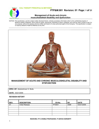
- 3. PHYSICAL THERAPY PRINCIPALS & METHODS PTP&M:001 Revision: 01 Page: 3 of 31 Management of Acute and chronic musculoskeletal disability and dysfunction NOTICE: This specification, and the subject matter disclosed therein, embody proprietary information which is the confidential property of Mullsons Health & Wellness, which shall be copied, reproduced, disclosed to others, published, and could be used in whole or part, for any purpose, without the express advance written permission of a duly authorized agent of the Company. This specification is subject to recall by Mullsons Health & Wellness at any time. 1.0 MUSCULOSKELETAL SYSTEM: The musculoskeletal system provides form, stability, and movement to the human body. It consists of the body's bones (which make up the skeleton), muscles, tendons, ligaments, joints, cartilage, and other connective tissue. The term "connective tissue" is used to describe the tissue that supports and binds tissues and organs together. Its chief components are elastic fibers and collagen, a protein substance. 1.1 FRONT VIEW & REAR VIEW: Medicine: it’s a noble profession, it serves humanity 3
- 4. PHYSICAL THERAPY PRINCIPALS & METHODS PTP&M:001 Revision: 01 Page: 4 of 31 Management of Acute and chronic musculoskeletal disability and dysfunction NOTICE: This specification, and the subject matter disclosed therein, embody proprietary information which is the confidential property of Mullsons Health & Wellness, which shall be copied, reproduced, disclosed to others, published, and could be used in whole or part, for any purpose, without the express advance written permission of a duly authorized agent of the Company. This specification is subject to recall by Mullsons Health & Wellness at any time. 1.2 MUSCULOSKELETAL BONES & MUSCLES: BONES BONES MUSCLES MUSCLES Maxilla Ulnar bones Semitendinosus Gluteus Maximus Skull Pelvis Trapezius External Oblique Mandible Femur Deltoids Biceps Femoris Spine Patella Pectoralis Rectus Femoris Scapulars Tibia Triceps Hamstrings Clavicle Fibula Biceps Rectus Abdominis Humerus Calcaneus Latissimus Dorsi Gastrocnemius Ribs Quadriceps Brachioradialis Sartorius Radius Tibialis Extensor digitorum longus Medicine: it’s a noble profession, it serves humanity 4
- 5. PHYSICAL THERAPY PRINCIPALS & METHODS PTP&M:001 Revision: 01 Page: 5 of 31 Management of Acute and chronic musculoskeletal disability and dysfunction NOTICE: This specification, and the subject matter disclosed therein, embody proprietary information which is the confidential property of Mullsons Health & Wellness, which shall be copied, reproduced, disclosed to others, published, and could be used in whole or part, for any purpose, without the express advance written permission of a duly authorized agent of the Company. This specification is subject to recall by Mullsons Health & Wellness at any time. Medicine: it’s a noble profession, it serves humanity 5
- 6. PHYSICAL THERAPY PRINCIPALS & METHODS PTP&M:001 Revision: 01 Page: 6 of 31 Management of Acute and chronic musculoskeletal disability and dysfunction NOTICE: This specification, and the subject matter disclosed therein, embody proprietary information which is the confidential property of Mullsons Health & Wellness, which shall be copied, reproduced, disclosed to others, published, and could be used in whole or part, for any purpose, without the express advance written permission of a duly authorized agent of the Company. This specification is subject to recall by Mullsons Health & Wellness at any time. 1.3 MUSCULOSKELETAL BONES: Bone, although strong, is a constantly changing tissue that has several functions. Bones serve as rigid structures to the body and as shields to protect delicate internal organs. They provide housing for the bone marrow, where the blood cells are formed. Bones also maintain the body's reservoir of calcium. In children, some bones have areas called growth plates. Bones lengthen in these areas until the child reaches full height, at which time the growth plates close. Thereafter, bones grow in thickness rather than in length, based on the body's need for additional bone strength in certain areas. Bones have two shapes: flat (such as the plates of the skull and the vertebrae) and tubular (such as the thighbones and arm bones, which are called long bones). All bones have essentially the same structure. The hard outer part (cortical bone) consists largely of proteins, such as collagen, and a substance called hydroxyapatite, which is composed mainly of calcium and other minerals. Hydroxyapatite is largely responsible for the strength and density of bones. The inner part of bones (trabeculae bone) is softer and less dense than the hard outer part. Bone marrow is the tissue that fills the spaces in the trabeculae bone. Bone marrow contains specialized cells (including stem cells) that produce blood cells. Blood vessels supply blood to the bone, and nerves surround the bone. Bones undergo a continuous process known as remodelling (see Osteoporosis). In this process, old bone tissue is gradually replaced by new bone tissue. Every bone in the body is completely reformed about every 10 years. To maintain bone density and strength, the body requires an adequate supply of calcium, other minerals, and vitamin D and must produce the proper amounts of several hormones, such as parathyroid hormone, growth hormone, Calcitonin, estrogens, and testosterone. Activity (for example, weight-bearing exercises for the legs) helps bones strengthen by remodelling. With activity and optimal amounts of hormones, vitamins, and minerals, trabeculae bone develops into a complex lattice structure that is lightweight but strong. A thin membrane called the periosteum covers bones. Injury to bone transmits pain because of nerves located mostly in the periosteum. Blood enters bones through blood vessels that enter through the periosteum. Did You Know. Bone structure adjusts throughout life in response to activity and stress (for example, weight- bearing exercise). Medicine: it’s a noble profession, it serves humanity 6
- 7. PHYSICAL THERAPY PRINCIPALS & METHODS PTP&M:001 Revision: 01 Page: 7 of 31 Management of Acute and chronic musculoskeletal disability and dysfunction NOTICE: This specification, and the subject matter disclosed therein, embody proprietary information which is the confidential property of Mullsons Health & Wellness, which shall be copied, reproduced, disclosed to others, published, and could be used in whole or part, for any purpose, without the express advance written permission of a duly authorized agent of the Company. This specification is subject to recall by Mullsons Health & Wellness at any time. 1.4 MUSCULOSKELETAL MUSCLES: There are three types of muscles: • Skeletal, • Smooth, • Cardiac (heart). Skeletal and Smooth—are part of the musculoskeletal system. Skeletal muscle is what most people think of as muscle, the type that can be contracted to move the various parts of the body. Skeletal muscles are bundles of contractile fibres that are organized in a regular pattern, so that under a microscope they appear as stripes (hence, they are also called striped or striated muscles). Skeletal muscles vary in their speeds of contraction. Skeletal muscles, which are responsible for posture and movement, are attached to bones and arranged in opposing groups around joints. For example, muscles that bend the elbow (biceps) are countered by muscles that straighten it (triceps). These countering movements are balanced. The balance makes movements smooth, which helps prevent damage to the musculoskeletal system. Skeletal muscles are controlled by the brain and are considered voluntary muscles because they operate with a person's awareness. The size and strength of skeletal muscles are maintained or increased by regular exercise. In addition, growth hormone and testosterone help muscles grow in childhood and maintain their size in adulthood. Medicine: it’s a noble profession, it serves humanity 7
- 8. PHYSICAL THERAPY PRINCIPALS & METHODS PTP&M:001 Revision: 01 Page: 8 of 31 Management of Acute and chronic musculoskeletal disability and dysfunction NOTICE: This specification, and the subject matter disclosed therein, embody proprietary information which is the confidential property of Mullsons Health & Wellness, which shall be copied, reproduced, disclosed to others, published, and could be used in whole or part, for any purpose, without the express advance written permission of a duly authorized agent of the Company. This specification is subject to recall by Mullsons Health & Wellness at any time. 2.0 MANAGEMENT OF COMMON MUSCULOSKELETAL DISORDERS: 2.1 GUIDELINES FOR THE INITIAL EVALUATION OF THE ADULT PATIENT WITH ACUTE MUSCULOSKELETAL SYMPTOMS: Approximately 1 of 7 patient visits to a primary care provider is prompted by musculoskeletal pain or dysfunction. While many or even most patients with these symptoms have benign, self- limited conditions, arthritis and chronic musculoskeletal disorders are leading causes of disability and work absenteeism, and are occasionally life-threatening. Determining whether the symptom is from local injury or inflammation, a mechanical problem, or a systemic illness will direct subsequent evaluation and management. A systematic approach starts with a careful history and physical examination (1-3). Inadequate history and physical examination commonly lead to inappropriate diagnostic testing and treatment. Diagnostic testing to reassure patients is generally unnecessary and test results may be abnormal in the absence of rheumatic disease. The following guidelines were prepared by an interdisciplinary group of primary care physicians who also practice rheumatology, rheumatologists, and allied health professionals. These guidelines provide a framework for the initial evaluation of the adult patient with acute (i.e., less than 6 weeks' duration) musculoskeletal pain who is seen in primary care settings and are recommended with the understanding that systematic follow up will be undertaken. 2.2 HISTORY: Musculoskeletal emergencies may present with acute symptoms. These conditions include infection (for example, septic arthritis, subacute bacterial endocarditis and sepsis, osteomyelitis, narcotising fasciitis), systemic vasculitis, acute myelopathy or spinal cord compression, fracture, deep vein thrombosis, and anterior compartment syndrome or tumor, and are suggested by the ``red flag'' signs or symptoms in Table 1. One should not miss these diagnoses because delayed recognition may lead to permanent disability or death. Once these have been excluded, an orderly evaluation will sort out the major diagnostic possibilities. Table 1. “Red flags” suggesting the need for urgent evaluation and management of the patient with musculoskeletal symptoms Feature Differential diagnosis History of significant trauma Soft tissue injury, internal derangement, or fracture Hot, swollen joint Infection, systemic rheumatic disease, gout, pseudogout Constitutional signs and symptoms (e.g., fever, weight loss, malaise) Infection, sepsis, systemic rheumatic disease Weakness Focal nerve lesion (compartment syndrome, entrapment neuropathy, Focal mononeuritis multiplex, motor neuron disease, radiculopathy*) Myositis, metabolic myopathy, paraneoplastic syndrome, degenerative Diffuse neuromuscular disorder, toxin, myelopathy,* transverse myelitis Neurogenic pain (burning, numbness, paresthesia) Asymmetric Radiculopathy,* reflex sympathetic dystrophy, entrapment neuropathy Symmetric Myelopathy,* peripheral neuropathy Peripheral vascular disease, giant cell arteritis (jaw pain), lumbar spinal Claudication pain pattern stenosis Medicine: it’s a noble profession, it serves humanity 8
- 9. PHYSICAL THERAPY PRINCIPALS & METHODS PTP&M:001 Revision: 01 Page: 9 of 31 Management of Acute and chronic musculoskeletal disability and dysfunction NOTICE: This specification, and the subject matter disclosed therein, embody proprietary information which is the confidential property of Mullsons Health & Wellness, which shall be copied, reproduced, disclosed to others, published, and could be used in whole or part, for any purpose, without the express advance written permission of a duly authorized agent of the Company. This specification is subject to recall by Mullsons Health & Wellness at any time. * Radiculopathy and myelopathy may be due to infectious, neoplastic, or mechanical processes. Details concerning the character of the pain, such as the location and the quality of the pain, its time of onset, and factors which make it worse or better, are important clues. Pain vaguely described as numbness, ``falling asleep,'' burning, shooting, or pins and needles is often due to a neurologic problem (neurogenic pain), especially when involving a dermatome, peripheral nerve, or stocking-glove distribution. Pain from arterial insufficiency is brought on by use and relieved promptly with rest (claudication pain pattern). In contradistinction, neurogenic claudication from lumbar spinal stenosis presents with leg pain when walking that improves slowly with sitting or spinal flexion. • Pain that originates from articular structures should be improved by resting the joint and made worse by moving it or by weight bearing. • Pain vaguely localized to a joint in which a careful examination cannot identify a specific structure of origin may be due to referred pain or a bone lesion. Bone lesions will often cause unrelenting pain at night. Patients with systemic rheumatic diseases, such as rheumatoid arthritis (RA) and systemic lupus erythematosus (SLE), may have prolonged morning stiffness in multiple symptomatic joints (4), less stiffness after using their joints, and may have constitutional symptoms (malaise, fever, weight loss) or signs or symptoms of multisystem involvement. These latter symptoms include fatigue, rash, diffuse adenopathy, alopecia, oral and nasal ulcers, pleuritic chest pain, Raynaud's phenomenon, and dry eyes and mouth (sicca) (Table 2). Table 2. Diagnostically useful clinical features in the initial evaluation of the patient with acute musculoskeletal symptoms Peak period of discomfort, Focal, periarticular, or trigger Non inflammatory joint problems*Over Non inflammatory joint Systemic rheumatic entire exposed joint spaces Tendonitis/bursitis problems* disease Symptoms am. stiffness Focal, brief Focal, brief Significant, prolonged Constitutional symptoms Absent Absent Present After prolonged Peak period of discomfort With use After prolonged use inactivity Unusual, except rotator cuff tear, Implies loose body, internal Locking or instability trigger finger derangement, or weakness Uncommon Symmetry Uncommon Occasional Common Signs Focal, periarticular, or tender points Over entire exposed Tenderness (fibromyalgia) Unusual joint spaces Inflammation (fluid, pain warmth, erythema) Over tendon or bursa Unusual Common Instability Uncommon Occasional Uncommon Multisystem disease No No Often * For example, osteoarthritis or internal derangement. Individuals with local mechanical problems (bursitis, tendinitis, sprains, and strains) and osteoarthritis (OA) typically have little morning stiffness, only one or a few symptomatic areas, no Medicine: it’s a noble profession, it serves humanity 9
- 10. PHYSICAL THERAPY PRINCIPALS & METHODS PTP&M:001 Revision: 01 Page: 10 of 31 Management of Acute and chronic musculoskeletal disability and dysfunction NOTICE: This specification, and the subject matter disclosed therein, embody proprietary information which is the confidential property of Mullsons Health & Wellness, which shall be copied, reproduced, disclosed to others, published, and could be used in whole or part, for any purpose, without the express advance written permission of a duly authorized agent of the Company. This specification is subject to recall by Mullsons Health & Wellness at any time. pain at rest, worsened symptoms with or after sustained activity, and no symptoms or signs of a systemic illness. The patient with a loss of function of an extremity may have weakness, loss of motion, instability, or pain. Weakness suggests a neurologic or muscle problem. Weakness with altered sensation suggests a nerve root or peripheral nerve abnormality. Loss of motion may be due to structural damage of the joint, contracture of surrounding soft tissues, or both. A symptom of ``giving way'' or ``locking'' of a joint without warning suggests a mechanical derangement due to ligamentous disruption or muscle weakness. If the symptom is associated with pain, an internal derangement (cartilage or ligament tear) or loose body within the joint are possibilities. Locking of the joint may also result from extraarticular soft tissue block, as in a trigger finger resulting from a flexor tendon sheath nodule. The patient's functional ability should be assessed by asking ``What is hard to do now that you could do before, and how does this affect your daily life?'' The use of a cane or a walker or assistive devices should be noted; the duration of their use provides helpful information for assessing progress. A vocational history can identify repetitive physical tasks which may need to be modified to aid recovery. 2.3 REVIEW OF SYSTEMS: Concomitant medical problems may have musculoskeletal manifestations or may affect treatment. Previous traumas, fractures, or surgical procedures of the symptomatic joint should be documented. Symptoms involving multiple organ systems may suggest that the joint symptoms are part of a systemic illness. It is important to obtain a complete list of the patient's medications; some may cause musculoskeletal symptoms, and previous experience with treatment may affect future treatment choices and compliance. The history should include visits to other physicians, adherence to previous treatments, and use of non-traditional remedies. Frequent use of medical services, a psychiatric disorder, or pending litigation about the condition should be noted. 2.4 PSYCHOLOGICAL AND SOCIAL FACTORS: Because musculoskeletal conditions can have a deleterious effect on the patient's work, home, and leisure activities, assessment of psychological and socio cultural factors is important in judging the strengths and resources the patient brings to coping with the condition. Functional changes may lead to depression, anxiety, and loss of confidence. Barriers to treatment adherence should be addressed to maximize treatment effects. Referral to community resources, such as the Arthritis Foundation's self-help programs, psychological and vocational counselling, and other community services may facilitate the patient's adjustment. 2.5 PHYSICAL EXAMINATION: Guided by the history, the physical examination helps to distinguish between mechanical problems, soft tissue disease, and non-inflammatory and inflammatory joint disease. A major goal of the examination is to detect warmth over a joint, joint effusion, and pain on joint motion. These are the hallmarks of synovitis. Limitations in range of motion and instability are also important to assess. The combination of point tenderness, reduced active range of motion, and preserved passive range of motion suggests soft tissue disorders, including bursitis, tendonitis, or muscle injury. Medicine: it’s a noble profession, it serves humanity 10
- 11. PHYSICAL THERAPY PRINCIPALS & METHODS PTP&M:001 Revision: 01 Page: 11 of 31 Management of Acute and chronic musculoskeletal disability and dysfunction NOTICE: This specification, and the subject matter disclosed therein, embody proprietary information which is the confidential property of Mullsons Health & Wellness, which shall be copied, reproduced, disclosed to others, published, and could be used in whole or part, for any purpose, without the express advance written permission of a duly authorized agent of the Company. This specification is subject to recall by Mullsons Health & Wellness at any time. Having the patient imitate the examiner as she/he moves a joint through its key motions tests active range of motion. If both active and passive range of motion is limited, soft tissue contracture, synovitis, or a structural abnormality of the joint is possibilities. Tendonitis may be suggested by tenderness to palpation along the course of the tendon, or pain or rub produced when the tendon is stretched or stressed during active range of motion against resistance. Inability to actively abduct the shoulder fully is strongly suggestive of a rotator cuff tear. Crepitus (joint noises or palpable grinding during joint motion) may be due to articular surface abnormalities or synovitis. Crepitus not associated with pain or limitation of motion is generally of no clinical significance. Soft tissue swelling may be due to an effusion in the joint, synovial thickening, or edema in the surrounding soft tissues. A bulge sign and patellar ballottement are useful signs of small and moderate effusions in the knee, respectively. The stability of a joint is of particular concern in knee and ankle pain. The medial and lateral collateral ligaments of the knee can be assessed by Valgus and varus stress of the joint. Excess laxity of the knee on anterior drawer test may indicate an anterior cruciate ligament tear. Extra articular findings such as oral/nasal ulcers, iritic, rash, nodules, pericardial or pulmonary rub, enlargement of liver, spleen, or lymph nodes, and neurologic abnormalities suggest a systemic disease (Table 2). 2.6 CLINICAL SYNDROMES: Some distinct symptom patterns are useful in sorting out musculoskeletal symptoms and suggesting the diagnostic possibilities. Monarthralgia or oligoarthralgia. Joint symptoms of one and up to a few joints may be due to trauma, infection, crystal-induced inflammation (gout, pseudogout), or primary inflammatory arthritis (including spondylarthropathies and atypical presentation of RA). In acute monarthritis, it is essential that infection of a joint be diagnosed or excluded, and this can only be done by joint aspiration and synovial fluid culture. Chronic monarticular symptoms with little or no effusion are usually from OA. Tendinitis and bursitis generally involve one joint region, and the physical examination is usually diagnostic. Common syndromes include de Quervain's tenosynovitis, olecranon bursitis, medial and lateral epicondylitis, bicipital and rotator cuff tendinitis, rotator cuff tear, trochanteric bursitis, patellar bursitis and prepatellar bursitis, anserine bursitis, plantar fasciitis, posterior tibial tendinitis, and Achilles tendinitis. Polyarthralgia or polyarthritis. A careful history and complete physical examination are essential to the evaluation of polyarthritis because the differential diagnosis is extensive (Figure 2). The presence of prolonged morning stiffness, systemic symptoms, Raynaud's phenomenon, rash, or sicca symptoms, and manifestations of other organ involvement suggest a systemic rheumatic disease. The specific evaluation is guided by the clinical manifestations and should screen organ systems which can be involved without overt signs, such as the lung, heart, liver, kidney, and bowel, for potential involvement. Precise diagnosis and effective management require close followup as well as consultation and are beyond the scope of this guideline. An algorithm, which outlines the minimum data that should be obtained, is presented in Figure 1. Medicine: it’s a noble profession, it serves humanity 11
- 12. PHYSICAL THERAPY PRINCIPALS & METHODS PTP&M:001 Revision: 01 Page: 12 of 31 Management of Acute and chronic musculoskeletal disability and dysfunction NOTICE: This specification, and the subject matter disclosed therein, embody proprietary information which is the confidential property of Mullsons Health & Wellness, which shall be copied, reproduced, disclosed to others, published, and could be used in whole or part, for any purpose, without the express advance written permission of a duly authorized agent of the Company. This specification is subject to recall by Mullsons Health & Wellness at any time. Figure 1. An initial approach to the patient with polyarticular joint symptoms. CBC = complete blood cell count; ESR = erythrocyte sedimentation rate; RF = rheumatoid factor; ANA = antinuclear antibodies. Arthralgia and/or myalgia without physical findings has an extensive differential diagnosis. Often, no definitive diagnosis is possible at the initial presentation. Common causes include fibromyalgia, viral infection, an overuse syndrome (tendon strain associated with repetitive motion injuries or muscle fatigue), a neuropathy (e.g., carpal tunnel syndrome), or hypothyroidism. Rare causes include metabolic bone disease (e.g., osteomalacia, hyperparathyroidism). If the history and physical examination do not provide a diagnosis, symptomatic management and reassessment over several weeks is more productive initially than is laboratory testing or diagnostic imaging. Myalgia is a common musculoskeletal symptom. This symptom may be secondary to a localized problem (trauma or overuse) or a systemic disorder (acute or chronic infection, toxic or metabolic disorders) or, less commonly, it may reflect a primary muscle disease. In otherwise healthy patients, the findings of normal strength and multiple tender points in characteristic locations (10) should raise the possibility of fibromyalgia. Proximal weakness and elevated creatine phosphokinase enzyme levels suggest inflammatory myopathy. A patient 50 years or older with myalgias of the shoulder and hip girdle and normal strength should be evaluated for polymyalgia rheumatica, including measurement of the erythrocyte sedimentation rate (ESR). Medicine: it’s a noble profession, it serves humanity 12
- 13. PHYSICAL THERAPY PRINCIPALS & METHODS PTP&M:001 Revision: 01 Page: 13 of 31 Management of Acute and chronic musculoskeletal disability and dysfunction NOTICE: This specification, and the subject matter disclosed therein, embody proprietary information which is the confidential property of Mullsons Health & Wellness, which shall be copied, reproduced, disclosed to others, published, and could be used in whole or part, for any purpose, without the express advance written permission of a duly authorized agent of the Company. This specification is subject to recall by Mullsons Health & Wellness at any time. 2.7 LABORATORY STUDIES: In the initial evaluation of acute joint symptoms, diagnostic testing for rheumatic disease should be undertaken only after a careful history and physical examination, and is unnecessary when a mechanical problem or extraarticular source is diagnosed. Laboratory testing for monitoring an established disease or for obtaining prognostic information once a disease has been established is not addressed here. The frequency of abnormalities increases with age for the ESR, uric acid, antinuclear antibody (ANA), rheumatoid factor (RF), and imaging studies, even in the absence of disease. The Westergren ESR is elevated in infection, inflammatory states, and malignancy and is not, by itself, diagnostic of a specific disease (13). Although the ESR is diagnostically non-specific, in the setting of polyarthralgia and an equivocal joint examination, an elevated ESR suggests that an inflammatory arthritis is more likely. The ESR is almost always markedly elevated and, therefore, diagnostically useful in patients with giant cell arthritis and polymyalgia rheumatica. Serum RF should be ordered when there is at least a moderate suspicion of RA: symmetric, small joint, polyarticular joint pain with inflammatory symptoms or signs. The utility of this test is limited when the likelihood of RF-associated disease (most notably, RA and Sjogren's syndrome) is low (17,20). Patients with other inflammatory conditions (e.g., SLE, subacute bacterial endocarditis, vasculitis, viral infection) may also be RF-positive. Twenty-five percent or more of patients with RA never have a positive RF. Therefore, the diagnosis should never be based solely on the results of RF testing. The higher the RF titer, the more likely a positive RF is related to RA. ANA tests should not be ordered in patients with focal problems (e.g., back pain, localized tendinitis) who do not have systemic symptoms. Nearly all patients with SLE show ANA positivity on human cell line substrates (HEp-2 cells), but positive test results without SLE are common when few manifestations of SLE are present. While a patient with a positive ANA with few or no compatible clinical features is unlikely to have SLE, the higher the titer, the more likely the result is related to SLE or other ANA-associated disease. A positive ANA can be further sub classified by the pattern and the specific autoantibody detected (anti-double-stranded DNA, anti-Ro, anti-La, anti-Scl-70, anti-RNP, anti-Sm, etc.) and can be useful in suggesting a specific rheumatic disease (24), but should not be ordered routinely. A variety of serologic and biochemical tests have been bundled into ``arthritis panels,'' which increases the frequency of finding positive results unrelated to rheumatic disease. This may confuse the situation and lead to unnecessary or inappropriate further testing or treatment; therefore, panels are not recommended. Definitive diagnosis of gout is based on the demonstration of monosodium urate crystals by polarized microscopy of synovial fluid. However, a compelling clinical presentation, such as recurrent, acute, self-limited podagra, may be sufficient. An elevated serum uric acid level without clinical evidence of gout adds no diagnostic information and requires no treatment. Measurement of uric acid may provide prognostic information but has limited diagnostic value in acute gout because high or normal levels may be found. Measurement of serum uric acid is most useful for monitoring the treatment of chronic hyperuricemia and gout. Routine blood or urine tests such as a complete blood cell count with differential counts, urinalysis, and tests of renal and liver function should be performed if a multisystem disease is suspected. For patients with arthralgias and abnormal liver enzyme levels, hepatitis serologies should be ordered. When weakness or muscle pain is present, creatine phosphokinase should be measured to investigate for myositis. Other tests such as HLA-B27, antineutrophil cytoplasmic antibody, Lyme or parvovirus serologies, myositis-specific antibodies (anti-Jo-1), and antiphospholipid antibodies are only useful when the clinical suspicion is high for a spondylarthropathy, Wegener's granulomatosis, Lyme or Medicine: it’s a noble profession, it serves humanity 13
- 14. PHYSICAL THERAPY PRINCIPALS & METHODS PTP&M:001 Revision: 01 Page: 14 of 31 Management of Acute and chronic musculoskeletal disability and dysfunction NOTICE: This specification, and the subject matter disclosed therein, embody proprietary information which is the confidential property of Mullsons Health & Wellness, which shall be copied, reproduced, disclosed to others, published, and could be used in whole or part, for any purpose, without the express advance written permission of a duly authorized agent of the Company. This specification is subject to recall by Mullsons Health & Wellness at any time. parvovirus infection, inflammatory myositis, or the antiphospholipid antibody syndrome, respectively. They should not be routinely ordered. Synovial fluid analysis is indicated in evaluating an acute monarthritis or in the febrile patient with established arthritis with an acute flare, to rule out septic arthritis. Inspection of fresh fluid, determination of the white cell and differential counts, culture with appropriate stains, and polarized light microscopy are the most useful tests. Noninflammatory fluids generally have fewer than 2,000 white blood cells/mm3, with less than 75% polymorphonuclear leukocytes. Someone competent in the technique must perform analysis of synovial fluid by polarized light microscopy promptly, since studies show considerable variation in laboratory accuracy. Any inflammatory fluid without an explanation, particularly when fever is present, should be assumed to be infected until proven otherwise by appropriate culture. 2.8 IMAGING STUDIES: Imaging studies are indicated when the examination cannot localize the anatomic structure that is causing symptoms, especially after significant trauma, when there is loss of joint function (e.g., unable to bear weight), when pain continues despite conservative management, when a fracture or bone infection is suspected, or when there is a history of malignancy. Plain radiographs will be unrevealing or unhelpful (and are therefore not indicated) for most patients with acute and new symptoms of RA, SLE, gout, mechanical back pain, or tendinitis/bursitis. Radiographs may confirm the diagnosis of OA and assess its severity, but normal findings on radiographs do not rule out the presence of OA. The earliest radiographic changes in RA are nonspecific and include soft tissue swelling and periarticular osteoporosis, but these features are often absent at the initial presentation. In established RA or longstanding gout, erosions may be diagnostic: marginal erosions in the former, the ``overhanging edge,'' indicating reparative changes, in the latter. For patients with typical acute mechanical low back pain, a plain radiograph adds little to management decisions. An anteroposterior radiograph of the pelvis is a more specific test than an HLA-B27 but may be negative early in patients with sacroiliitis due to a seronegative spondylarthropathy. Calcification of fibrocartilage is often found in calcium pyrophosphate deposition disease, but is frequently an asymptomatic finding in elderly patients. Repeat radiographs after 7-10 days are appropriate when a fracture is suspected despite an unrevealing initial evaluation, because callus formation or abnormal alignment may be evident. Repeated imaging in patients with established rheumatic disease may be useful in assessing structural damage. More specialized imaging such as MRI or radionuclide bone scanning is useful when specific disorders are suspected and the management would be altered according to the findings. An MRI may reveal the presence of a rotator cuff tear, spinal stenosis, avascular necrosis of bone, or mechanical derangement of the knee. A bone scan may be useful when osteomyelitis, stress fracture, or bony metastases are a concern. In general, MRI is better for assessing soft tissue and spinal cord elements, whereas nuclear medicine studies are best for assessing bone turnover. MRI and bone scanning are expensive, and the latter exposes the patient to significant radiation. In older patients, MRI of the shoulder and back commonly show rotator cuff degeneration and disc abnormalities, respectively; these may be incidental findings. These studies should be reserved for patients in whom specific disorders are suspected, when the diagnosis cannot be made in a less costly manner, and only after a thorough history and physical examination. Medicine: it’s a noble profession, it serves humanity 14
- 15. PHYSICAL THERAPY PRINCIPALS & METHODS PTP&M:001 Revision: 01 Page: 15 of 31 Management of Acute and chronic musculoskeletal disability and dysfunction NOTICE: This specification, and the subject matter disclosed therein, embody proprietary information which is the confidential property of Mullsons Health & Wellness, which shall be copied, reproduced, disclosed to others, published, and could be used in whole or part, for any purpose, without the express advance written permission of a duly authorized agent of the Company. This specification is subject to recall by Mullsons Health & Wellness at any time. 2.9 REFERRAL CRITERIA: To increase the likelihood of an optimal outcome, consultation is recommended in patients who have the following conditions: • Suspected septic arthritis • Undiagnosed multisystem or systemic rheumatic disease • Acute myelopathy or mononeuritis multiplex • Undiagnosed synovitis, in which arthrocentesis or synovial biopsy may be needed • Musculoskeletal pain undiagnosed after 6 weeks • Unexplained immunochemical test abnormalities suggestive of an underlying rheumatic disease • Musculoskeletal pain not adequately controlled with therapy • Musculoskeletal pain associated with severe or progressive loss of function or work productivity • Conditions for which treatment with steroids or immunosuppressive drugs is considered • Systemic rheumatic disease in a pregnant or postpartum patient • Dysfunction out of proportion to objective findings • Suspected acute tendon/muscle rupture • Acute internal derangement with severe pain, poor function, or instability • End-stage joint disease 2.10 CONCLUSION: The most useful information in evaluating musculoskeletal pain comes from the history and physical examination, with reassessment as necessary. When the diagnosis and proper management are obscure, selective ordering of tests and/or consultation may be the most cost- effective approach. Medicine: it’s a noble profession, it serves humanity 15
- 16. PHYSICAL THERAPY PRINCIPALS & METHODS PTP&M:001 Revision: 01 Page: 16 of 31 Management of Acute and chronic musculoskeletal disability and dysfunction NOTICE: This specification, and the subject matter disclosed therein, embody proprietary information which is the confidential property of Mullsons Health & Wellness, which shall be copied, reproduced, disclosed to others, published, and could be used in whole or part, for any purpose, without the express advance written permission of a duly authorized agent of the Company. This specification is subject to recall by Mullsons Health & Wellness at any time. 2.11 CHRONIC MUSCULOSKELETAL PAIN: Musculoskeletal disorders are the most common cause of chronic incapacity in industrialised countries. Fortunately, some of the most important advances in physiotherapy have been in the management of these problems, particularly chronic low back pain, osteoarthritis of the hip and knee, and rheumatoid arthritis. There is strong evidence from recent studies that simple interventions provided soon after onset of symptoms can prevent the development of chronic back pain. For example, reassuring patients about the self-limiting nature of most low back pain and advice to return to normal activity as soon as possible increases the rate of return to work for workers with low back pain. On the other hand, encouraging rest is probably harmful. Traditionally, the mainstays of physiotherapy management of musculoskeletal pain have been massage, manual therapy (that is, manipulation and joint mobilisation), electrotherapy (such as therapeutic ultrasound, short-wave diathermy, and low energy laser), and therapeutic exercise. Current evidence paints a mixed picture of the effects of these interventions. There has been little rigorous research into the effects of massage, so the clinical benefits of massage, if any, remain unsubstantiated. Manual therapy is more effective than placebo in relieving low back pain, but it is not clear if it is more effective than other physiotherapy treatments. Most electrotherapies probably have little more than placebo effects. The most positive findings come from recent studies of therapeutic exercise. Many trials and several systematic reviews have shown that exercise can produce clinically worthwhile reductions in the disability and handicap associated with chronic low back pain, osteoarthritis of the hip and knee, and rheumatoid arthritis. The exercise programmes most often shown to be effective in contemporary clinical trials differ from traditional exercise in two important ways. Firstly, formal exercise programmes are considered to be part of a more global process of "activity prescription" involving a structured return to normal home, work, and social activities. Secondly, the programmes are based on evidence from behavioural sciences as well as the biological sciences. Many exercise programmes now explicitly incorporate principles of cognitive-behavioural therapy. With this approach, patients are taught to exercise to quotas rather than as symptoms permit, specific rewards are provided when exercise quotas or activity goals are met, and pain behaviours are not rewarded by attention from the therapist. Medicine: it’s a noble profession, it serves humanity 16
- 17. PHYSICAL THERAPY PRINCIPALS & METHODS PTP&M:001 Revision: 01 Page: 17 of 31 Management of Acute and chronic musculoskeletal disability and dysfunction NOTICE: This specification, and the subject matter disclosed therein, embody proprietary information which is the confidential property of Mullsons Health & Wellness, which shall be copied, reproduced, disclosed to others, published, and could be used in whole or part, for any purpose, without the express advance written permission of a duly authorized agent of the Company. This specification is subject to recall by Mullsons Health & Wellness at any time. 2.12 URINARY INCONTINENCE: About a quarter of women experience involuntary loss of urine, and about 2.5% report this causes much bother or a great problem. The problem of urinary incontinence in women, including genuine stress urinary incontinence, can be treated effectively with training of the pelvic floor muscles. For example, training pelvic floor muscles with weighted vaginal cones (weights inserted into the vagina) substantially increases the probability of cure or improvement compared with no exercise. An important recent trial of the effects of training pelvic floor muscles in women with genuine stress urinary incontinence has shown that doing eight near maximal pelvic floor muscle contractions three times a day for six months produces large reductions in the risk of incontinence related problems with social life, sex life, and physical activity. In this sample (mean duration of symptoms 10 years), absolute reductions in risk of each of these problems exceeded 35%, implying that at least a third of women experience each of these benefits from exercise. It is not clear if training with weighted cones, biofeedback (electromyographic feedback of pelvic floor muscle activity) or electrical stimulation produces better outcomes than pelvic floor muscle training alone. Medicine: it’s a noble profession, it serves humanity 17
- 18. PHYSICAL THERAPY PRINCIPALS & METHODS PTP&M:001 Revision: 01 Page: 18 of 31 Management of Acute and chronic musculoskeletal disability and dysfunction NOTICE: This specification, and the subject matter disclosed therein, embody proprietary information which is the confidential property of Mullsons Health & Wellness, which shall be copied, reproduced, disclosed to others, published, and could be used in whole or part, for any purpose, without the express advance written permission of a duly authorized agent of the Company. This specification is subject to recall by Mullsons Health & Wellness at any time. 1.0 The Pelvic Floor Muscles The Three Lower Diaphragms of the Pelvic Floor. The most superficial muscles are shown in green, the muscles of the urogenital diaphragm are in yellow, and the puboccygeus is coloured red. Palpation of the Pubococcygeus for Tone and Function. The index finger introduced to about the second joint and moved about normally meets resistance in all directions (left). When the pubococcygeus is atrophic, the middle third of the vagina is roomy; the walls are thin and feel as though detached from the surrounding structures, particularly anteriorly and laterally (right). Normal patients can voluntarily contract the pubococcygeus firmly about the palpating finger. When atrophy has occurred, no such contractions can be elicited. Medicine: it’s a noble profession, it serves humanity 18
- 19. PHYSICAL THERAPY PRINCIPALS & METHODS PTP&M:001 Revision: 01 Page: 19 of 31 Management of Acute and chronic musculoskeletal disability and dysfunction NOTICE: This specification, and the subject matter disclosed therein, embody proprietary information which is the confidential property of Mullsons Health & Wellness, which shall be copied, reproduced, disclosed to others, published, and could be used in whole or part, for any purpose, without the express advance written permission of a duly authorized agent of the Company. This specification is subject to recall by Mullsons Health & Wellness at any time. 2.12.1 KEGEL EXERCISES FOR WOMEN: Firm pressure on the posterior Kegel exercises are designed to strengthen the pelvic floor muscles that segment of the puboccygeus (or the surround the openings of the urethra, vagina and rectum. levator ani plate) may produce an Often, seemingly simple body functions can be quite complex. One example of this is urination, which is controlled only if a series of physical and behavioral dominoes fall into place each time a person needs to urinate. The bladder has muscles that let it to expand to hold urine arriving from the kidneys. When the bladder is full, nerves signal the need to urinate. If there is no appropriate place to do so, the brain overrides this signal. The brain keeps closed the muscular valve, or sphincter around the urethra, which is the tube that carries urine away from the bladder to the outside of the body. Muscles within the bladder that help squeeze it must stay relaxed, too, to prevent urination. When conditions are right, these muscles tighten, the valve relaxes and urine starts to flow. Damage to the pelvic floor muscles that help support the uterus, bladder, urethra and rectum can allow those body parts to sag. Pelvic muscles are most often damaged or weakened by childbirth and aging. As the problem worsens, incontinence can occur. 2.12.1.1 PELVIC EXAMS CAN DETERMINE IF THERE IS: • A cystocoele, which is a weakened muscle between vagina and bladder • A rectocele, or a weakened muscle between vagina and rectum • A prolasped uterus • Urinary stress incontinence, which is the loss of urine when a person sneezes, coughs, or laughs Weight loss can reduce incontinence Kegel exercises can be used for the urinary stress incontinence condition. The in obese women exercises can also enhance sexual pleasure. After childbirth, the exercises promote healing of the perinium. The perineum is the area between the vagina and rectum that is sometimes is cut or torn during childbirth. Because weak pelvic floor muscles contribute to incontinence, exercises to strengthen these muscles may lessen or sometimes prevent incontinence. To do Kegel exercises, a woman squeezes or tightens the vaginal muscles normally used to stop urination. Some women have poor sensation in the lower pelvis. They may incorrectly contract their abdominal muscles instead of their pelvic floor muscles. To find the right muscles, a woman can try one or more of these approaches: • Sit on the toilet to urinate. As the stream of urine slows, try to stop it by contracting the pelvic floor muscles. • Ask a healthcare provider to help by inserting two fingers into the vagina during a pelvic exam. Squeeze down on the fingers. The provider can tell when the muscles are tightening properly. • Place two fingers inside the vagina. Squeeze down as if stopping the flow of urine. Pressure should then be felt on the two fingers. Repeat the chosen approach several times until the feel of squeezing and releasing the right muscles is familiar. Urinary reflux can result from multiple times of holding urine. Medicine: it’s a noble profession, it serves humanity 19
- 20. PHYSICAL THERAPY PRINCIPALS & METHODS PTP&M:001 Revision: 01 Page: 20 of 31 Management of Acute and chronic musculoskeletal disability and dysfunction NOTICE: This specification, and the subject matter disclosed therein, embody proprietary information which is the confidential property of Mullsons Health & Wellness, which shall be copied, reproduced, disclosed to others, published, and could be used in whole or part, for any purpose, without the express advance written permission of a duly authorized agent of the Company. This specification is subject to recall by Mullsons Health & Wellness at any time. 2.12.1.2 TO DO KEGEL EXERCISES: • Empty the bladder. • Tighten the pelvic muscles for 10 seconds. • Relax the muscles for 10 seconds. • Repeat this sequence a total of 20 times, at least 3 to 5 times a day. Do not overdo it, though. Tiring the muscles may make incontinence worse. • Do not contract abdominal, thigh, leg or buttocks muscles while doing the exercises. This makes them less effective. • Keep breathing during the exercises. Learn to relax while doing the exercises and concentrate on isolating the right muscles. A woman can ask a healthcare provider about using special weighted vaginal cones or balloons inside the vagina. Working to hold the weights inside can also help isolate and strengthen pelvic floor muscles. And if none of these steps work, a woman can seek further help from a healthcare provider. The provider may suggest electrical stimulation of the correct muscles if the muscles cannot be identified in any other way. Electrical stimulation can be done in the office by the healthcare provider or with a home unit. Electrical stimulation is usually performed daily, for 15 to 20 minutes. When Kegel exercises are done 150-200 times per day, 15 to 30 percent (15% to 30%) of women with stress incontinence report satisfactory improvement. Another 30 to 40 percent (30% to 40%) find that symptoms get somewhat better. After doing Kegel exercises regularly for at least six weeks, a woman can expect to see some improvement. Just as occasional sit-ups will not give a woman a flat stomach, occasional Kegel exercises will not cure incontinence. If incontinence does not get better with regular exercise, options such as weight loss, medicine or surgery may be necessary. Medicine: it’s a noble profession, it serves humanity 20
- 21. PHYSICAL THERAPY PRINCIPALS & METHODS PTP&M:001 Revision: 01 Page: 21 of 31 Management of Acute and chronic musculoskeletal disability and dysfunction NOTICE: This specification, and the subject matter disclosed therein, embody proprietary information which is the confidential property of Mullsons Health & Wellness, which shall be copied, reproduced, disclosed to others, published, and could be used in whole or part, for any purpose, without the express advance written permission of a duly authorized agent of the Company. This specification is subject to recall by Mullsons Health & Wellness at any time. 2.12.1.3 MORE EXERCISES: Step 1: Lie Flat After finding the position of pelvic floor muscles, you can start the Kegel exercise. Before you wake up in the morning, take a nap in the afternoon, or go to bed at night, lie flat, constrict your pelvic–muscle for 5 seconds, and then relax for 5 seconds. Repeat the constrict-relax exercise for 10 to 20 minutes. Step 2: Anytime When you are used to doing the constrict-relax exercise for the pelvic floor- muscles, you can do it at anytime. You can practice the pelvic floor muscle constriction whenever sitting, standing, walking, cleaning, waiting for a bus or watching TV. Step 3: Buttock-raising Constriction You can use this position to constrict the pelvic floor-muscles, and beautify the shape of your buttock as well. Lie flat, bend your legs as illustrated and open them slightly. Then you constrict the pelvic floor-muscles, raise your waist and buttock as illustrated. Then constrict those muscles for 5 seconds, lower your buttock slightly, and relax those muscles for 5 seconds. Repeat this exercise for 5 to 10 times. Step 4: Heaving objects Exercise When you carry heaving objects, your bladder receives a lot pressure from the abdomen. Therefore, when the pelvic floor muscles are weak, the annoying urine leakage occurs. You can try to practice the Kegel exercise by carrying a heaving object. First, you constrict the pelvic floor-muscles, bend your knees, straighten your back, slightly open your feet, and then carry the heaving object. When carrying it, you constrict those muscles more. If you can prevent urine leakage when carrying heaving objects by constricting the pelvic floor-muscles, you have learned how to constrict those muscles effectively. Step 5: Jumping Exercise When you are jumping or running, your bladder also receives a lot of pressure from the abdomen. Therefore, by fixed-point jumping, you can exercise pelvic floor muscles effectively. First, you constrict pelvic floor-muscles, start to jump, bend your knees, and then lay your feet flat on the ground. During the whole jumping process, you have to constrict those muscles (if urine leakage occurs, stop the jumping exercise If you can control urine leakage successfully in those 5 steps, try to do more Kegel exercises with different positions every day to prevent urine leakage completely. After doing the pelvic floor-muscle exercise for 6 consecutive weeks, you will feel the urine leakage condition has considerably improved. Immediately!). Medicine: it’s a noble profession, it serves humanity 21
- 22. PHYSICAL THERAPY PRINCIPALS & METHODS PTP&M:001 Revision: 01 Page: 22 of 31 Management of Acute and chronic musculoskeletal disability and dysfunction NOTICE: This specification, and the subject matter disclosed therein, embody proprietary information which is the confidential property of Mullsons Health & Wellness, which shall be copied, reproduced, disclosed to others, published, and could be used in whole or part, for any purpose, without the express advance written permission of a duly authorized agent of the Company. This specification is subject to recall by Mullsons Health & Wellness at any time. 2.12.2 KEGEL OR PELVIC FLOOR MUSCLE EXERCISES FOR MEN: Kegel or pelvic floor muscle exercises are to help strengthen weak muscles around the bladder. When these muscles are weak, urine can leak from the bladder. The Pelvic Floor is a ''hammock'' of muscles that supports the internal abdominal and pelvic organs. This is shown in the picture below. These muscles run in different directions and are different sizes. The job of these muscles is to support, lift, and control the muscles that close the urethra (tube that urine passes through). How to Exercise These Muscles: Exercise these muscles by squeezing and relaxing them. This takes effort and practice. 2.12.1.1 FINDING THE RIGHT MUSCLES: To make sure that you are exercising the right muscles, try starting and stopping your urine stream. This exercise will help you find the correct muscles. Repeat this exercise once a week to check whether or not you are using the right muscles. Do not tighten your buttock or thigh muscles when doing these exercises. Relax your stomach muscles as much as possible. When you are standing and squeeze your pelvic floor muscles, you should see your penis move slightly. 2.12.1.2 EXERCISE PROCEDURE: • Begin by squeezing the muscles for a count of four (4), then relax for a count of four (4). At first, you can only squeeze the muscles for 1-2 seconds, but as your muscles get stronger, you will be able to hold to the count of four (4). • Work up to repeating these exercises for five (5) minutes twice a day. • Remember to relax between each squeeze and just let the muscles go loose. Do not push down. 2.12.1.3 PLACE OF EXERCISE: Set aside time when the exercise can be done and must be done without interruption. Once the exercise has been done for a while, practice these exercises any time and anywhere. Remember: • Always squeeze your pelvic floor muscles when you: • Sit up from lying down • Stand from a sitting position • Lift something heavy • You can practice squeezing these muscles when you are watching TV, standing in line, or driving a car. Since these muscles are inside your body, people will not know you are doing exercises. It usually takes 6-12 weeks to see results. Do these exercises regularly. Medicine: it’s a noble profession, it serves humanity 22
- 23. PHYSICAL THERAPY PRINCIPALS & METHODS PTP&M:001 Revision: 01 Page: 23 of 31 Management of Acute and chronic musculoskeletal disability and dysfunction NOTICE: This specification, and the subject matter disclosed therein, embody proprietary information which is the confidential property of Mullsons Health & Wellness, which shall be copied, reproduced, disclosed to others, published, and could be used in whole or part, for any purpose, without the express advance written permission of a duly authorized agent of the Company. This specification is subject to recall by Mullsons Health & Wellness at any time. 2.12.1.4 PRECAUTIONS: • Some people exercise more than they should, hope that they would regain bladder control quicker. If you exercise too much or too soon, your bladder control may get worse for a while. Start slowing and increase the amount of exercise slowly. Follow the guidelines that your health care team has given you. • Be sure to breathe during the exercises. Holding your breath may put extra pressure on your pelvic muscles 2.13 MOVEMENT DYSFUNCTION RESULTING FROM STROKE: Stroke causes disabilities. Besides the paralysis or problems controlling movement; problems using or understanding language; problems with thinking and memory and emotional disturbance, there’s another functional loss that people don’t often mention: the sensory disturbance. Sensory dysfunction Stroke patients may lose the ability to feel touch, pain, temperature, or position. Sensory deficits may also hinder the ability to recognize objects that patients are holding and can even be severe enough to cause loss of recognition of one’s own limb. Recent studies have provided evidence of the widespread incidence of sensory dysfunction following stroke. The incidence of sensory deficits in stroke is high ranging from 50% to 74%. The importance of these findings lies in the association between sensory loss post-stroke and poorer outcomes in motor capacity, functional abilities, length of inpatient stay, and quality of life. Since literature suggests that clinicians can use information about patients’ sensory status to predict rehabilitation outcomes and select appropriate interventions, the accuracy of the sensory system assessment is extremely relevant. There are several measurement methods employed in the recovery of sensory disturbance, for instance, QST: “Quantitative sensory tests”, which are psychophysical in nature and the tests require cooperation from the patient. That means the patient must be cognitively competent, able to follow instructions and respond to the test stimuli. QST tests are not only an alternative or complementary study for the detection of sensory nerve abnormalities, but also techniques employed to measure the intensity of stimuli needed to produce specific sensory perceptions. QST systems are separable into devices that generate specific physical vibratory or thermal stimuli and those that deliver electrical impulses at specific frequencies. The objective is to test the sensory threshold as follows for instance: a thermode (thermal stimuli surface) contacts the skin and the subject is asked to report sensation of temperature change or heat pain. An alternative stimulation modality utilizes electrical stimuli of variable frequency and intensity to determine sensory thresholds. QST could contribute and has the potential to further contribute to research of sensory dysfunction. Apart from the loss of abilities, there’s another consequence that could not be ignored which seems to be quite on the contrary, however comes from the same origin. Some stroke patients experience pain, numbness or odd sensations of tingling or prickling in paralysed or weakened limbs, a condition known as paresthesia, a neurological skin disease. Medicine: it’s a noble profession, it serves humanity 23
- 24. PHYSICAL THERAPY PRINCIPALS & METHODS PTP&M:001 Revision: 01 Page: 24 of 31 Management of Acute and chronic musculoskeletal disability and dysfunction NOTICE: This specification, and the subject matter disclosed therein, embody proprietary information which is the confidential property of Mullsons Health & Wellness, which shall be copied, reproduced, disclosed to others, published, and could be used in whole or part, for any purpose, without the express advance written permission of a duly authorized agent of the Company. This specification is subject to recall by Mullsons Health & Wellness at any time. Stroke survivors frequently have a variety of chronic pain syndromes resulting from stroke-induced damage to the nervous system (neuropathic pain). Patients who have a seriously weakened or paralysed arm commonly experience moderate to severe pain that radiates outward from the shoulder. Most often, the pain results from a joint becoming immobilized due to lack of movement and the tendons and ligaments around the joint become fixed in one position. This is commonly called a “frozen” joint; “passive” movement at the joint in a paralysed limb is essential to prevent painful “freezing” and to allow easy movement if and when voluntary motor strength returns. In some stroke patients, pathways for sensation in the brain are damaged, causing the transmission of false signals that result in the sensation of pain in a limb or side of the body that has the sensory deficit. The loss of urinary continence is fairly common immediately after a stroke and often results from a combination of sensory and motor deficits. Stroke survivors may lose the ability to sense the need to urinate or the ability to control muscles of the bladder. Some may lack enough mobility to reach a toilet in time. Loss of bowel control or constipation may also occur. Permanent incontinence after a stroke is uncommon. But even a temporary loss of bowel or bladder control can be emotionally difficult for stroke survivors. Moreover, sensory disturbance means loss of sight, hearing or the ability to communicate clearly; the results can be the same: a sense of isolation and loss. This section lists organisations working to help people deal with these feelings and find practical ways to carry on with their lives, in spite of their disabilities. 2.13.1 PHYSIOTHERAPY FOR MOVEMENT DYSFUNCTIONAL PATIENTS RESULTING FROM STROKE: 2.13.1.1 AIMS OF TREATMENT: • Identify the patient’s level of postural stability and areas of inefficiency, that is, isolate the weak or ineffective components and facilitate dynamic posture and balance to access functional selective antigravity activity. • Modify the effort required by changing the task and/or the environment to allow for functional activity without triggering associated reactions • Facilitate changes in the hypersensitive response by augmenting lateral inhibition through specific handling. • Alter biomechanical changes in muscle through specific mobilisation of muscle to realign muscle fibres, increase circulation, augment axoplasmic flow and optimise muscle firing. (This is an active process) • Educate the patient with reference to his or her own functional movement and activity, whilst for example, minimising the need for compensatory activity. Medicine: it’s a noble profession, it serves humanity 24
- 25. PHYSICAL THERAPY PRINCIPALS & METHODS PTP&M:001 Revision: 01 Page: 25 of 31 Management of Acute and chronic musculoskeletal disability and dysfunction NOTICE: This specification, and the subject matter disclosed therein, embody proprietary information which is the confidential property of Mullsons Health & Wellness, which shall be copied, reproduced, disclosed to others, published, and could be used in whole or part, for any purpose, without the express advance written permission of a duly authorized agent of the Company. This specification is subject to recall by Mullsons Health & Wellness at any time. 2.13.1.2 OTHER CONSIDERATIONS: • The patient’s environment, with particular reference to seating, to support postural activity and maintenance of body schema. • Positioning in other postural sets and the length of time the patient is exposed to a position Handling by caretakers • The early use of botulinum toxin and other mechanisms such as medication and to maintain muscle length and viability. • Manipulation of afferent input can affect a change in the structural organisation of the nervous system, through spatial and temporal summation and the facilitation of pre and post-synaptic inhibition. In treatment, the challenge is to facilitate the patient to regain control over their aberrant motor behaviour, incorporating selective and appropriate strengthening techniques and channel the deviant recruitment of tone through appropriate therapy, thereby directing more appropriate plastic adaptation leading to more efficient movement. pre and post-synaptic inhibition BOTULINUM TOXIN Medicine: it’s a noble profession, it serves humanity 25