2. 338
SALEHI and REZAEIAN DOLOEI / Turk J Vet Anim Sci
gain when present in the feed at 1–3 ppm, especially in
swine. Cattle and poultry can tolerate much higher levels
of DON than pigs. The US Food and Drug Administration
has recommended that total feed levels of DON not exceed
5 ppm for cattle and chicken and 1 ppm for swine. DON
levels for human food should be less than 1 ppm (10).
Since there have been no studies regarding corn and
barley contamination with F. graminearum in industrial
dairy farms in Iran, and especially the northeast thus far,
this research was conducted as the first study to investigate
the moisture content and health of corn and barley grains
used in animal feed in terms of contamination with seed-
borne Fusarium spp. in different seasons and to detect
DON and NIV toxin-producing species by the PCR
method.
2. Materials and methods
2.1. Sampling
In this study, 128 samples including 64 corn and 64 barley
samples were collected from 16 industrial dairy farms
in Khorasan-Razavi Province in the northeast of Iran
during winter, spring, summer, and fall (16 samples in
each season). Considering the capacity of the warehouse,
sampling was randomly performed from the middle part
of the barn and the samples did not have apparent signs of
mold contamination. The collected samples were kept in
sterile plastic bags and were immediately transferred to the
laboratory at 4 °C.
2.2. Determination of moisture content of samples
To determine the moisture content, the samples were first
fully ground, and then 5 ± 0.1 g of the sample was weighed
in a dry glass plate and kept in an oven at 100 °C for 2 h to
completely dry out (11). For further assurance, moisture of
the samples was also simultaneously determined at 110 °C
using a hygrometer (PCE Instruments, UK).
2.3. Determination of mycoflora and identification of F.
graminearum
To determine the fungal contamination of samples, 10 g
of each ground sample was completely mixed in 90 mL of
diluent solution to prepare serial dilutions from 10–1
to 10–6
.
Each of the obtained dilutions was then cultured in potato
dextrose agar (PDA) medium and incubated at 25 °C for
5–7 days (12). Fungal flora of samples was characterized
and identified based on morphologic and microscopic
characteristics using the keys of Pitt and Hocking (13)
and Raper and Fennell (14). Grains samples were surface-
sterilized for 1 min with a 5% sodium hypochlorite
solution, rinsed twice with sterile distilled water, and dried
in a laminar flow cabinet. The grains were then incubated
in PDA at 28 °C in the dark for 7 days. All Fusarium spp.
isolates were subcultured on PDA and incubated at 25 °C
for 2–4 weeks (7). Isolates were also grown in carnation
leaf agar (CLA, Merck) according to Fisher et al. (15). The
morphology of macroconidia and chlamydospores was
assessed from cultures grown on CLA. F. graminearum
was identified according to Leslie and Summerell (7).
2.4. Molecular identification of F. graminearum isolates
and chemotype determination
F. graminearum isolates were also identified by PCR with
species-specific primers. To prepare the mycelial mass,
all the F. graminearum isolates were cultured in potato
dextrose broth (Merck) medium and incubated at 25
°C for 7 days. Fungal mycelium was then isolated with
Whatman filter paper and freeze-dried. Genomic DNA
of isolates was extracted with a commercial kit (Bioneer,
Korea). The primer pairs of ITS-F/ITS-R and Fg16N-F/R
for identification of Fusarium spp. and F. graminearum,
which amplified a fragment containing 431 and 280 bp,
respectively, were used (5,16).
To identify chemotypes of F. graminearum isolates,
Tri5-F/R-specific primers related to tracking the Tri5 gene
(17) and Tri13DONR/Tri13F and Tri13NIVF/Tri13R
primers related to the Tril3 gene (18) and responsible for
controlling DON and NIV production, which produced
fragments containing 380, 282, and 312 bp, respectively,
were used (Table 1). The thermal schedule of the thermal
cycler related to PCR reactions is presented in Table 2.
PCR products were electrophoresed by agarose gel (1.2%)
and were visualized under UV light of 260 nm in gel
documentation (Vilber Lourmat).
2.5. Statistical analysis
Data were analyzed using descriptive statistics as mean ±
SD. One-way analysis of variance (ANOVA) was used to
compare the fungal contamination at different seasons.
To evaluate differences between two seasons, we used the
Duncan test in post hoc multiple comparisons. In order to
compare contamination with different types of Fusarium
in barley and corn samples, the chi-square test was used,
which led to obtaining significant results. The significance
level of the tests was 5%.
3. Results
3.1. Determination of moisture content and fungal
contamination of samples
Mean moisture of the barley and corn samples in different
seasons is shown in Table 3. Mean moisture of the barley
samples in winter, spring, summer, and fall was 11.23 ±
1.21%, 9.45 ± 1.18%, 8.61 ± 1.17%, and 10.93 ± 1.51% and
that of corn samples was 13.7 ± 1.46%, 10.22 ± 1.21%,
9.12 ± 0.84 %, and 13.6 ± 1.36 %, respectively (Table 3).
The mean moisture of corn and barley samples in winter
and fall were statistically higher than that of the samples
collected in spring and summer (P < 0.05). There were no
differences between the mean moisture content of winter
and fall samples (P < 0.05).
3. 339
SALEHI and REZAEIAN DOLOEI / Turk J Vet Anim Sci
Results obtained from culturing of the samples are
presented in Table 4. The most common molds isolated
from corn samples were Penicillium spp. (90.62%),
Fusarium spp. (85.93%), Aspergillus spp. (82.81%), Mucor
(75%), and Rhizopus (51.56%). Among the studied barley
samples, the maximum level of contamination was related
to Penicillium spp. (89.06%), Aspergillus spp. (73.43%),
Mucor (68.75%), and Rhizopus (50%) (Table 4).
3.2. Determination of Fusarium spp. and F. graminearum
isolates
Out of a total of 128 samples of corn and barley, Fusarium
spp. were isolated from 55 (85.93%) and 17 (26.56%)
samples, respectively. All of the isolates (100%) that
were detected as Fusarium spp. using the culturing
method produced a fragment containing 431 bp in PCR
reaction with the ITS-F/R pair primer (Figure 1a). Of
the 72 Fusarium isolates, 8 (11.11%) were identified as F.
graminearum by Fg16N-F/R, which produced a fragment
containing 280 bp (Figure 1b).
3.3. Chemotype determination of F. graminearum
isolates
Chemotyping of F. graminearum isolates was done by
PCR assay using a set of Tri5-F/R-specific primers for the
presence of trichothecene-producing genes. The results
Table 1. Primers sequences used in this study.
Target species Primer destination Sequence (5’→3’) References Size (bp)
Fusarium spp.
ITS-F
ITS-R
AACTCCCAAACCCCTGTGAACATA
TTTAACGGCGTGGCCGC
16 431
F. graminearum
Fg16N-F
Fg16N-R
ACAGATGACAAGATTCAGGCACA
TTCTTTGACATCTGTTCAACCCA
5 280
DON-producing
F. graminearum
Tri13DONR
Tri13F
GCTAGATCGATTGTTGCATTGAG
CATCATGAGACTTGTKCRAGTTTGGG
18 282
NIV-producing
F. graminearum
Tri13NIVF
Tri13R
CCAAATCCGAAAACCGCAG
TTGAAAGCTCCAATGTCGTG
18 312
Toxigenic
Fusarium spp.
Tri5-F
Tri5-R
GGGATGCTGGATTGAGCAG
CATCACCTGAGGGTCCTTGT
17 380
Table 2. PCR conditions in this study.
Primer designation Initial denaturation Cycle Denaturation Annealing Extension Final extension
ITS F/R 94 °C, 5 min 35 94 °C, 60 s 58 °C, 60 s 72 °C, 60 s 72 °C, 10 min
Fg16N-F/R 94 °C, 5 min 35 94 °C, 60 s 60 °C, 30 s 72 °C, 60 s 72 °C, 5 min
Tri5-F/R 94 °C, 5 min 35 94 °C, 30s 60 °C, 45 s 72 °C, 30 s 72 °C, 7 min
Tri13DONR/Tri13F 94 °C, 5 min 35 94 °C, 30 s 58 °C, 45 s 60 °C, 30 s 72 °C, 7 min
Tri13NIVF/Tri13R 94 °C, 5 min 35 94 °C, 30 s 58 °C, 45 s 60 °C, 30 s 72 °C, 7 min
Table 3. Mean moisture content (%) of barley and corn samples.
Feedstuff No.
Mean ± SD (%)
Winter (n = 16) Spring (n = 16) Summer (n = 16) Fall (n = 16)
Barley 64 11.23a,
* ± 1.15 9.45b
± 1.12 8.61c
± 0.82 10.93a
± 1.07
Corn 64 13.71a
± 1.2 10.22b
± 1.19 9.12c
± 0.8 13.66a
± 1.15
*Values with different letters are significantly different (P < 0.05).
4. 340
SALEHI and REZAEIAN DOLOEI / Turk J Vet Anim Sci
showed that 5 out of 8 F. graminearum isolates were
positive for trichothecene-producing genes (Figure 2a).
Three and 2 out of 5 F. graminearum isolates were positive
for DON- and NIV-producing genes, respectively, using
Tri13DONR/Tri13F and Tri13NIVF/Tri13R primers
(Figure 2b and 2c).
4. Discussion
Corn and barley are considered the major components of
animal feed and might be infected with different types of
fungi during the harvesting, collecting, and warehousing
stages. Under optimal conditions, such as high
temperature (25–35 °C), moisture content (13%–16%),
Table 4. Frequency of different genera of fungi present in corn and barley samples used in animal feed.
Fungal genus
Corn samples (n = 64) Barley samples (n = 64)
Winter Spring Summer Fall Winter Spring Summer Fall
Aspergillus spp. 14 15 10 14 9 15 8 15
Fusarium spp. 16 13 13 13 2 3 4 8
Penicillium spp. 13 16 11 14 14 15 13 15
Cladosporium 1 8 2 1 5 7 6 10
Rhizopus 11 6 9 7 10 11 4 7
Mucor 13 9 12 14 9 8 13 14
Geotrichum 0 0 0 0 3 1 1 0
Alternaria 1 0 0 0 5 2 11 5
Monilinia 3 2 0 0 2 0 0 2
Syncephalastrum spp. 4 3 0 1 3 1 3 3
Nocardia 0 0 0 1 2 1 1 0
Yeast 6 5 10 10 9 12 6 5
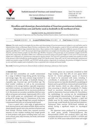
Figure 1. Agarose gel electrophoresis of species-specific PCR products. (a) M: 100-bp plus marker; Lane 1: Standard F. graminearum
isolate (positive control); Lane 2: negative control (without genomic DNA); Lane 3: Aspergillus flavus with ITS primers; Lane 4: A. niger
with ITS primers; Lanes 5–15: Fusarium spp. isolates. (b) M: 100-bp plus marker; Lane 1: standard F. graminearum isolate (positive
control); Lane 2: negative control (without genomic DNA); Lane 3: F. culmorum with Fg16N primers; Lane 4: F. oxysporum Fg16N
primers; Lane 5: F. proliferatum with Fg16N primers; Lanes 6–13: F. graminearum isolates.
5. 341
SALEHI and REZAEIAN DOLOEI / Turk J Vet Anim Sci
and water activity (0.70–0.90), toxigenic fungi can produce
mycotoxins. Mycotoxins are fungal secondary metabolites
produced by some fungi such as Aspergillus, Penicillium,
Fusarium, and Alternaria, which are detrimental to both
humans and animals (19). Trichothecenes are compounds
with a stable chemical structure and are considered one of
the most important food contaminant mycotoxins in the
world.
According to official Iranian standards, moisture
content of corn and barley is below 14% and 12%,
respectively (20,21). In the present study, the highest level
of moisture in the corn and barley samples was found in
winter and the lowest value was found in samples collected
in summer, which was in agreement with the study by
Jouany (22).
Mean total moisture content levels higher than 15% in
cereals are usually required for the growth of fungi (22).
In the present study there were a significant difference
between corn and barley samples collected in winter,
spring, and summer seasons separately (P < 0.05), but there
were no differences between winter and fall seasons (P <
0.05). According to Birzele et al. (23), DON concentrations
increase significantly in the presence of Fusarium spores if
moisture level exceeds 17%–20% at 20 °C.
Results obtained from culturing barley samples in
PDA medium demonstrated that the highest level of
contamination was related to Penicillium (89.06%) and
then Aspergillus (73.43%), both of which are capable of
producing toxins. Among the studied corn samples, the
highest level of contamination was related to Penicillium
(90.62%), Fusarium (85.93%), and Aspergillus (82.81%),
respectively, which can produce mycotoxins (Table 4).
Results of investigating fungal flora in corn and barley
samples in this study were in agreement with those of
Figure 2: (a) PCR products of trichothecene-producing F. graminearum. M: 100-bp plus marker; Lane 1: positive control (trichothecene-
producingF.graminearum);Lane2:negativecontrol(trichothecene-nonproducingF.graminearum);Lanes3–7:trichothecene-producing
F. graminearum isolates. (b) PCR products of DON-producing F. graminearum. M: 100-bp plus marker; Lane 1: negative control; Lane 2:
positive control; Lanes 3–5: DON-nonproducing F. graminearum isolates; Lanes 6–8: DON-producing F. graminearum isolates. (c) M:
100-bp plus marker; Lane 1: negative control; Lane 2: positive control; Lanes 3, 4, 6, 8–10: NIV-nonproducing F. graminearum; Lanes 5,
7: NIV-producing F. graminearum isolates.
6. 342
SALEHI and REZAEIAN DOLOEI / Turk J Vet Anim Sci
Roige et al. (24), Gerbaldo et al. (25), and Ghiasian and
Maghsood (26). Roige et al. (24) concluded that the highest
numbers of fungi infecting corn seed used in animal feed
in Argentina were Penicillium (70%), Fusarium (47%), and
Aspergillus (34%).
The study conducted by Ghiasian and Maghsood (26)
on samples of wheat, alfalfa, and barley used as animal
feed for 93 dairy farms in Hamadan Province in different
seasons demonstrated that the most widespread isolated
fungi were Aspergillus (37.4%), Penicillium (23.7%),
Fusarium (17.5%), Cladosporium (9.1%), Alternaria
(4.3%), Rhizopus (3.9%), and Mucor (3.4%), respectively.
According to the results obtained in this study,
Fusarium spp. was isolated in 55 (85.93%) of 64 corn
samples and 17 (26.56%) of 64 barley samples.
Fusarium contamination in grains can be produced
by different factors including physicochemical properties
of seeds, sensitivity to fungus, plant variety, and moisture
content of seeds in different phases of harvesting,
collecting, and warehousing. Corn is more sensitive to
various environmental damaging factors than barley, due
to being less thick; thus, it is more prone to fungal damage.
The results of this study also showed that corn samples
contained a higher level of moisture than barley samples,
which can be the cause of higher contamination rates with
seed-borne fungi such as Fusarium.
All isolates (100%) that were determined as Fusarium
spp. by culturing method were confirmed by PCR method
(Figure 1a). In order to determine the species of F.
graminearum, Fg16N-F/R-specific primers were applied
(27,28). Eight (11.11%) of 72 detected Fusarium isolates
were confirmed as F. graminearum (Figure 1b). Yoruk
and Albayrak (28) investigated 33 F. graminearum strains
isolated from Turkish wheat and barley farms with Fg16N-
F/R-specific primers. Twelve of 33 studied isolates were
confirmed as F. graminearum.
In this study, 5 of 8 F. graminearum isolates amplified
a 380-bp fragment using Tri5-F/R-specific primers and
were positive for trichothecene-producing genes (Figure
2a). In order to quickly detect Fusarium spp. with the
purpose of preventing the entrance of mycotoxins into the
food chain via agricultural products such as wheat, Amar
et al. (17) used a Tri5-specific primer, which targets the
route of trichothecene biosynthesis, to directly identify
toxin-producing F. culmorum. In the PCR assay, 3 of 5
trichothecene-producing F. graminearum isolates were
positive as DON producers (Figure 2b). Two out of 5 toxin-
producing F. graminearum isolates were also positive as
NIV producers, one of which was isolated from barley and
the other from the corn samples (Figure 2c). Qu et al. (29)
investigated the detection of DON- and NIV-producing
F. graminearum isolates in grain samples collected from
China, Nepal, the United States, and Europe based on
the PCR method. A Tri13 primer was used to amplify a
fragment with 583 bp, which indicated the presence of
a DON-producing gene, and a fragment with 644 bp,
which indicated the presence of a NIV-producing gene.
Results demonstrated that this method was faster and
more reliable than other detection methods in detecting
mycotoxins in Fusarium species and could control the
safety of food products. Using the PCR method, Astolfi
et al. (30) conducted a study on barley seeds collected
from some Brazilian farms to evaluate their capability in
producing 13-AcDON, 15-AcDON, and NIV mycotoxins
by F. graminearum. Results showed that 66%, 29.3%, and
4.4% of F. graminearum isolates were capable of producing
15-AcDON, NIV, and 13-AcDON, respectively.
In conclusion, this study showed a high level
contamination of corn and barley samples with toxigenic
fungi.Althoughthepresenceoftoxigenicfungiinfeedstuffs
does not certainly indicate that mycotoxins are naturally
occurring in the feed, it alerts us to the potential risk of
contamination. The presence of trichothecene-producing
strains of Fusarium in Iranian grain samples indicates the
possibility of large-scale contamination of the grains with
these toxins and thus the need for proper screening of food
and feed commodities for the detection of these toxins or
toxigenic fungi.
Acknowledgments
The authors wish to acknowledge financial support from
the Islamic Azad University Research Council of Mashhad
during this work. We would also like to thank Dr M Jabbari
Nooghabi for assistance with statistical analysis.
References
1. Nakai VK, Rocha LO, Goncalez E, Fonseca H, Ortega EMM,
Correa B. Distribution of fungi and aflatoxins in a stored
peanut variety. Food Chem 2008; 106: 285(290.
2. Rahimi P, Sharifnabi B, Bahar M. Detection of aflatoxin in
AspergillusspeciesisolatedfrompistachioinIran.JPhytopathol
2008; 156: 15(20.
3. Lacmanova I, Pazlarova J, Kostelanska M, Hajslova J. PCR
based identification of toxinogenic Fusarium species. Czech J
Food Sci 2009; 27: 90(94.
4. Sacchi C, Gonalez HHL, Broggi LE, Pacin A, Resnik SL, Cano
G, Taglieri D. Fungal contamination and mycotoxin natural
occurrence in oats for race horses feeding in Argentina. Anim
Feed Science Technol 2009; 152: 330(335.
7. 343
SALEHI and REZAEIAN DOLOEI / Turk J Vet Anim Sci
5. Nicholson P, Simpson DR, Weston G, Rezanoor HN, Lees AK,
Parry DW, Joyce D. Detection and quantification of Fusarium
culmorum and Fusarium graminearum in cereals using PCR
assays. Physiol Mol Plant Pathol 1998; 53: 17–37.
6. Desjardins A, Hohn TM. Mycotoxins in plant pathogenesis.
Mol Plant-Microbe Interact 1997; 10: 147–152.
7. Leslie JF, Summerell BA. The Fusarium Laboratory Manual.
Ames, IA, USA: Blackwell Publishing Professional; 2006.
8. Moss MO, Thrane U. Fusarium taxonomy with relation to
trichothecene formation. Toxicol Let 2004; 153: 23–28.
9. Kimura M, Takeshi T, Naoko TA, Shuichi O, Makoto F.
Molecular and genetic studies of Fusarium trichothecene
biosynthesis: pathways, genes, and evolution. Biosci Biotech
Bioch 2007; 71: 2105–2123.
10. Pleadin J, Zadravec M, Persi, N, Vulic A, Jaki V, Mitak M.
Mould and mycotoxin contamination of pig feed in northwest
Croatia. Mycotoxin Res 2012; 28: 157–162.
11. International Organization for Standardization. ISO 6540.
Maize. Determination of Moisture Content (on Milled Grains
and on Whole Grains). Geneva, Switzerland: International
Organization for Standardization; 1980.
12. International Organization for Standardization. ISO 21527-2.
Microbiology of Food and Animal Feeding Stuffs – Horizontal
Method for the Enumeration of Yeasts and Moulds – Part 1:
Colony Count Technique in Products with Water Activity
Less Than or Equal to 0.95. Geneva, Switzerland: International
Organization for Standardization; 2008.
13. Pitt JI, Hocking AD. Fungi and Food Spoilage. New York, NY,
USA: Springer USA; 1997.
14. Raper KB, Fennell DI. The Genus Aspergillus. Philadelphia, PA,
USA: Williams and Wilkins; 1965.
15. Fisher JH, Jansen R, Tai TA, Witt M, Lin WS, Pestka JJ.
Carnation leaves as a substrate and for preserving cultures of
Fusarium species. Phytopathology 1982; 72: 151–153.
16. Bluhm BH, Cousin MA, Woloshuk CP. Multiplex real-time
PCR detection of fumonisin-producing and trichothecene-
producing groups of Fusarium species. J Food Protec 2004; 67:
536–543.
17. Amar AB, Oueslati S, Ghorbel A, Mliki A. Prediction and early
detection of mycotoxigenic Fusarium culmorum in wheat by
direct PCR based procedure. Food Control 2012; 23: 506–510.
18. Chandler E, Simpson D, Thomsett M, Nichotson P.
Development of PCR assays to Tri7 and Tri13 trichothecene
biosynthetic genes and characterization of chemotypes of
Fusarium graminearum, Fusarium culmorum and Fusarium
cerealis, Physiol Mol Plant Pathol 2003; 62: 355–367.
19. Moss MO. Recent studies of mycotoxins. J Appl Microbiol
Symp Supp 1998; 84: 62S–76S.
20. Institute of Standards and Industrial Research of I.R. Iran.
ISIRI 47. Cereal and Cereal Products-Barley Specifications
and Test Methods. National Standard. Tehran, Iran: Institute of
Standards and Industrial Research of I.R. Iran; 2002.
21. Institute of Standard and Industrial Research of I.R. Iran. ISIRI
10690. Maize - Specifications and Test Methods. National
Standard. Tehran, Iran: Institute of Standards and Industrial
Research of I.R. Iran; 2008.
22. Jouany JP. Methods for preventing, decontaminating and
minimizing the toxicity of mycotoxins in feeds. Anim Feed Sci
Technol 2007; 137: 342–362.
23. Birzele B, Prange A, Kramer J. Deoxynivalenol and ochratoxin
A in German wheat and changes of level in relation to storage
parameters. Food Addit Contam 2000; 12: 1027–1035.
24. Roige MB, Aranguren SM, Riccio MB, Pereyra S, Soraci AL,
Tapia MO. Mycobiota and mycotoxins in fermented feed,
wheat grains and corn grains in Southeastern Buenos Aires
Province, Argentina. Rev Iberoam Micol 2009; 26: 233–237.
25. Gerbaldo GA, Pereyra CM, Cavaglieri LR, Ruiz F, Pascual
L, Dalcero AM, Barberis IL. Surveillance of aflatoxin and
microbiota related to brewer’s grain destined for swine feed in
Argentina. Vet Med Inter 2011; 2011: 912480.
26. Ghiasian SA, Maghsood AH. Occurrence of aflatoxigenic
fungi in cow feeds during the summer and winter season in
Hamadan, Iran. Afr J Microbiol Res 2011; 5: 516–521.
27. Prodi A, Tonti S, Nipoti P, Pancaldi D, Pisi A. Identification
of deoxynivalenol and nivalenol producing chemotypes
of Fusarium graminearum isolated from durum wheat in
restricted area of northern Italy. J Plant Pathol 2009; 91: 727–
731.
28. Yoruk E, Albayrak G. Chemotyping of Fusarium graminearum
and F. culmorum isolates from Turkey by PCR assay.
Mycopathologia 2012; 173: 53–61.
29. Qu B, Li HP, Zhang JB, Huang T, Carter J, Liao YC, Nicholson
P. Comparison of genetic diversity and pathogenicity of
Fusarium head blight pathogens from China and Europe by
SSCP and seedling assays on wheat. Plant Pathol 2008; 57:
642–651.
30. Astolfi P, dos Santos J, Schneider L, Gomes LB, Silva CN,
Tessmann DJ, Del Ponte EM. Molecular survey of trichothecene
genotypes of Fusarium graminearum species complex from
barley in Southern Brazil, Int J Food Microbiol 2009; 148: 197–
201.