SlideShare ist ein Scribd-Unternehmen logo
1 von 35
Radiological imaging
of ectopic pregnancy.
Dr/ ABD ALLAH NAZEER. MD.
Ectopic pregnancy is a major health problem for women
of childbearing age and a leading cause of pregnancy-
related death in the first trimester. Untreated, ectopic
pregnancy can lead to massive hemorrhage, infertility and
death. With the advent of high-resolution transvaginal
sonography, in conjunction with serum assays for the ß-
subunit of human chorionic gonadotropin (ß-hCG), rapid and
accurate diagnosis of this entity is now routinely possible.
Ectopic pregnancy is defined as implantation of a fertilized
ovum outside the endometrial lining of the uterus. Based on
data from the Centers for Disease
Control and Prevention, ectopic pregnancy has an incidence
of approximately 2% of all reported pregnancies
and accounts for 9% of pregnancy related deaths.
Locations of ectopic pregnancy. (A) ampullary/isthmic, (B) infundibulum, (C)
fimbria, (D) interstitial, (E) intra-abdominal, (F) ovarian and (G) cervical.
Etiology of ectopic pregnancy.
Congenital.
Tubal hypoplasia, Tubal turtuosity, Congenital diverticulae,
Accessory ostia, Partial stenosis, Elongation, Intra-mural
polyp and Entrap the ovum on its way.
Acquired.
Pelvic inflammatory disease.
Chlamydia trachomatis is most common.
Contraceptive failure.
Smoking.
Multiple sexual partners.
Endometriosis.
History of tubal surgery.
Endometriosis and leiomyoma.
Recent use of in vitro fertilization.
Magnetic Resonance Imaging.
MRI findings that can suggest an ectopic pregnancy
include the presence of a tubal gestational sac; a
tubal hematoma, which is a hematoma suggested by
the ring sign (peripheral hyperintensity) on T1-
weighted images; tubal wall enhancement; and an
adnexal mass with hemorrhagic fluid in the
peritoneum. Blood is suggested by the presence of
high-signal-intensity fluid on T1-weighted images.
The MRI characteristics of an ectopic pregnancy and
its rupture are seen in the T2-weighted images.
Live cervical ectopic pregnancy at 6 weeks gestational age. Sagittal view of
the cervix shows the gestational sac with an embryo (arrow) within the cervix.
Heterotopic pregnancy in an in vitro fertilization patient at 15 weeks gestational age. (a)
Transabdominal view shows no myometrium (arrows) around the ovarian ectopic pregnancy.
(b) Coronal T2-weighted magnetic resonance (MR) image IUP and ectopic pregnancy (EP). P =
placenta. MR was used for surgical planning to show the blood flow to the heterotopic
pregnancy. Note the flow void in vessels (arrows) supplying the ovarian ectopic pregnancy.
After surgical removal of the ectopic pregnancy, the IUP continued to term.
Tubal ring of ectopic pregnancy. (a) Transvaginal transverse view of the left adnexa shows
an echogenic ringlike mass (arrows) medial to the left ovary. Within the left ovary is a
thick-walled corpus luteum cyst (arrowheads). Note that the wall of the ectopic pregnancy
is more echogenic than the wall of the corpus luteum cyst. (b) Color Doppler image shows
more blood flow (arrowheads) to the corpus luteum than to the ectopic pregnancy (arrow).
Note that the “ring of fire” (hypervascular ring) appearance in the adnexa can be seen with
both ectopic pregnancy and corpus luteum.
Interstitial ectopic pregnancy 8 weeks after LMP. Sagittal transvaginal view
of the uterus (UT) demonstrates a thick-walled mass arising from the
interstitial portion of the uterus. The embryo is measured with calipers.
Ectopic pregnancy. Abdominal/pelvic CT. Image compatible with gestational sac in
the left adnexal region (arrow on A), separated from the uterine image (stars on A
and B) and from the ipsilateral ovary identified by visualization of the corpus
luteum (hollow arrow on B). Also, note the presence moderate amount of fluid in
the peritoneal cavity with foci of high density characterizing hematic content (L).
(A,B: contrast-enhanced axial sections in the portal phase).
Axial T2-weighted fast spin-echo magnetic resonance image of the pelvis.
This image shows an abnormal fluid-containing fallopian tube (red arrow)
on the right side. A simple right ovarian cyst (white arrow) is also present.
Ectopic pregnancy. Pelvic MRI. The presence of a gestational sac is observed in the
left adnexal region (arrows on A and B), in association with a heterogeneous mass
(arrow on C). Note the uterine and ovarian images (stars on A and D) separated
from the adnexal mass, as well as the presence of placenta showing contrast
enhancement (hollow arrow on D). (A: axial T2-weighted; B: coronal T2-weighted;
C: sagittal T2- weighted; D: post-contrast axial T1-weighted images).
Ectopic pregnancy. Pelvic MRI. Expansile, heterogeneous mass in the right adnexal
region (arrows on A to D), separated from the uterine (star on B) and ovarian
(hollow arrows on A and B) images. Also, note the presence of moderate amount of
free fluid in the pelvis, with intermediate signal intensity on T1-weighted images,
suggesting hematic contents (stars on A and C). (A,B: axial T2- weighted image with
fat saturation; C,D: axial T1-weighted, in phase and out-phase images).
Cornual ectopic pregnancy. Pelvic MRI. The presence of a heterogeneous mass is
observed in the cornual segment of the left uterine tube (white arrows on A to D).
Note the communication with the uterine cavity (white hollow arrows on B and C). The
myometrium is indicated by the black hollow arrow on C. (A,B: axial T1- weighted and
T2-weighted images, respectively; C: post-contrast, axial T1-weighted image with fat
saturation; D: coronal T2-weighted image).
Ectopic pregnancy with enhancing papillary solid components with a nonenhancing
necrotic portion and high signal intensity on outer surface on sagittal T1-weighted
turbo spin echo spectral presaturation with inversion recovery with contrast imaging
(A), on coronal T1-weighted fast field echo spectral presaturation with inversion
recovery with contrast imaging (B), on transverse T1-weighted fast field echo in-
phase with contrast imaging (C) (black arrows).
Tubal pregnancy in a 41-
year-old woman with acute
abdominal pain and
positive results of a
pregnancy test.
CONCLUSION.
Sonography plays a central role in the diagnosis and management
of ectopic pregnancy. If an extraovarian mass is present in a
pregnant patient with pain and bleeding, and no intrauterine
gestational sac is seen, the diagnosis of ectopic pregnancy should be
considered until proved otherwise. Therapy is determined by a
combination of clinical symptoms, sonographic findings, and serum
β-hCG values. While an intrauterine gestational sac is typically seen
when the β-hCG value is greater than 2000 mIU/mL (IRP), this value
should be used as a guideline and not an absolute threshold. Since
the fallopian tube is the most common location of ectopic
pregnancy, care should be taken while scanning to search between
the uterus and ovary for a tubal mass. Surgery is being performed
less often for ectopic pregnancy since the alternative treatments of
expectant management, methotrexate, and percutaneous injection
are now available. With these conservative treatments, there is an
increased role for sonography in patient follow-up.
Thank You.

Weitere ähnliche Inhalte

Was ist angesagt?

2nd trimester ultrasound..
2nd trimester ultrasound..2nd trimester ultrasound..
2nd trimester ultrasound..Soumitra Halder
 
Fetal biometry parameters lk
Fetal biometry parameters lkFetal biometry parameters lk
Fetal biometry parameters lkLALIT KARKI
 
First trimester ultrasound
First trimester ultrasoundFirst trimester ultrasound
First trimester ultrasoundRoshan Valentine
 
Ultrasound in Obstetric Emergencies by Dr Wannanee Meennuch
Ultrasound in Obstetric Emergencies by Dr Wannanee MeennuchUltrasound in Obstetric Emergencies by Dr Wannanee Meennuch
Ultrasound in Obstetric Emergencies by Dr Wannanee MeennuchRathachai Kaewlai
 
Ultrasonography of the ovary
Ultrasonography of the ovaryUltrasonography of the ovary
Ultrasonography of the ovaryAboubakr Elnashar
 
role of Imaging in female infertility
role of Imaging in  female infertilityrole of Imaging in  female infertility
role of Imaging in female infertilitycharusmita chaudhary
 
Presentation1.pptx, radilogical imaging of ovarian lesions.
Presentation1.pptx, radilogical imaging of ovarian lesions.Presentation1.pptx, radilogical imaging of ovarian lesions.
Presentation1.pptx, radilogical imaging of ovarian lesions.Abdellah Nazeer
 
Presentation11, radiological imaging of ovarian torsion.
Presentation11, radiological imaging of ovarian torsion.Presentation11, radiological imaging of ovarian torsion.
Presentation11, radiological imaging of ovarian torsion.Abdellah Nazeer
 
Obstetric Colour Doppler Study
Obstetric Colour Doppler Study Obstetric Colour Doppler Study
Obstetric Colour Doppler Study Sandeep Awal
 
Placenta ultrasound
Placenta ultrasoundPlacenta ultrasound
Placenta ultrasoundDoaa Gadalla
 
Obstetrics doppler ultrasound
Obstetrics doppler ultrasoundObstetrics doppler ultrasound
Obstetrics doppler ultrasoundBharti Gahtori
 
Doppler interpretation in pregnancy
Doppler interpretation in pregnancyDoppler interpretation in pregnancy
Doppler interpretation in pregnancyAboubakr Elnashar
 
Fetal anomaly scan
Fetal anomaly scanFetal anomaly scan
Fetal anomaly scanLALIT KARKI
 
3rd trimester us
3rd trimester us3rd trimester us
3rd trimester usmagdy abdel
 
USG AND DOPPLER IN DIAGNOSIS AND MANAGEMENT OF IUGR
USG AND DOPPLER IN DIAGNOSIS AND MANAGEMENT OF IUGRUSG AND DOPPLER IN DIAGNOSIS AND MANAGEMENT OF IUGR
USG AND DOPPLER IN DIAGNOSIS AND MANAGEMENT OF IUGRshiv lasune
 
Ectopic pregnancy
Ectopic pregnancyEctopic pregnancy
Ectopic pregnancyDurre Sabih
 

Was ist angesagt? (20)

2nd trimester ultrasound..
2nd trimester ultrasound..2nd trimester ultrasound..
2nd trimester ultrasound..
 
Doppler in pregnancy
Doppler in pregnancyDoppler in pregnancy
Doppler in pregnancy
 
Fetal biometry parameters lk
Fetal biometry parameters lkFetal biometry parameters lk
Fetal biometry parameters lk
 
First trimester ultrasound
First trimester ultrasoundFirst trimester ultrasound
First trimester ultrasound
 
Ultrasound in Obstetric Emergencies by Dr Wannanee Meennuch
Ultrasound in Obstetric Emergencies by Dr Wannanee MeennuchUltrasound in Obstetric Emergencies by Dr Wannanee Meennuch
Ultrasound in Obstetric Emergencies by Dr Wannanee Meennuch
 
Imaging of placenta
Imaging of placentaImaging of placenta
Imaging of placenta
 
Ultrasonography of the ovary
Ultrasonography of the ovaryUltrasonography of the ovary
Ultrasonography of the ovary
 
role of Imaging in female infertility
role of Imaging in  female infertilityrole of Imaging in  female infertility
role of Imaging in female infertility
 
Presentation1.pptx, radilogical imaging of ovarian lesions.
Presentation1.pptx, radilogical imaging of ovarian lesions.Presentation1.pptx, radilogical imaging of ovarian lesions.
Presentation1.pptx, radilogical imaging of ovarian lesions.
 
Presentation11, radiological imaging of ovarian torsion.
Presentation11, radiological imaging of ovarian torsion.Presentation11, radiological imaging of ovarian torsion.
Presentation11, radiological imaging of ovarian torsion.
 
Obstetric Colour Doppler Study
Obstetric Colour Doppler Study Obstetric Colour Doppler Study
Obstetric Colour Doppler Study
 
Placenta ultrasound
Placenta ultrasoundPlacenta ultrasound
Placenta ultrasound
 
Obstetrics doppler ultrasound
Obstetrics doppler ultrasoundObstetrics doppler ultrasound
Obstetrics doppler ultrasound
 
Doppler interpretation in pregnancy
Doppler interpretation in pregnancyDoppler interpretation in pregnancy
Doppler interpretation in pregnancy
 
Fetal anomaly scan
Fetal anomaly scanFetal anomaly scan
Fetal anomaly scan
 
Doppler in pregnancy
Doppler in pregnancyDoppler in pregnancy
Doppler in pregnancy
 
3rd trimester us
3rd trimester us3rd trimester us
3rd trimester us
 
USG AND DOPPLER IN DIAGNOSIS AND MANAGEMENT OF IUGR
USG AND DOPPLER IN DIAGNOSIS AND MANAGEMENT OF IUGRUSG AND DOPPLER IN DIAGNOSIS AND MANAGEMENT OF IUGR
USG AND DOPPLER IN DIAGNOSIS AND MANAGEMENT OF IUGR
 
Ectopic pregnancy
Ectopic pregnancyEctopic pregnancy
Ectopic pregnancy
 
Ultrasound evaluation of fetal thorax
Ultrasound evaluation of fetal thoraxUltrasound evaluation of fetal thorax
Ultrasound evaluation of fetal thorax
 

Andere mochten auch (9)

USS in Ectopic Pregnancy
USS in Ectopic PregnancyUSS in Ectopic Pregnancy
USS in Ectopic Pregnancy
 
Ultra sound imaging general presentation
Ultra sound imaging general presentationUltra sound imaging general presentation
Ultra sound imaging general presentation
 
Talk on obstetric emergencies dr.pradeep
Talk on obstetric emergencies dr.pradeepTalk on obstetric emergencies dr.pradeep
Talk on obstetric emergencies dr.pradeep
 
Normal early pregnancy imaging
Normal early pregnancy  imagingNormal early pregnancy  imaging
Normal early pregnancy imaging
 
Ectopic Pregnancy
Ectopic PregnancyEctopic Pregnancy
Ectopic Pregnancy
 
Gynecological causes of acute abdominal pain
Gynecological causes of  acute abdominal painGynecological causes of  acute abdominal pain
Gynecological causes of acute abdominal pain
 
Cervical pregnancy
Cervical pregnancyCervical pregnancy
Cervical pregnancy
 
ASER 2009
ASER 2009ASER 2009
ASER 2009
 
Ectopic pregnancy
Ectopic pregnancyEctopic pregnancy
Ectopic pregnancy
 

Ähnlich wie Presentation1.pptx, radiological imaging of ectopic pregancy.

Imaging abdomen trauma uterine trauma part 11 Dr Ahmed Esawy
Imaging abdomen trauma  uterine trauma part 11 Dr Ahmed EsawyImaging abdomen trauma  uterine trauma part 11 Dr Ahmed Esawy
Imaging abdomen trauma uterine trauma part 11 Dr Ahmed EsawyAHMED ESAWY
 
Ectopic Pregnancy in the Rudimentary Horn of a Unicornuate Uterus: The Trila...
Ectopic Pregnancy in the Rudimentary Horn of a Unicornuate Uterus: The Trila...Ectopic Pregnancy in the Rudimentary Horn of a Unicornuate Uterus: The Trila...
Ectopic Pregnancy in the Rudimentary Horn of a Unicornuate Uterus: The Trila...WCER 2021
 
Presentation1, radiological imaging of undescended testis.
Presentation1, radiological imaging of undescended testis.Presentation1, radiological imaging of undescended testis.
Presentation1, radiological imaging of undescended testis.Abdellah Nazeer
 
23205065
2320506523205065
23205065radgirl
 
New microsoft office power point presentation
New microsoft office power point presentationNew microsoft office power point presentation
New microsoft office power point presentationRiyadhWaheed
 
first trimester ultrasound and overview of obs imaging
first trimester ultrasound and overview of obs imagingfirst trimester ultrasound and overview of obs imaging
first trimester ultrasound and overview of obs imagingBharat Jain
 
first trimester ultrasound and overview of obs imaging
first trimester ultrasound and overview of obs imagingfirst trimester ultrasound and overview of obs imaging
first trimester ultrasound and overview of obs imagingBharat Jain
 
Imaging of the placenta
Imaging of the placentaImaging of the placenta
Imaging of the placentathambimbbs
 
Paget’s Disease of Breast (PDB)
Paget’s Disease of Breast (PDB)Paget’s Disease of Breast (PDB)
Paget’s Disease of Breast (PDB)Mohammed Fathy
 
Presentation1, radiological imaging of internal abdominal hernia.
Presentation1, radiological imaging of internal abdominal hernia.Presentation1, radiological imaging of internal abdominal hernia.
Presentation1, radiological imaging of internal abdominal hernia.Abdellah Nazeer
 
ectopic pregnancy.pptx
ectopic pregnancy.pptxectopic pregnancy.pptx
ectopic pregnancy.pptxrochisha
 
Anorectal malformations
Anorectal malformationsAnorectal malformations
Anorectal malformationsDr. Mohit Goel
 
Sonographic Evaluation of Pelvic Masses
Sonographic Evaluation of Pelvic MassesSonographic Evaluation of Pelvic Masses
Sonographic Evaluation of Pelvic MassesAboubakr Elnashar
 
A Case of Abdominal Pregnancy, Primary vs Secondary – Radiological Workup
A Case of Abdominal Pregnancy, Primary vs Secondary – Radiological WorkupA Case of Abdominal Pregnancy, Primary vs Secondary – Radiological Workup
A Case of Abdominal Pregnancy, Primary vs Secondary – Radiological Workupiosrjce
 
23205062
2320506223205062
23205062radgirl
 
OBSTETRICS EMERGENCIES
OBSTETRICS EMERGENCIESOBSTETRICS EMERGENCIES
OBSTETRICS EMERGENCIESLohith Varma
 
A case report of cervical ectopic pregnancy misdiagnosed as cervical miscarriage
A case report of cervical ectopic pregnancy misdiagnosed as cervical miscarriageA case report of cervical ectopic pregnancy misdiagnosed as cervical miscarriage
A case report of cervical ectopic pregnancy misdiagnosed as cervical miscarriageApollo Hospitals
 
Artigo selecionado 3 vasos e sling ap
Artigo selecionado 3 vasos e sling apArtigo selecionado 3 vasos e sling ap
Artigo selecionado 3 vasos e sling apgisa_legal
 

Ähnlich wie Presentation1.pptx, radiological imaging of ectopic pregancy. (20)

Imaging abdomen trauma uterine trauma part 11 Dr Ahmed Esawy
Imaging abdomen trauma  uterine trauma part 11 Dr Ahmed EsawyImaging abdomen trauma  uterine trauma part 11 Dr Ahmed Esawy
Imaging abdomen trauma uterine trauma part 11 Dr Ahmed Esawy
 
Ectopic Pregnancy in the Rudimentary Horn of a Unicornuate Uterus: The Trila...
Ectopic Pregnancy in the Rudimentary Horn of a Unicornuate Uterus: The Trila...Ectopic Pregnancy in the Rudimentary Horn of a Unicornuate Uterus: The Trila...
Ectopic Pregnancy in the Rudimentary Horn of a Unicornuate Uterus: The Trila...
 
Presentation1, radiological imaging of undescended testis.
Presentation1, radiological imaging of undescended testis.Presentation1, radiological imaging of undescended testis.
Presentation1, radiological imaging of undescended testis.
 
23205065
2320506523205065
23205065
 
New microsoft office power point presentation
New microsoft office power point presentationNew microsoft office power point presentation
New microsoft office power point presentation
 
first trimester ultrasound and overview of obs imaging
first trimester ultrasound and overview of obs imagingfirst trimester ultrasound and overview of obs imaging
first trimester ultrasound and overview of obs imaging
 
first trimester ultrasound and overview of obs imaging
first trimester ultrasound and overview of obs imagingfirst trimester ultrasound and overview of obs imaging
first trimester ultrasound and overview of obs imaging
 
Imaging of the placenta
Imaging of the placentaImaging of the placenta
Imaging of the placenta
 
Paget’s Disease of Breast (PDB)
Paget’s Disease of Breast (PDB)Paget’s Disease of Breast (PDB)
Paget’s Disease of Breast (PDB)
 
Presentation1, radiological imaging of internal abdominal hernia.
Presentation1, radiological imaging of internal abdominal hernia.Presentation1, radiological imaging of internal abdominal hernia.
Presentation1, radiological imaging of internal abdominal hernia.
 
ectopic pregnancy.pptx
ectopic pregnancy.pptxectopic pregnancy.pptx
ectopic pregnancy.pptx
 
Anorectal malformations
Anorectal malformationsAnorectal malformations
Anorectal malformations
 
anal canal.pptx
anal canal.pptxanal canal.pptx
anal canal.pptx
 
Sonographic Evaluation of Pelvic Masses
Sonographic Evaluation of Pelvic MassesSonographic Evaluation of Pelvic Masses
Sonographic Evaluation of Pelvic Masses
 
A Case of Abdominal Pregnancy, Primary vs Secondary – Radiological Workup
A Case of Abdominal Pregnancy, Primary vs Secondary – Radiological WorkupA Case of Abdominal Pregnancy, Primary vs Secondary – Radiological Workup
A Case of Abdominal Pregnancy, Primary vs Secondary – Radiological Workup
 
23205062
2320506223205062
23205062
 
OBSTETRICS EMERGENCIES
OBSTETRICS EMERGENCIESOBSTETRICS EMERGENCIES
OBSTETRICS EMERGENCIES
 
A case report of cervical ectopic pregnancy misdiagnosed as cervical miscarriage
A case report of cervical ectopic pregnancy misdiagnosed as cervical miscarriageA case report of cervical ectopic pregnancy misdiagnosed as cervical miscarriage
A case report of cervical ectopic pregnancy misdiagnosed as cervical miscarriage
 
A case report of cervical ectopic pregnancy misdiagnosed as cervical miscarriage
A case report of cervical ectopic pregnancy misdiagnosed as cervical miscarriageA case report of cervical ectopic pregnancy misdiagnosed as cervical miscarriage
A case report of cervical ectopic pregnancy misdiagnosed as cervical miscarriage
 
Artigo selecionado 3 vasos e sling ap
Artigo selecionado 3 vasos e sling apArtigo selecionado 3 vasos e sling ap
Artigo selecionado 3 vasos e sling ap
 

Mehr von Abdellah Nazeer

Muculoskeletal Pediatic Imaging..pptx
Muculoskeletal Pediatic Imaging..pptxMuculoskeletal Pediatic Imaging..pptx
Muculoskeletal Pediatic Imaging..pptxAbdellah Nazeer
 
Presentation1, Ultrasound of the bowel loops and the lymph nodes..pptx
Presentation1, Ultrasound of the bowel loops and the lymph nodes..pptxPresentation1, Ultrasound of the bowel loops and the lymph nodes..pptx
Presentation1, Ultrasound of the bowel loops and the lymph nodes..pptxAbdellah Nazeer
 
Presentation1 Short cases MD..pptx
Presentation1 Short cases MD..pptxPresentation1 Short cases MD..pptx
Presentation1 Short cases MD..pptxAbdellah Nazeer
 
Presentation1, MD MCQ Cases..pptx
Presentation1, MD MCQ Cases..pptxPresentation1, MD MCQ Cases..pptx
Presentation1, MD MCQ Cases..pptxAbdellah Nazeer
 
Presentation1, Short Cases Quiz..pptx
Presentation1, Short Cases Quiz..pptxPresentation1, Short Cases Quiz..pptx
Presentation1, Short Cases Quiz..pptxAbdellah Nazeer
 
Presentation1, radiological imaging of lateral hindfoot impingement.
Presentation1, radiological imaging of lateral hindfoot impingement.Presentation1, radiological imaging of lateral hindfoot impingement.
Presentation1, radiological imaging of lateral hindfoot impingement.Abdellah Nazeer
 
Presentation2, radiological anatomy of the liver and spleen.
Presentation2, radiological anatomy of the liver and spleen.Presentation2, radiological anatomy of the liver and spleen.
Presentation2, radiological anatomy of the liver and spleen.Abdellah Nazeer
 
Presentation1, artifacts and pitfalls of the wrist and elbow joints.
Presentation1, artifacts and pitfalls of the wrist and elbow joints.Presentation1, artifacts and pitfalls of the wrist and elbow joints.
Presentation1, artifacts and pitfalls of the wrist and elbow joints.Abdellah Nazeer
 
Presentation1, artifact and pitfalls of the knee, hip and ankle joints.
Presentation1, artifact and pitfalls of the knee, hip and ankle joints.Presentation1, artifact and pitfalls of the knee, hip and ankle joints.
Presentation1, artifact and pitfalls of the knee, hip and ankle joints.Abdellah Nazeer
 
Presentation1, radiological imaging of artifact and pitfalls in shoulder join...
Presentation1, radiological imaging of artifact and pitfalls in shoulder join...Presentation1, radiological imaging of artifact and pitfalls in shoulder join...
Presentation1, radiological imaging of artifact and pitfalls in shoulder join...Abdellah Nazeer
 
Presentation1, musculoskeletal anatomy.
Presentation1, musculoskeletal anatomy.Presentation1, musculoskeletal anatomy.
Presentation1, musculoskeletal anatomy.Abdellah Nazeer
 
Presentation1, new mri techniques in the diagnosis and monitoring of multiple...
Presentation1, new mri techniques in the diagnosis and monitoring of multiple...Presentation1, new mri techniques in the diagnosis and monitoring of multiple...
Presentation1, new mri techniques in the diagnosis and monitoring of multiple...Abdellah Nazeer
 
Presentation1, radiological application of diffusion weighted mri in neck mas...
Presentation1, radiological application of diffusion weighted mri in neck mas...Presentation1, radiological application of diffusion weighted mri in neck mas...
Presentation1, radiological application of diffusion weighted mri in neck mas...Abdellah Nazeer
 
Presentation1, radiological application of diffusion weighted images in breas...
Presentation1, radiological application of diffusion weighted images in breas...Presentation1, radiological application of diffusion weighted images in breas...
Presentation1, radiological application of diffusion weighted images in breas...Abdellah Nazeer
 
Presentation1, radiological application of diffusion weighted images in abdom...
Presentation1, radiological application of diffusion weighted images in abdom...Presentation1, radiological application of diffusion weighted images in abdom...
Presentation1, radiological application of diffusion weighted images in abdom...Abdellah Nazeer
 
Presentation1, radiological application of diffusion weighted imges in neuror...
Presentation1, radiological application of diffusion weighted imges in neuror...Presentation1, radiological application of diffusion weighted imges in neuror...
Presentation1, radiological application of diffusion weighted imges in neuror...Abdellah Nazeer
 
Presentation1, mr physics.
Presentation1, mr physics.Presentation1, mr physics.
Presentation1, mr physics.Abdellah Nazeer
 
Presentation1. ct physics.
Presentation1. ct physics.Presentation1. ct physics.
Presentation1. ct physics.Abdellah Nazeer
 
Presentation1, ultrasound physics.
Presentation1, ultrasound physics.Presentation1, ultrasound physics.
Presentation1, ultrasound physics.Abdellah Nazeer
 
Presentation4, x ray physics
Presentation4, x ray physicsPresentation4, x ray physics
Presentation4, x ray physicsAbdellah Nazeer
 

Mehr von Abdellah Nazeer (20)

Muculoskeletal Pediatic Imaging..pptx
Muculoskeletal Pediatic Imaging..pptxMuculoskeletal Pediatic Imaging..pptx
Muculoskeletal Pediatic Imaging..pptx
 
Presentation1, Ultrasound of the bowel loops and the lymph nodes..pptx
Presentation1, Ultrasound of the bowel loops and the lymph nodes..pptxPresentation1, Ultrasound of the bowel loops and the lymph nodes..pptx
Presentation1, Ultrasound of the bowel loops and the lymph nodes..pptx
 
Presentation1 Short cases MD..pptx
Presentation1 Short cases MD..pptxPresentation1 Short cases MD..pptx
Presentation1 Short cases MD..pptx
 
Presentation1, MD MCQ Cases..pptx
Presentation1, MD MCQ Cases..pptxPresentation1, MD MCQ Cases..pptx
Presentation1, MD MCQ Cases..pptx
 
Presentation1, Short Cases Quiz..pptx
Presentation1, Short Cases Quiz..pptxPresentation1, Short Cases Quiz..pptx
Presentation1, Short Cases Quiz..pptx
 
Presentation1, radiological imaging of lateral hindfoot impingement.
Presentation1, radiological imaging of lateral hindfoot impingement.Presentation1, radiological imaging of lateral hindfoot impingement.
Presentation1, radiological imaging of lateral hindfoot impingement.
 
Presentation2, radiological anatomy of the liver and spleen.
Presentation2, radiological anatomy of the liver and spleen.Presentation2, radiological anatomy of the liver and spleen.
Presentation2, radiological anatomy of the liver and spleen.
 
Presentation1, artifacts and pitfalls of the wrist and elbow joints.
Presentation1, artifacts and pitfalls of the wrist and elbow joints.Presentation1, artifacts and pitfalls of the wrist and elbow joints.
Presentation1, artifacts and pitfalls of the wrist and elbow joints.
 
Presentation1, artifact and pitfalls of the knee, hip and ankle joints.
Presentation1, artifact and pitfalls of the knee, hip and ankle joints.Presentation1, artifact and pitfalls of the knee, hip and ankle joints.
Presentation1, artifact and pitfalls of the knee, hip and ankle joints.
 
Presentation1, radiological imaging of artifact and pitfalls in shoulder join...
Presentation1, radiological imaging of artifact and pitfalls in shoulder join...Presentation1, radiological imaging of artifact and pitfalls in shoulder join...
Presentation1, radiological imaging of artifact and pitfalls in shoulder join...
 
Presentation1, musculoskeletal anatomy.
Presentation1, musculoskeletal anatomy.Presentation1, musculoskeletal anatomy.
Presentation1, musculoskeletal anatomy.
 
Presentation1, new mri techniques in the diagnosis and monitoring of multiple...
Presentation1, new mri techniques in the diagnosis and monitoring of multiple...Presentation1, new mri techniques in the diagnosis and monitoring of multiple...
Presentation1, new mri techniques in the diagnosis and monitoring of multiple...
 
Presentation1, radiological application of diffusion weighted mri in neck mas...
Presentation1, radiological application of diffusion weighted mri in neck mas...Presentation1, radiological application of diffusion weighted mri in neck mas...
Presentation1, radiological application of diffusion weighted mri in neck mas...
 
Presentation1, radiological application of diffusion weighted images in breas...
Presentation1, radiological application of diffusion weighted images in breas...Presentation1, radiological application of diffusion weighted images in breas...
Presentation1, radiological application of diffusion weighted images in breas...
 
Presentation1, radiological application of diffusion weighted images in abdom...
Presentation1, radiological application of diffusion weighted images in abdom...Presentation1, radiological application of diffusion weighted images in abdom...
Presentation1, radiological application of diffusion weighted images in abdom...
 
Presentation1, radiological application of diffusion weighted imges in neuror...
Presentation1, radiological application of diffusion weighted imges in neuror...Presentation1, radiological application of diffusion weighted imges in neuror...
Presentation1, radiological application of diffusion weighted imges in neuror...
 
Presentation1, mr physics.
Presentation1, mr physics.Presentation1, mr physics.
Presentation1, mr physics.
 
Presentation1. ct physics.
Presentation1. ct physics.Presentation1. ct physics.
Presentation1. ct physics.
 
Presentation1, ultrasound physics.
Presentation1, ultrasound physics.Presentation1, ultrasound physics.
Presentation1, ultrasound physics.
 
Presentation4, x ray physics
Presentation4, x ray physicsPresentation4, x ray physics
Presentation4, x ray physics
 

Presentation1.pptx, radiological imaging of ectopic pregancy.

  • 1. Radiological imaging of ectopic pregnancy. Dr/ ABD ALLAH NAZEER. MD.
  • 2. Ectopic pregnancy is a major health problem for women of childbearing age and a leading cause of pregnancy- related death in the first trimester. Untreated, ectopic pregnancy can lead to massive hemorrhage, infertility and death. With the advent of high-resolution transvaginal sonography, in conjunction with serum assays for the ß- subunit of human chorionic gonadotropin (ß-hCG), rapid and accurate diagnosis of this entity is now routinely possible. Ectopic pregnancy is defined as implantation of a fertilized ovum outside the endometrial lining of the uterus. Based on data from the Centers for Disease Control and Prevention, ectopic pregnancy has an incidence of approximately 2% of all reported pregnancies and accounts for 9% of pregnancy related deaths.
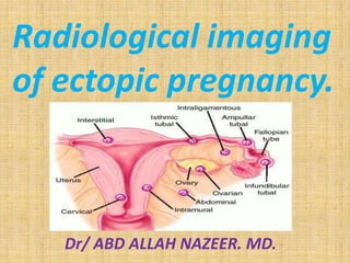
  • 3. Locations of ectopic pregnancy. (A) ampullary/isthmic, (B) infundibulum, (C) fimbria, (D) interstitial, (E) intra-abdominal, (F) ovarian and (G) cervical.
  • 4.
  • 5.
  • 6.
  • 7. Etiology of ectopic pregnancy. Congenital. Tubal hypoplasia, Tubal turtuosity, Congenital diverticulae, Accessory ostia, Partial stenosis, Elongation, Intra-mural polyp and Entrap the ovum on its way. Acquired. Pelvic inflammatory disease. Chlamydia trachomatis is most common. Contraceptive failure. Smoking. Multiple sexual partners. Endometriosis. History of tubal surgery. Endometriosis and leiomyoma. Recent use of in vitro fertilization.
  • 8.
  • 9.
  • 10.
  • 11.
  • 12.
  • 13.
  • 14. Magnetic Resonance Imaging. MRI findings that can suggest an ectopic pregnancy include the presence of a tubal gestational sac; a tubal hematoma, which is a hematoma suggested by the ring sign (peripheral hyperintensity) on T1- weighted images; tubal wall enhancement; and an adnexal mass with hemorrhagic fluid in the peritoneum. Blood is suggested by the presence of high-signal-intensity fluid on T1-weighted images. The MRI characteristics of an ectopic pregnancy and its rupture are seen in the T2-weighted images.
  • 15.
  • 16. Live cervical ectopic pregnancy at 6 weeks gestational age. Sagittal view of the cervix shows the gestational sac with an embryo (arrow) within the cervix.
  • 17.
  • 18.
  • 19.
  • 20.
  • 21.
  • 22.
  • 23. Heterotopic pregnancy in an in vitro fertilization patient at 15 weeks gestational age. (a) Transabdominal view shows no myometrium (arrows) around the ovarian ectopic pregnancy. (b) Coronal T2-weighted magnetic resonance (MR) image IUP and ectopic pregnancy (EP). P = placenta. MR was used for surgical planning to show the blood flow to the heterotopic pregnancy. Note the flow void in vessels (arrows) supplying the ovarian ectopic pregnancy. After surgical removal of the ectopic pregnancy, the IUP continued to term.
  • 24. Tubal ring of ectopic pregnancy. (a) Transvaginal transverse view of the left adnexa shows an echogenic ringlike mass (arrows) medial to the left ovary. Within the left ovary is a thick-walled corpus luteum cyst (arrowheads). Note that the wall of the ectopic pregnancy is more echogenic than the wall of the corpus luteum cyst. (b) Color Doppler image shows more blood flow (arrowheads) to the corpus luteum than to the ectopic pregnancy (arrow). Note that the “ring of fire” (hypervascular ring) appearance in the adnexa can be seen with both ectopic pregnancy and corpus luteum.
  • 25. Interstitial ectopic pregnancy 8 weeks after LMP. Sagittal transvaginal view of the uterus (UT) demonstrates a thick-walled mass arising from the interstitial portion of the uterus. The embryo is measured with calipers.
  • 26.
  • 27. Ectopic pregnancy. Abdominal/pelvic CT. Image compatible with gestational sac in the left adnexal region (arrow on A), separated from the uterine image (stars on A and B) and from the ipsilateral ovary identified by visualization of the corpus luteum (hollow arrow on B). Also, note the presence moderate amount of fluid in the peritoneal cavity with foci of high density characterizing hematic content (L). (A,B: contrast-enhanced axial sections in the portal phase).
  • 28. Axial T2-weighted fast spin-echo magnetic resonance image of the pelvis. This image shows an abnormal fluid-containing fallopian tube (red arrow) on the right side. A simple right ovarian cyst (white arrow) is also present.
  • 29. Ectopic pregnancy. Pelvic MRI. The presence of a gestational sac is observed in the left adnexal region (arrows on A and B), in association with a heterogeneous mass (arrow on C). Note the uterine and ovarian images (stars on A and D) separated from the adnexal mass, as well as the presence of placenta showing contrast enhancement (hollow arrow on D). (A: axial T2-weighted; B: coronal T2-weighted; C: sagittal T2- weighted; D: post-contrast axial T1-weighted images).
  • 30. Ectopic pregnancy. Pelvic MRI. Expansile, heterogeneous mass in the right adnexal region (arrows on A to D), separated from the uterine (star on B) and ovarian (hollow arrows on A and B) images. Also, note the presence of moderate amount of free fluid in the pelvis, with intermediate signal intensity on T1-weighted images, suggesting hematic contents (stars on A and C). (A,B: axial T2- weighted image with fat saturation; C,D: axial T1-weighted, in phase and out-phase images).
  • 31. Cornual ectopic pregnancy. Pelvic MRI. The presence of a heterogeneous mass is observed in the cornual segment of the left uterine tube (white arrows on A to D). Note the communication with the uterine cavity (white hollow arrows on B and C). The myometrium is indicated by the black hollow arrow on C. (A,B: axial T1- weighted and T2-weighted images, respectively; C: post-contrast, axial T1-weighted image with fat saturation; D: coronal T2-weighted image).
  • 32. Ectopic pregnancy with enhancing papillary solid components with a nonenhancing necrotic portion and high signal intensity on outer surface on sagittal T1-weighted turbo spin echo spectral presaturation with inversion recovery with contrast imaging (A), on coronal T1-weighted fast field echo spectral presaturation with inversion recovery with contrast imaging (B), on transverse T1-weighted fast field echo in- phase with contrast imaging (C) (black arrows).
  • 33. Tubal pregnancy in a 41- year-old woman with acute abdominal pain and positive results of a pregnancy test.
  • 34. CONCLUSION. Sonography plays a central role in the diagnosis and management of ectopic pregnancy. If an extraovarian mass is present in a pregnant patient with pain and bleeding, and no intrauterine gestational sac is seen, the diagnosis of ectopic pregnancy should be considered until proved otherwise. Therapy is determined by a combination of clinical symptoms, sonographic findings, and serum β-hCG values. While an intrauterine gestational sac is typically seen when the β-hCG value is greater than 2000 mIU/mL (IRP), this value should be used as a guideline and not an absolute threshold. Since the fallopian tube is the most common location of ectopic pregnancy, care should be taken while scanning to search between the uterus and ovary for a tubal mass. Surgery is being performed less often for ectopic pregnancy since the alternative treatments of expectant management, methotrexate, and percutaneous injection are now available. With these conservative treatments, there is an increased role for sonography in patient follow-up.