SlideShare ist ein Scribd-Unternehmen logo
1 von 85
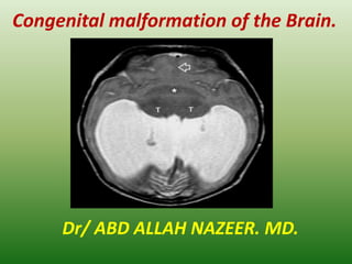
Congenital malformation of the Brain.
Dr/ ABD ALLAH NAZEER. MD.
Congenital birth Abnormalities:
Changing Interpretations Across Time.
More than 2000 different congenital malformations of the
brain have been described in the literature, and their
incidence is reported to be about 1 percent of all live
births. Since the congenital anomalies of the brain are
commonly encountered in day to day practice, it is very
important for every radiologist to be familiar with the
basic imaging findings of common congenital anomalies
to make a correct diagnosis necessary for optimum
management of these conditions. Magnetic resonance
imaging (MRI) is very useful in studying these
malformations. The aim of this chapter is to provide an
overview of all important and routinely encountered
congenital malformations of the brain.
Introduction:
Normal brain development:
Congenital anomalies of the brain are extremely complex and are best
studied by correlating with embryological development. Basic events in
normal brain development includes the following four stages:1
Stage 1: Dorsal Induction: Formation and closure of the neural tube
- Occurs at 3-5 weeks
- Three phases: Neurulation, canalization, retrogressive differentiation
- Failure: Neural tube defects (Anencephaly, Cephalocele, Chiari
malformations) and Spinal dysraphic disorders.
Stage 2: Ventral Induction: Formation of the brain segments and face
- Occurs at 5-10 weeks of gestation
- Three vesicles (prosencephalon, mesencephalon, and
rhombencephalon) form the cerebral hemispheres/thalamus, midbrain,
and cerebellum/brain stem respectively.
- Development of face
- Failure: Holoprosencephalies, Dandy Walker malformation, Cerebellar
hypoplasia/dysplasia (Chiari-IV), Joubert syndrome,
Rhombencephalosynapsis, Septo-optic dysplasia, and facial anomalies.
Stage 3: Migration and Histogenesis
- Occurs at 2-5 months of gestation
- Neuronal migration from germinal matrix to the cortex
- Cortical organization
- Failure of Migration: Heterotopias, Agyria-pachygyria,
Polymicrogyria, Lissencephaly,
Schizencephaly, Corpus callosal agenesis, Lhermitte-Duclos disease.
- Failure of Histogenesis: Aqueductal stenosis, Arachnoid cyst,
Megalencephaly,
Micoencephaly, Neurocutaneous syndrome (phakomatoses),
Congenital vascular malformation, and Congenital neoplasms.
Stage 4: Myelination
- Begins at 6 months of gestation; matures by 3 years.
-Progresses from caudal to cephalad, from dorsal to ventral, and
from central to peripheral
- Failure: Dismyelinating diseases, Metabolic disorders.
Classification of congenital malformation of brain.
A number of classification systems have been proposed, but with regards
to our basic purpose, simplified classification of brain malformations has
been taken into account, which is as follows:
a. Disorders of Organogenesis
- Neural tube closure
- Diverticulation and cleavage
- Sulcation/cellular migration
- Cerebellar hypoplasia/dysplasia
b. Disorders of Histogenesis
- Neurocutaneous syndromes (Phakomatoses)
c. Disorders of Cytogenesis
- Congenital vascular malformations
- Congenital neoplasms of brain
d. Disorders of Myelination
- Leukodystrophies
Imaging features of congenital malformations of the brain:
Disorders of organogenesis.
The Chiari malformations:
Termed as tonsillar ectopia.
- Herniation of elongated peg like cerebellar tonsils into the upper cervical
canal through the foramen magna.
-Abnormal tonsillar descent below the opisthion-basion line
- At least 6mm in first decade
-5mm in the 2nd /3rd decade
4mm between 4th -8th decade, and
3mm by 9th decade
-Causes of abnormal tonsillar descent
- Congenital asymptomatic tonsillar ectopia
Intracranial hypotension due to chronic CSF leak (sagging brain)
Long-standing compensated hydrocephalus
Craniovertebral anomalies (Basilar invagination, Platybasia, Atlantooccipital
assimilation)
-Associated anomalies
- Syringohydromyelia(30-60%)
Chiari I Malformation. Sagittal T1W MR scan of brain shows peg like low
lying tonsil in upper posterior cervical canal (thin white arrow). The
cerebellar tonsils lie more than 10mm below the foramen magnum. Note
mild enlargement of supratentorial ventricular system.
Sagittal MRI images shows a
classic Chiari I malformation
with “peglike” tonsils
extending inferiorly through
the foramen magnum.
Chiari II Malformation:
- Complex anomaly involving skull, dura, brain, spine and the cord
-Skull and dural involvement
- Luckenschadel (lacunar skull), concave clivus and petrous ridges
Small and shallow posterior fossa with low lying transverse sinuses and
torcular Herophilli
Hypoplastic tentorium cerebelli with gaping (heart shaped) incisura
Hypoplastic, fenestrated falx cerebri with interdigitating gyri
Gaping foramen magnum
-Brain involvement
- Cascading protrusion of vermian nodulus, fourth ventricle choroid
plexus and the medulla into the spinal canal with formation of
cervicomedullary spur and kink upward herniation of cerebellar
hemispheres and vermis through gaping incisura (towering cerebellum)
producing tectal deformity (beaked tectum) cerebellar hemispheres
creep around to engulf the brain stem.
-Large massa intermedia
Hydrocephalus (90%) with serrated appearance of lateral ventricles
Chiari 11
malformation.
Chiari III Malformation:
- Features of Chiari II malformation with a low occipital or high cervical
encephalocele. The encephalocele may contain meninges, cerebellum, occipital
lobe or brain stem. Cisterns and dural sinuses may also be present.
Chiari III Malformation. Sagittal T1w MR scan shows features of ACM II,
evidenced as a large massa intermedia (asterix), tectal beaking (thin black
arrow), inferior herniation of the cerebellar tissue through the foramen magnum
into the upper posterior cervical canal (thin white arrow), and a low occipital
meningoencephalocele (thick white arrow).
Chiari 111
malformation.
Chiari IV Malformation:
-Severe cerebellar hypoplasia or dysplasia, small brain stem
and large posterior fossa CSF space.
- No hydrocephalus and other CNS anomalies
Cephaloceles:
- Characterized by protrusion of intracranial contents through a congenital defect
in the dura and skull
- Usually located at or near the midline.
-Pathological classification of cephalocele (based on the contents of the
herniated sac)
- Meningocele (leptomeninges and CSF)
Meningoencephalocele (leptomeninges, CSF and brain)
Meningoencephalocystocele (leptomeninges, CSF, brain and ventricles)
Atretic cephalocele (small nodule of fibrous fatty tissue)
Gliocele (CSF lined by glial tissue).
Anatomic classification of cephalocele (based on location)
Occipital - most common in America, Europe
Parietal, Temporal , Frontal or Frontoethmoidal (Sincipital) - most common in
Asia
Transsphenoidal – uncommon Nasal
- The herniated brain dysgenetic and non-functional
- Absence or erosion of the crista galli with enlargement of foramen cecum is a
constant
feature of a nasal cephalocele.
Corpus Callosum Agenesis:
-Corpus callosum, a midline commissure connects two
cerebral hemispheres
-Develops in cephalocaudal direction, beginning with genu
then followed by the body and splenium. The rostrum is
last to develop
- Two types: complete or partial
-Complete callosal agenesis
- Absence of entire corpus callosum, cingulate gyrus and sulcus
high riding 3rd ventricle with spoke like orientation of gyri
around it widely separated, parallel and non-converging lateral
ventricles.
Probst bundles (longitudinal white matter tracts) indent
superomedial lateral ventricles Colpocephaly (dilated occipital
horns) common, frontal horns small and pointed
High incidence of dorsal interhemispheric cyst.
Partial callosal agenesis:
Splenium and rostrum absent or hypoplastic
Genu and body present to various degrees
-Associated anomalies.
- Migration disorders (heterotopias,
lissencephaly, schizencephaly).
Chiari II malformation.
Dandy-Walker malformation.
Holoprosencephaly.
Corpus callosal lipoma.
Disorders of diverticulation and cleavage:
Holoprosencephaly:
- Characterized by complete or partial failure of cleavage and
differentiation of developing cerebrum (prosencephalon) into
hemispheres and lobes.
- Cerebellum and brain stem relatively normal
-Classified into three types on the basis of degree of severity
- Alobar (most severe).
Semilobar ( moderately severe).
Lobar (mildest form).
- Precise boundaries among these three groups does not exist,
intermediate cases may be identified
-Alobar Holoprosencephaly
- Near complete lack of hemispheric cleavage, Cresent-shaped
monoventricle
Absence of septum pellucidum, corpus callosum, falx cerebri, and
interhemispheric fissure fused thalami and basal ganglia
Associated anomalies: Dorsal interhemispheric cyst,
severe craniofacial anomaly
-Semilobar Holoprosencephaly
- Partial brain diverticulation
H-shaped holoventricle with rudimentary occipital and
temporal horns.
Rudimentary third ventricle may be present.
Septum pellucidum absent, callosal splenium may be
formed, falx cerebri and interhemispheric fissure
partially developed posteriorly.
Thalami and basal ganglia partially separated
Associated anomalies: Dorsal interhemispheric cyst,
variable craniofacial anomalies.
CT of Alobar Holoprosencephaly.
CT and MRI of semilobar Holoprosencephaly
Lobar holoprosencephaly.
Septooptic Dysplasia (de Morsier syndrome)
- Milder form of lobar holoprosencephaly
- Characterized by hypoplastic optic nerves and optic chiasma, and absent
septum pellucidum
- Associated hypoplasia of hypothalamic-pituitary axis seen in 2/3rd cases
- MR picture of small optic nerves and chiasma, widened anterior recess of 3rd
ventricle and suprasellar cistern, and squared frontal horns
- Associated anomalies: Scizencephaly (most common).
Disorders of sulcation/cellular migration
Lissencephaly (Agyria-Pachygyria).
- Refers to “smooth brain” with absent or poor sulcation.
- Can be complete (agyria) or incomplete (pachygyria).
Intermediate features of agyria and pachygyria may coexist
www.intechopen.com
Congenital Malformation of the Brain
- Three types: Type I, II, and III
-Type I (classical) lissencephaly
- Typical figure eight configuration of brain with oblique and
shallow sylvian fissures.
Thickened cortex with flat broad gyri and smooth gray-white
matter interface.
Colpocephaly associated with Miller-Dieker syndrome.
Type II(Cobblestone) lissencephaly:
Neuroimaging:
Thickened cortex with polymicrogyric appearance. Concurrent
hypomyelination of underlying white matter present.
Associated with Fukuyama congenital muscular dystrophy, Walker-
Warburg syndrome and muscle-eye-brain syndrome.
-Type III(cerebrocerebellar) lissencephaly
- Microcephaly with moderately thickened cortex and hypoplastic
cerebellum and brain stem
Non lissencephalic Cortical Dysplasia:
- Two types: Polymicrogyria and pachygyria
-Polymicrogyria.
- characterized by diffusely thickened cortex with irregular, bumpy
gyral pattern
MRI: thick cortex with flat surface, irregular gray-white matter
junction.
Pachygyria:
Focal areas of thickened and flattened cortex with blurred gray-white matter junction
-Both types of cortical dysplasia show relative paucity and marked T2 prolongation of
underlying white matter.
Focal cortical dysplasia. Axial NCCT brain shows focal cortical
dysplasia seen as localized thickening and infolding of the cortex
(arrow) in left fronto-parietal junction region with paucity of
underlying white matter and prominence of overlying sulcus.
Heterotopias:
- Characterized by the presence of normal neurons at abnormal sites
- Result of arrested neuronal migration from periventricular
germinal zone to the cortex along the radial glial fibers
- Two types: Nodular type(common), band (laminar) type
(uncommon)
-Nodular type:
- Multiple masses of gray matter which are of variable size
Common location: subependymal or subcortical
Focal or diffuse subependymal focal nodules indent the ventricular
wall, whereas diffuse heterotopias border the walls of the lateral
ventricle and are X-linked.
Heterotopias are isointense to normal gray matter in all pulse
sequences and do not enhance on administration of intravenous
contrast. They are best appreciated on medium tau inversion
recovery sequences.
Schizencephaly (split brain) .
- Characterized by presence of heterotopic gray matter lined cleft that
extends from the ventricular (ependyma) to the periphery (pial
surface) of the brain, traversing through the white matter.
- Can be unilateral or bilateral
- Two types: Closed lip (type I) or open lip (type II)
-Closed lip (type I) schizencephaly
- Walls of the cleft oppose each other and there is no intervening CSF
Imaging shows an outpouching or nippling at the ependymal surface
of the cleft
-Open lip (type II) schizencephaly
- Walls of the cleft are widely separated and the cleft is occupied by
CSF. Severe form of open lip schizencephaly has an appearance which
is called “basket brain”.
Closest differential is porencephalic cyst in which CSF space is lined by
gliotic white matter, in contrast to gray matter lined Schizencephaly.
Open lip schizencephaly.
Dandy-Walker Complex :
- Includes Dandy-Walker Malformation and Dandy-Walker Variant
-Dandy-Walker Malformation Characterized by
- Large posterior fossa with cystic dilatation of the fourth ventricle; that elevates
the tentorium, torcular Herophili and the transverse sinuses above the lamdoid
suture (lamdoid torcula inversion)
Mega Cistern Magna:
- Characterized by variable size dilatation of the cistern magna which, however, freely
communicates with both the fourth ventricle and the adjacent subarachnoid
spaces(can extend up to the straight sinus superiorly and C1-C2 level inferiorly).
Posterior Fossa Arachnoid Cyst:
- The posterior fossa arachnoid cyst is a CSF collection within the layers
of arachnoid membrane which does not communicates fully with the
fourth ventricle or adjacent subarachnoid spaces.
Joubert’s Syndrome (Congenital Vermian Hypoplasia).
- Characterized by inherited vermian dysgenesis, enlarged
superior cerebellar peduncles and a high riding fourth ventricle.
- On MR imaging, the vermis is completely or partially absent.
Superior fourth ventricle is bat-wing or umbrella shaped (on
axial image) and has a convex roof (on sagittal image). The
superior cerebellar peduncles are elongated, thin, running
parallel to each other. Isthmus (area of pontomesencephalic
junction) is narrow. Midbrain has the typical “molar tooth”
appearance.
- Hydrocephalus is absent
- All patients of Joubert’s syndrome should be screened for
occipital encephalocele (30%), callosal dysgenesis, cortical
dysplasia, hypothalamic hamartoma, and ocular, hepatic &
renal diseases.
Rhombencephalosynapsis:
- Rare abnormality characterized by vermian agenesis or hypogenesis combined with
midline fusion of cerebellar hemispheres, peduncles and dentate nuclei.
- Associated anomalies: ventriculomegaly (common), callosal dysgenesis, absent septum
pellucidum, cephalocele and schizencephaly.
Lhermitte-Duclos Disease:
- Also known as dysplastic gangliocytoma of the cerebellum
- Uncommon cerebellar dysplasia characterized by gross thickening of the
cerebellar folias with or without mass effect.
Disorders of histogenesis:
“Neurocutaneous syndromes” or “Phakomatoses” constitute a group of
congenital malformations which are characterized by cutaneous lesions
associated with CNS anomalies. Some of the common neurocutaneous
syndromes are described below.
Neurofibromatosis :
Neurofibromatosis type 1 (NF 1)
- Also known as Von Recklinghausen disease or peripheral neurofibromatosis
- Accounts for > 90% of all NF cases
- Incidence = 1:2000 to 3000 live births
-Diagnostic criteria: two or more of the following findings are present
- Six or more café-au-lait spots(≥5mm in pre-pubertal children and ≥15mm in
post pubertal period). One plexiform neurofibroma or two or more
neurofibromas of any type two or more pigmented iris hamartomas (Lisch
nodules) ,optic nerve glioma ,axillary or inguinal freckling
Osseous lesions such as dysplasia of greater wing of sphenoid,
pseudoarthrosis
First degree relative with NF-1
CNS lesions present in 15-20% cases. These include
Optic nerve glioma (most common CNS lesion), may extend to involve the optic
chiasma, optic tract, optic radiation and the lateral geniculate bodies.
Nonoptic gliomas may involve the brain stem, tectum, and periaqueductal region.
Plexiform neurofibroma is a hallmark of NF-1. It is an unencapsulated
neurofibroma along the path of major cutaneous nerve of the scalp and neck,
which commonly involves the first (ophthalmic) division of trigeminal nerve. It is
often associated with dysplasia of sphenoid bone and bony orbit.
Non-neoplastic hamartomatous lesions (80%) of basal ganglia and white matter.
Majority of lesions show no mass effect or contrast enhancement. These lesions
may increase in size or number in early childhood, diminishes with age and rarely
observed into adulthood.
Other intracranial lesions include astrocytic proliferation of the retina, intracranial
aneurysms, vascular ectasia and a progressive cerebral arterial occlusion disease
akin to moya-moya pattern.
Spinal lesions may include cord astrocytoma / hamartoma, dural ectasia and
lateral/anterior intrathoracic meningoceles.
Skeletal dysplasias may include hypoplasia of sphenoid bone and bony orbit,
kyphoscoliosis, scalloping of posterior aspect of the vertebral bodies.
Neurofibromatosis type 1:
Plexiform neurofibroma with
sphenoid wing dysplasia.
Neurofibromatosis type 1: Vascular
abnormalities. Axial T2W MR image
of brain with left internal carotid
artery aneurysm .
Neurofibromatosis type 2 (NF 2):
- Also known as central neurofibromatosis
- Incidence = 1:50,000 live births
-Cutaneous manifestations rare.
-CNS lesions present in 100% cases. These include
-Bilateral acoustic schwannomas, hallmark of NF-2
Schwannomas of other cranial nerves. Trigeminal nerve is
next most frequently involved nerve, albeit, any cranial
nerve may be affected (with the exception of the
olfactory and optic nerves).
Meningiomas, often multiple choroid plexus calcification
Spinal lesions include cord ependymomas, meningiomas,
or multilevel bulky schwannomas of exiting roots.
Neurofibromatosis type 11.
Tuberous sclerosis:
- Also known as Bourneville disease or multiple
hamartomatous syndrome
- Incidence=1:10,000 to 50,000 live births
- Classical triad of popular facial lesions (adenoma
sebaceum), seizure disorder and mental retardation
-CNS lesions include
- Cortical tubers or hamartomas present in 95% cases of
TS, are characteristic lesions at pathologic examination.
On MR imaging, these lesions may expand and distort
the affected gyri and show age-related signal changes.
Enhancement following contrast administration occurs in
less than 5% cases. Calcification of theses lesions
increases with age.
Tuberous
sclerosis.
Sturge-Weber Syndrome:
- Also known as encephalotrigeminal angiomatosis
- Characterized by facial port wine vascular nevus flammeus
in the trigeminal nerve distribution (1st division most
commonly involved), leptomeningeal venous angiomatosis of
ipsilateral brain, hemiparesis, homonymous hemianopia and
seizure.
Usually unilateral, rarely bilateral
- Occipital and posterior parietal lobe on the side of facial
angioma is most commonly involved.
- Tram-track or gyriform pattern of cortical calcification
underlying the leptomeningeal angioma is diagnostic of the
syndrome. The calcification is unusual before two years of
age. Calcifications are best seen on plain CT, T2W and GRE
image.
Von Hippel-Lindau syndrome:
- Is a multisystem disease characterized by presence of cysts,
angiomas, and neoplasms of the CNS and abdominal viscera.
-CNS lesions include:
-Cerebellar (75%), retinal (50%) and spinal cord (25%)
hemangioblatomas.
Supratentorial hemangioblatomas are extremely rare.
On MR imaging, majority of hemangioblastomas have cystic
appearance with intensely enhancing mural nodule. Between
20-40% tumors are solid. Contrast enhanced MRI has increased
sensitivity for detection of small lesions. Flow voids in the
afferent and efferent vessels supplying the tumor can often be
detected. The angiographic appearance of the
hemangioblastomas is highly characteristic, showing tangles of
tightly packed vessels that become opacified in the early arterial
phase.
Hemangioblastoma.
Presentation1.pptx, congenital malformation of the brain.

Weitere ähnliche Inhalte

Was ist angesagt?

Presentation2.pptx. posterior fossa tumour
Presentation2.pptx. posterior fossa tumourPresentation2.pptx. posterior fossa tumour
Presentation2.pptx. posterior fossa tumourAbdellah Nazeer
 
Holoprosencephaly
HoloprosencephalyHoloprosencephaly
HoloprosencephalyVikram Patil
 
Normal CT BRAIN
Normal CT BRAINNormal CT BRAIN
Normal CT BRAINNeurologyKota
 
Hypoxic ischemic encephalopathy modified
Hypoxic ischemic encephalopathy  modifiedHypoxic ischemic encephalopathy  modified
Hypoxic ischemic encephalopathy modifiedAnish Choudhary
 
Diagnostic Imaging of Central Nervous System Infections
Diagnostic Imaging of Central Nervous System InfectionsDiagnostic Imaging of Central Nervous System Infections
Diagnostic Imaging of Central Nervous System InfectionsMohamed M.A. Zaitoun
 
Dandy-Walker Malformation: Classification and Management
Dandy-Walker Malformation: Classification and ManagementDandy-Walker Malformation: Classification and Management
Dandy-Walker Malformation: Classification and ManagementDr. Shahnawaz Alam
 
Imaging neurology spotters
Imaging   neurology spottersImaging   neurology spotters
Imaging neurology spottersNeurologyKota
 
1.schizencephaly 2.holoprosencephaly 3.porencephaly
1.schizencephaly 2.holoprosencephaly 3.porencephaly1.schizencephaly 2.holoprosencephaly 3.porencephaly
1.schizencephaly 2.holoprosencephaly 3.porencephalyairwave12
 
Diagnostic Imaging of Brain Tumors
Diagnostic Imaging of Brain TumorsDiagnostic Imaging of Brain Tumors
Diagnostic Imaging of Brain TumorsMohamed M.A. Zaitoun
 
HYPOXIC ISCHEMIC ENCEPHALOPATHY-RADIOLOGICAL APPROACH
HYPOXIC ISCHEMIC ENCEPHALOPATHY-RADIOLOGICAL APPROACHHYPOXIC ISCHEMIC ENCEPHALOPATHY-RADIOLOGICAL APPROACH
HYPOXIC ISCHEMIC ENCEPHALOPATHY-RADIOLOGICAL APPROACHNEHA NUPUR
 
DISORDERS OF MYELINATION
DISORDERS OF MYELINATIONDISORDERS OF MYELINATION
DISORDERS OF MYELINATIONManideep Malaka
 
MRI brain; Basics and Radiological Anatomy
MRI brain; Basics and Radiological AnatomyMRI brain; Basics and Radiological Anatomy
MRI brain; Basics and Radiological AnatomyImran Rizvi
 
Intradural extramedullary mass - a case on MRI
Intradural extramedullary mass - a case on  MRIIntradural extramedullary mass - a case on  MRI
Intradural extramedullary mass - a case on MRIREKHAKHARE
 
Cns infections radiology.
Cns infections radiology.Cns infections radiology.
Cns infections radiology.Raeez Basheer
 
Presentation1.pptx. radiological imaging of epilepsy.
Presentation1.pptx. radiological imaging of epilepsy.Presentation1.pptx. radiological imaging of epilepsy.
Presentation1.pptx. radiological imaging of epilepsy.Abdellah Nazeer
 
Radiological vascular anatomy of brain
Radiological vascular anatomy of brainRadiological vascular anatomy of brain
Radiological vascular anatomy of brainDev Lakhera
 
Ring enhancing lesions
Ring enhancing lesionsRing enhancing lesions
Ring enhancing lesionsAbhijeet Deshmukh
 
Presentation1.pptx, radiological imaging of skeletal dysplasia
Presentation1.pptx, radiological imaging of skeletal dysplasiaPresentation1.pptx, radiological imaging of skeletal dysplasia
Presentation1.pptx, radiological imaging of skeletal dysplasiaAbdellah Nazeer
 
IMAGING OF INTRAVENTRICULAR TUMORS
IMAGING OF INTRAVENTRICULAR TUMORS IMAGING OF INTRAVENTRICULAR TUMORS
IMAGING OF INTRAVENTRICULAR TUMORS Ameen Rageh
 

Was ist angesagt? (20)

Presentation2.pptx. posterior fossa tumour
Presentation2.pptx. posterior fossa tumourPresentation2.pptx. posterior fossa tumour
Presentation2.pptx. posterior fossa tumour
 
Holoprosencephaly
HoloprosencephalyHoloprosencephaly
Holoprosencephaly
 
Normal CT BRAIN
Normal CT BRAINNormal CT BRAIN
Normal CT BRAIN
 
Hypoxic ischemic encephalopathy modified
Hypoxic ischemic encephalopathy  modifiedHypoxic ischemic encephalopathy  modified
Hypoxic ischemic encephalopathy modified
 
Diagnostic Imaging of Central Nervous System Infections
Diagnostic Imaging of Central Nervous System InfectionsDiagnostic Imaging of Central Nervous System Infections
Diagnostic Imaging of Central Nervous System Infections
 
Dandy-Walker Malformation: Classification and Management
Dandy-Walker Malformation: Classification and ManagementDandy-Walker Malformation: Classification and Management
Dandy-Walker Malformation: Classification and Management
 
Imaging neurology spotters
Imaging   neurology spottersImaging   neurology spotters
Imaging neurology spotters
 
1.schizencephaly 2.holoprosencephaly 3.porencephaly
1.schizencephaly 2.holoprosencephaly 3.porencephaly1.schizencephaly 2.holoprosencephaly 3.porencephaly
1.schizencephaly 2.holoprosencephaly 3.porencephaly
 
Diagnostic Imaging of Brain Tumors
Diagnostic Imaging of Brain TumorsDiagnostic Imaging of Brain Tumors
Diagnostic Imaging of Brain Tumors
 
HYPOXIC ISCHEMIC ENCEPHALOPATHY-RADIOLOGICAL APPROACH
HYPOXIC ISCHEMIC ENCEPHALOPATHY-RADIOLOGICAL APPROACHHYPOXIC ISCHEMIC ENCEPHALOPATHY-RADIOLOGICAL APPROACH
HYPOXIC ISCHEMIC ENCEPHALOPATHY-RADIOLOGICAL APPROACH
 
DISORDERS OF MYELINATION
DISORDERS OF MYELINATIONDISORDERS OF MYELINATION
DISORDERS OF MYELINATION
 
MRI brain; Basics and Radiological Anatomy
MRI brain; Basics and Radiological AnatomyMRI brain; Basics and Radiological Anatomy
MRI brain; Basics and Radiological Anatomy
 
Intradural extramedullary mass - a case on MRI
Intradural extramedullary mass - a case on  MRIIntradural extramedullary mass - a case on  MRI
Intradural extramedullary mass - a case on MRI
 
Cns infections radiology.
Cns infections radiology.Cns infections radiology.
Cns infections radiology.
 
Presentation1.pptx. radiological imaging of epilepsy.
Presentation1.pptx. radiological imaging of epilepsy.Presentation1.pptx. radiological imaging of epilepsy.
Presentation1.pptx. radiological imaging of epilepsy.
 
Radiological vascular anatomy of brain
Radiological vascular anatomy of brainRadiological vascular anatomy of brain
Radiological vascular anatomy of brain
 
Ring enhancing lesions
Ring enhancing lesionsRing enhancing lesions
Ring enhancing lesions
 
Presentation1.pptx, radiological imaging of skeletal dysplasia
Presentation1.pptx, radiological imaging of skeletal dysplasiaPresentation1.pptx, radiological imaging of skeletal dysplasia
Presentation1.pptx, radiological imaging of skeletal dysplasia
 
IMAGING OF INTRAVENTRICULAR TUMORS
IMAGING OF INTRAVENTRICULAR TUMORS IMAGING OF INTRAVENTRICULAR TUMORS
IMAGING OF INTRAVENTRICULAR TUMORS
 
Meningioma of brain
Meningioma of brainMeningioma of brain
Meningioma of brain
 

Andere mochten auch

Congenital malformation of cns
Congenital malformation of cnsCongenital malformation of cns
Congenital malformation of cnsPS Deb
 
Congenital Anomalies of Nervous System
Congenital Anomalies of Nervous SystemCongenital Anomalies of Nervous System
Congenital Anomalies of Nervous SystemDhaval Shukla
 
cns embryology and anomalies
 cns embryology and anomalies cns embryology and anomalies
cns embryology and anomaliesHussein Abdeldayem
 
Imaging in developmental malformations of the brain
Imaging in developmental malformations of the brainImaging in developmental malformations of the brain
Imaging in developmental malformations of the brainChakradhar Reddy
 
Saurabh neuroscience. final
Saurabh neuroscience. finalSaurabh neuroscience. final
Saurabh neuroscience. finalsaurabh jadhav
 
3D&4D brain anomalies
3D&4D brain anomalies 3D&4D brain anomalies
3D&4D brain anomalies Tarek Mansour
 
Ms vs adem BASIC DIFFERENCES AND APPROACH
Ms vs adem BASIC DIFFERENCES AND APPROACHMs vs adem BASIC DIFFERENCES AND APPROACH
Ms vs adem BASIC DIFFERENCES AND APPROACHSatyendra Raghuwanshi
 
Osmotic demyelination syndrome
Osmotic demyelination syndromeOsmotic demyelination syndrome
Osmotic demyelination syndromeUbaidur Rahaman
 
Neural tube defects: Importance of Folic Acid and Vitamin B12 intake
Neural tube defects: Importance of Folic Acid and Vitamin B12 intakeNeural tube defects: Importance of Folic Acid and Vitamin B12 intake
Neural tube defects: Importance of Folic Acid and Vitamin B12 intakeVijaya Sawant,PMP, OCP
 
Mri evaluation of pediatric white matter lesions
Mri evaluation of pediatric white matter lesions Mri evaluation of pediatric white matter lesions
Mri evaluation of pediatric white matter lesions DrBhishm Sevendra
 
Presentation1.pptx white matter disorder in pediatric
Presentation1.pptx white matter disorder in pediatricPresentation1.pptx white matter disorder in pediatric
Presentation1.pptx white matter disorder in pediatricAbdellah Nazeer
 
Inherited white matter diseases
Inherited white matter diseasesInherited white matter diseases
Inherited white matter diseasesNavdeep Shah
 
Traumatic Brain Injury Power Point
Traumatic Brain Injury Power PointTraumatic Brain Injury Power Point
Traumatic Brain Injury Power Pointctrythall
 
Multiple sclerosis
Multiple sclerosisMultiple sclerosis
Multiple sclerosisKapil Dhital
 
Malformations of Cerebellum
Malformations of CerebellumMalformations of Cerebellum
Malformations of CerebellumNawras AlHalabi
 
Glioneuronal tumours
Glioneuronal tumoursGlioneuronal tumours
Glioneuronal tumoursMayurakshi D
 
Radiological approach to a child with chest infection
Radiological approach to a child with chest infectionRadiological approach to a child with chest infection
Radiological approach to a child with chest infectionAhmed Bahnassy
 

Andere mochten auch (20)

Congenital malformation of cns
Congenital malformation of cnsCongenital malformation of cns
Congenital malformation of cns
 
Congenital Anomalies of Nervous System
Congenital Anomalies of Nervous SystemCongenital Anomalies of Nervous System
Congenital Anomalies of Nervous System
 
cns embryology and anomalies
 cns embryology and anomalies cns embryology and anomalies
cns embryology and anomalies
 
Imaging in developmental malformations of the brain
Imaging in developmental malformations of the brainImaging in developmental malformations of the brain
Imaging in developmental malformations of the brain
 
Congenital anomalies ppt
Congenital anomalies pptCongenital anomalies ppt
Congenital anomalies ppt
 
Saurabh neuroscience. final
Saurabh neuroscience. finalSaurabh neuroscience. final
Saurabh neuroscience. final
 
3D&4D brain anomalies
3D&4D brain anomalies 3D&4D brain anomalies
3D&4D brain anomalies
 
Ms vs adem BASIC DIFFERENCES AND APPROACH
Ms vs adem BASIC DIFFERENCES AND APPROACHMs vs adem BASIC DIFFERENCES AND APPROACH
Ms vs adem BASIC DIFFERENCES AND APPROACH
 
Osmotic demyelination syndrome
Osmotic demyelination syndromeOsmotic demyelination syndrome
Osmotic demyelination syndrome
 
Neural tube defects: Importance of Folic Acid and Vitamin B12 intake
Neural tube defects: Importance of Folic Acid and Vitamin B12 intakeNeural tube defects: Importance of Folic Acid and Vitamin B12 intake
Neural tube defects: Importance of Folic Acid and Vitamin B12 intake
 
Mri evaluation of pediatric white matter lesions
Mri evaluation of pediatric white matter lesions Mri evaluation of pediatric white matter lesions
Mri evaluation of pediatric white matter lesions
 
Ms
MsMs
Ms
 
Presentation1.pptx white matter disorder in pediatric
Presentation1.pptx white matter disorder in pediatricPresentation1.pptx white matter disorder in pediatric
Presentation1.pptx white matter disorder in pediatric
 
Inherited white matter diseases
Inherited white matter diseasesInherited white matter diseases
Inherited white matter diseases
 
Traumatic Brain Injury Power Point
Traumatic Brain Injury Power PointTraumatic Brain Injury Power Point
Traumatic Brain Injury Power Point
 
Multiple sclerosis
Multiple sclerosisMultiple sclerosis
Multiple sclerosis
 
Malformations of Cerebellum
Malformations of CerebellumMalformations of Cerebellum
Malformations of Cerebellum
 
Glioneuronal tumours
Glioneuronal tumoursGlioneuronal tumours
Glioneuronal tumours
 
Surgery 5th year, 2nd lecture (Dr. Mazin)
Surgery 5th year, 2nd lecture (Dr. Mazin)Surgery 5th year, 2nd lecture (Dr. Mazin)
Surgery 5th year, 2nd lecture (Dr. Mazin)
 
Radiological approach to a child with chest infection
Radiological approach to a child with chest infectionRadiological approach to a child with chest infection
Radiological approach to a child with chest infection
 

Ă„hnlich wie Presentation1.pptx, congenital malformation of the brain.

Imaging of congenital CNS lesions
Imaging of congenital CNS lesionsImaging of congenital CNS lesions
Imaging of congenital CNS lesionsMilan Silwal
 
Imaging of congenital cns lesions
Imaging of congenital cns lesionsImaging of congenital cns lesions
Imaging of congenital cns lesionsGobardhan Thapa
 
Congenital CNS lesions
Congenital CNS lesionsCongenital CNS lesions
Congenital CNS lesionsMilan Silwal
 
Disorders of neural tube closure and neuronal migration
Disorders of neural tube closure and neuronal migrationDisorders of neural tube closure and neuronal migration
Disorders of neural tube closure and neuronal migrationdrnaveent
 
Embyrogenesis of the CNS and its disorders
Embyrogenesis of the CNS and its disordersEmbyrogenesis of the CNS and its disorders
Embyrogenesis of the CNS and its disordersAmr Hassan
 
Disorders of neural tube closure and neuronal migration
Disorders of neural tube closure and neuronal migrationDisorders of neural tube closure and neuronal migration
Disorders of neural tube closure and neuronal migrationdrnaveent
 
Radiological findings of congenital anomalies of the spine and spinal cord
Radiological findings of congenital anomalies of the spine and spinal cordRadiological findings of congenital anomalies of the spine and spinal cord
Radiological findings of congenital anomalies of the spine and spinal cordDr. Armaan Singh
 
Embryology of brain
Embryology of brainEmbryology of brain
Embryology of brainAnady Eleccion
 
Annormalities of head size and shape
Annormalities of head size and shapeAnnormalities of head size and shape
Annormalities of head size and shapeRiyaz Khan
 
Cerebral tumors 1
Cerebral tumors 1Cerebral tumors 1
Cerebral tumors 1Sabir Ahmed
 
Cortical dysplasia and epilepsy
Cortical dysplasia and epilepsyCortical dysplasia and epilepsy
Cortical dysplasia and epilepsyDr-Ashraf Abdou
 
Disorders of neuronal migration
Disorders of neuronal migrationDisorders of neuronal migration
Disorders of neuronal migrationShasidhar Reddy
 
FETAL CENTRAL NERVOUS SYSTEM ANAOMALIES PRESENTATION
FETAL CENTRAL NERVOUS SYSTEM ANAOMALIES PRESENTATIONFETAL CENTRAL NERVOUS SYSTEM ANAOMALIES PRESENTATION
FETAL CENTRAL NERVOUS SYSTEM ANAOMALIES PRESENTATIONkumarramalakshmi
 
Neural tube defects and Craniosynostosis
Neural tube defects and CraniosynostosisNeural tube defects and Craniosynostosis
Neural tube defects and CraniosynostosisThe Medical Post
 
Corpus callosum with disconnection syndromes
Corpus callosum with disconnection syndromes Corpus callosum with disconnection syndromes
Corpus callosum with disconnection syndromes Amruta Rajamanya
 
Microcephaly - For medical students
Microcephaly - For medical studentsMicrocephaly - For medical students
Microcephaly - For medical studentsfaculty of medicine
 
Congenitalbrainmalformations 150913122145-lva1-app6891
Congenitalbrainmalformations 150913122145-lva1-app6891Congenitalbrainmalformations 150913122145-lva1-app6891
Congenitalbrainmalformations 150913122145-lva1-app6891Battulga Munkhtsetseg
 

Ă„hnlich wie Presentation1.pptx, congenital malformation of the brain. (20)

Imaging of congenital CNS lesions
Imaging of congenital CNS lesionsImaging of congenital CNS lesions
Imaging of congenital CNS lesions
 
Imaging of congenital cns lesions
Imaging of congenital cns lesionsImaging of congenital cns lesions
Imaging of congenital cns lesions
 
Congenital CNS lesions
Congenital CNS lesionsCongenital CNS lesions
Congenital CNS lesions
 
Disorders of neural tube closure and neuronal migration
Disorders of neural tube closure and neuronal migrationDisorders of neural tube closure and neuronal migration
Disorders of neural tube closure and neuronal migration
 
Embyrogenesis of the CNS and its disorders
Embyrogenesis of the CNS and its disordersEmbyrogenesis of the CNS and its disorders
Embyrogenesis of the CNS and its disorders
 
Disorders of neural tube closure and neuronal migration
Disorders of neural tube closure and neuronal migrationDisorders of neural tube closure and neuronal migration
Disorders of neural tube closure and neuronal migration
 
Radiological findings of congenital anomalies of the spine and spinal cord
Radiological findings of congenital anomalies of the spine and spinal cordRadiological findings of congenital anomalies of the spine and spinal cord
Radiological findings of congenital anomalies of the spine and spinal cord
 
Embryology of brain
Embryology of brainEmbryology of brain
Embryology of brain
 
Annormalities of head size and shape
Annormalities of head size and shapeAnnormalities of head size and shape
Annormalities of head size and shape
 
Cerebral tumors 1
Cerebral tumors 1Cerebral tumors 1
Cerebral tumors 1
 
Cortical dysplasia and epilepsy
Cortical dysplasia and epilepsyCortical dysplasia and epilepsy
Cortical dysplasia and epilepsy
 
Disorders of neuronal migration
Disorders of neuronal migrationDisorders of neuronal migration
Disorders of neuronal migration
 
FETAL CENTRAL NERVOUS SYSTEM ANAOMALIES PRESENTATION
FETAL CENTRAL NERVOUS SYSTEM ANAOMALIES PRESENTATIONFETAL CENTRAL NERVOUS SYSTEM ANAOMALIES PRESENTATION
FETAL CENTRAL NERVOUS SYSTEM ANAOMALIES PRESENTATION
 
Case record...Cerebral palsy
Case record...Cerebral palsyCase record...Cerebral palsy
Case record...Cerebral palsy
 
Neural tube defects and Craniosynostosis
Neural tube defects and CraniosynostosisNeural tube defects and Craniosynostosis
Neural tube defects and Craniosynostosis
 
Corpus callosum with disconnection syndromes
Corpus callosum with disconnection syndromes Corpus callosum with disconnection syndromes
Corpus callosum with disconnection syndromes
 
Case record... Septo-optic dysplasia
Case record... Septo-optic dysplasiaCase record... Septo-optic dysplasia
Case record... Septo-optic dysplasia
 
Neural tube defects
Neural tube defectsNeural tube defects
Neural tube defects
 
Microcephaly - For medical students
Microcephaly - For medical studentsMicrocephaly - For medical students
Microcephaly - For medical students
 
Congenitalbrainmalformations 150913122145-lva1-app6891
Congenitalbrainmalformations 150913122145-lva1-app6891Congenitalbrainmalformations 150913122145-lva1-app6891
Congenitalbrainmalformations 150913122145-lva1-app6891
 

Mehr von Abdellah Nazeer

Muculoskeletal Pediatic Imaging..pptx
Muculoskeletal Pediatic Imaging..pptxMuculoskeletal Pediatic Imaging..pptx
Muculoskeletal Pediatic Imaging..pptxAbdellah Nazeer
 
Presentation1, Ultrasound of the bowel loops and the lymph nodes..pptx
Presentation1, Ultrasound of the bowel loops and the lymph nodes..pptxPresentation1, Ultrasound of the bowel loops and the lymph nodes..pptx
Presentation1, Ultrasound of the bowel loops and the lymph nodes..pptxAbdellah Nazeer
 
Presentation1 Short cases MD..pptx
Presentation1 Short cases MD..pptxPresentation1 Short cases MD..pptx
Presentation1 Short cases MD..pptxAbdellah Nazeer
 
Presentation1, MD MCQ Cases..pptx
Presentation1, MD MCQ Cases..pptxPresentation1, MD MCQ Cases..pptx
Presentation1, MD MCQ Cases..pptxAbdellah Nazeer
 
Presentation1, Short Cases Quiz..pptx
Presentation1, Short Cases Quiz..pptxPresentation1, Short Cases Quiz..pptx
Presentation1, Short Cases Quiz..pptxAbdellah Nazeer
 
Presentation1, radiological imaging of lateral hindfoot impingement.
Presentation1, radiological imaging of lateral hindfoot impingement.Presentation1, radiological imaging of lateral hindfoot impingement.
Presentation1, radiological imaging of lateral hindfoot impingement.Abdellah Nazeer
 
Presentation2, radiological anatomy of the liver and spleen.
Presentation2, radiological anatomy of the liver and spleen.Presentation2, radiological anatomy of the liver and spleen.
Presentation2, radiological anatomy of the liver and spleen.Abdellah Nazeer
 
Presentation1, artifacts and pitfalls of the wrist and elbow joints.
Presentation1, artifacts and pitfalls of the wrist and elbow joints.Presentation1, artifacts and pitfalls of the wrist and elbow joints.
Presentation1, artifacts and pitfalls of the wrist and elbow joints.Abdellah Nazeer
 
Presentation1, artifact and pitfalls of the knee, hip and ankle joints.
Presentation1, artifact and pitfalls of the knee, hip and ankle joints.Presentation1, artifact and pitfalls of the knee, hip and ankle joints.
Presentation1, artifact and pitfalls of the knee, hip and ankle joints.Abdellah Nazeer
 
Presentation1, radiological imaging of artifact and pitfalls in shoulder join...
Presentation1, radiological imaging of artifact and pitfalls in shoulder join...Presentation1, radiological imaging of artifact and pitfalls in shoulder join...
Presentation1, radiological imaging of artifact and pitfalls in shoulder join...Abdellah Nazeer
 
Presentation1, radiological imaging of internal abdominal hernia.
Presentation1, radiological imaging of internal abdominal hernia.Presentation1, radiological imaging of internal abdominal hernia.
Presentation1, radiological imaging of internal abdominal hernia.Abdellah Nazeer
 
Presentation11, radiological imaging of ovarian torsion.
Presentation11, radiological imaging of ovarian torsion.Presentation11, radiological imaging of ovarian torsion.
Presentation11, radiological imaging of ovarian torsion.Abdellah Nazeer
 
Presentation1, musculoskeletal anatomy.
Presentation1, musculoskeletal anatomy.Presentation1, musculoskeletal anatomy.
Presentation1, musculoskeletal anatomy.Abdellah Nazeer
 
Presentation1, new mri techniques in the diagnosis and monitoring of multiple...
Presentation1, new mri techniques in the diagnosis and monitoring of multiple...Presentation1, new mri techniques in the diagnosis and monitoring of multiple...
Presentation1, new mri techniques in the diagnosis and monitoring of multiple...Abdellah Nazeer
 
Presentation1, radiological application of diffusion weighted mri in neck mas...
Presentation1, radiological application of diffusion weighted mri in neck mas...Presentation1, radiological application of diffusion weighted mri in neck mas...
Presentation1, radiological application of diffusion weighted mri in neck mas...Abdellah Nazeer
 
Presentation1, radiological application of diffusion weighted images in breas...
Presentation1, radiological application of diffusion weighted images in breas...Presentation1, radiological application of diffusion weighted images in breas...
Presentation1, radiological application of diffusion weighted images in breas...Abdellah Nazeer
 
Presentation1, radiological application of diffusion weighted images in abdom...
Presentation1, radiological application of diffusion weighted images in abdom...Presentation1, radiological application of diffusion weighted images in abdom...
Presentation1, radiological application of diffusion weighted images in abdom...Abdellah Nazeer
 
Presentation1, radiological application of diffusion weighted imges in neuror...
Presentation1, radiological application of diffusion weighted imges in neuror...Presentation1, radiological application of diffusion weighted imges in neuror...
Presentation1, radiological application of diffusion weighted imges in neuror...Abdellah Nazeer
 
Presentation1, mr physics.
Presentation1, mr physics.Presentation1, mr physics.
Presentation1, mr physics.Abdellah Nazeer
 
Presentation1. ct physics.
Presentation1. ct physics.Presentation1. ct physics.
Presentation1. ct physics.Abdellah Nazeer
 

Mehr von Abdellah Nazeer (20)

Muculoskeletal Pediatic Imaging..pptx
Muculoskeletal Pediatic Imaging..pptxMuculoskeletal Pediatic Imaging..pptx
Muculoskeletal Pediatic Imaging..pptx
 
Presentation1, Ultrasound of the bowel loops and the lymph nodes..pptx
Presentation1, Ultrasound of the bowel loops and the lymph nodes..pptxPresentation1, Ultrasound of the bowel loops and the lymph nodes..pptx
Presentation1, Ultrasound of the bowel loops and the lymph nodes..pptx
 
Presentation1 Short cases MD..pptx
Presentation1 Short cases MD..pptxPresentation1 Short cases MD..pptx
Presentation1 Short cases MD..pptx
 
Presentation1, MD MCQ Cases..pptx
Presentation1, MD MCQ Cases..pptxPresentation1, MD MCQ Cases..pptx
Presentation1, MD MCQ Cases..pptx
 
Presentation1, Short Cases Quiz..pptx
Presentation1, Short Cases Quiz..pptxPresentation1, Short Cases Quiz..pptx
Presentation1, Short Cases Quiz..pptx
 
Presentation1, radiological imaging of lateral hindfoot impingement.
Presentation1, radiological imaging of lateral hindfoot impingement.Presentation1, radiological imaging of lateral hindfoot impingement.
Presentation1, radiological imaging of lateral hindfoot impingement.
 
Presentation2, radiological anatomy of the liver and spleen.
Presentation2, radiological anatomy of the liver and spleen.Presentation2, radiological anatomy of the liver and spleen.
Presentation2, radiological anatomy of the liver and spleen.
 
Presentation1, artifacts and pitfalls of the wrist and elbow joints.
Presentation1, artifacts and pitfalls of the wrist and elbow joints.Presentation1, artifacts and pitfalls of the wrist and elbow joints.
Presentation1, artifacts and pitfalls of the wrist and elbow joints.
 
Presentation1, artifact and pitfalls of the knee, hip and ankle joints.
Presentation1, artifact and pitfalls of the knee, hip and ankle joints.Presentation1, artifact and pitfalls of the knee, hip and ankle joints.
Presentation1, artifact and pitfalls of the knee, hip and ankle joints.
 
Presentation1, radiological imaging of artifact and pitfalls in shoulder join...
Presentation1, radiological imaging of artifact and pitfalls in shoulder join...Presentation1, radiological imaging of artifact and pitfalls in shoulder join...
Presentation1, radiological imaging of artifact and pitfalls in shoulder join...
 
Presentation1, radiological imaging of internal abdominal hernia.
Presentation1, radiological imaging of internal abdominal hernia.Presentation1, radiological imaging of internal abdominal hernia.
Presentation1, radiological imaging of internal abdominal hernia.
 
Presentation11, radiological imaging of ovarian torsion.
Presentation11, radiological imaging of ovarian torsion.Presentation11, radiological imaging of ovarian torsion.
Presentation11, radiological imaging of ovarian torsion.
 
Presentation1, musculoskeletal anatomy.
Presentation1, musculoskeletal anatomy.Presentation1, musculoskeletal anatomy.
Presentation1, musculoskeletal anatomy.
 
Presentation1, new mri techniques in the diagnosis and monitoring of multiple...
Presentation1, new mri techniques in the diagnosis and monitoring of multiple...Presentation1, new mri techniques in the diagnosis and monitoring of multiple...
Presentation1, new mri techniques in the diagnosis and monitoring of multiple...
 
Presentation1, radiological application of diffusion weighted mri in neck mas...
Presentation1, radiological application of diffusion weighted mri in neck mas...Presentation1, radiological application of diffusion weighted mri in neck mas...
Presentation1, radiological application of diffusion weighted mri in neck mas...
 
Presentation1, radiological application of diffusion weighted images in breas...
Presentation1, radiological application of diffusion weighted images in breas...Presentation1, radiological application of diffusion weighted images in breas...
Presentation1, radiological application of diffusion weighted images in breas...
 
Presentation1, radiological application of diffusion weighted images in abdom...
Presentation1, radiological application of diffusion weighted images in abdom...Presentation1, radiological application of diffusion weighted images in abdom...
Presentation1, radiological application of diffusion weighted images in abdom...
 
Presentation1, radiological application of diffusion weighted imges in neuror...
Presentation1, radiological application of diffusion weighted imges in neuror...Presentation1, radiological application of diffusion weighted imges in neuror...
Presentation1, radiological application of diffusion weighted imges in neuror...
 
Presentation1, mr physics.
Presentation1, mr physics.Presentation1, mr physics.
Presentation1, mr physics.
 
Presentation1. ct physics.
Presentation1. ct physics.Presentation1. ct physics.
Presentation1. ct physics.
 

KĂĽrzlich hochgeladen

Call Girls Kanakapura Road Just Call 7001305949 Top Class Call Girl Service A...
Call Girls Kanakapura Road Just Call 7001305949 Top Class Call Girl Service A...Call Girls Kanakapura Road Just Call 7001305949 Top Class Call Girl Service A...
Call Girls Kanakapura Road Just Call 7001305949 Top Class Call Girl Service A...narwatsonia7
 
Call Girls Service In Shyam Nagar Whatsapp 8445551418 Independent Escort Service
Call Girls Service In Shyam Nagar Whatsapp 8445551418 Independent Escort ServiceCall Girls Service In Shyam Nagar Whatsapp 8445551418 Independent Escort Service
Call Girls Service In Shyam Nagar Whatsapp 8445551418 Independent Escort Serviceparulsinha
 
Hemostasis Physiology and Clinical correlations by Dr Faiza.pdf
Hemostasis Physiology and Clinical correlations by Dr Faiza.pdfHemostasis Physiology and Clinical correlations by Dr Faiza.pdf
Hemostasis Physiology and Clinical correlations by Dr Faiza.pdfMedicoseAcademics
 
College Call Girls Vyasarpadi Whatsapp 7001305949 Independent Escort Service
College Call Girls Vyasarpadi Whatsapp 7001305949 Independent Escort ServiceCollege Call Girls Vyasarpadi Whatsapp 7001305949 Independent Escort Service
College Call Girls Vyasarpadi Whatsapp 7001305949 Independent Escort ServiceNehru place Escorts
 
Russian Call Girls in Pune Riya 9907093804 Short 1500 Night 6000 Best call gi...
Russian Call Girls in Pune Riya 9907093804 Short 1500 Night 6000 Best call gi...Russian Call Girls in Pune Riya 9907093804 Short 1500 Night 6000 Best call gi...
Russian Call Girls in Pune Riya 9907093804 Short 1500 Night 6000 Best call gi...Miss joya
 
VIP Call Girls Lucknow Nandini 7001305949 Independent Escort Service Lucknow
VIP Call Girls Lucknow Nandini 7001305949 Independent Escort Service LucknowVIP Call Girls Lucknow Nandini 7001305949 Independent Escort Service Lucknow
VIP Call Girls Lucknow Nandini 7001305949 Independent Escort Service Lucknownarwatsonia7
 
Call Girl Service Bidadi - For 7001305949 Cheap & Best with original Photos
Call Girl Service Bidadi - For 7001305949 Cheap & Best with original PhotosCall Girl Service Bidadi - For 7001305949 Cheap & Best with original Photos
Call Girl Service Bidadi - For 7001305949 Cheap & Best with original Photosnarwatsonia7
 
Dwarka Sector 6 Call Girls ( 9873940964 ) Book Hot And Sexy Girls In A Few Cl...
Dwarka Sector 6 Call Girls ( 9873940964 ) Book Hot And Sexy Girls In A Few Cl...Dwarka Sector 6 Call Girls ( 9873940964 ) Book Hot And Sexy Girls In A Few Cl...
Dwarka Sector 6 Call Girls ( 9873940964 ) Book Hot And Sexy Girls In A Few Cl...rajnisinghkjn
 
call girls in Connaught Place DELHI 🔝 >༒9540349809 🔝 genuine Escort Service ...
call girls in Connaught Place  DELHI 🔝 >༒9540349809 🔝 genuine Escort Service ...call girls in Connaught Place  DELHI 🔝 >༒9540349809 🔝 genuine Escort Service ...
call girls in Connaught Place DELHI 🔝 >༒9540349809 🔝 genuine Escort Service ...saminamagar
 
Low Rate Call Girls Mumbai Suman 9910780858 Independent Escort Service Mumbai
Low Rate Call Girls Mumbai Suman 9910780858 Independent Escort Service MumbaiLow Rate Call Girls Mumbai Suman 9910780858 Independent Escort Service Mumbai
Low Rate Call Girls Mumbai Suman 9910780858 Independent Escort Service Mumbaisonalikaur4
 
Call Girls Frazer Town Just Call 7001305949 Top Class Call Girl Service Avail...
Call Girls Frazer Town Just Call 7001305949 Top Class Call Girl Service Avail...Call Girls Frazer Town Just Call 7001305949 Top Class Call Girl Service Avail...
Call Girls Frazer Town Just Call 7001305949 Top Class Call Girl Service Avail...narwatsonia7
 
Ahmedabad Call Girls CG Road 🔝9907093804 Short 1500 💋 Night 6000
Ahmedabad Call Girls CG Road 🔝9907093804  Short 1500  💋 Night 6000Ahmedabad Call Girls CG Road 🔝9907093804  Short 1500  💋 Night 6000
Ahmedabad Call Girls CG Road 🔝9907093804 Short 1500 💋 Night 6000aliya bhat
 
Housewife Call Girls Bangalore - Call 7001305949 Rs-3500 with A/C Room Cash o...
Housewife Call Girls Bangalore - Call 7001305949 Rs-3500 with A/C Room Cash o...Housewife Call Girls Bangalore - Call 7001305949 Rs-3500 with A/C Room Cash o...
Housewife Call Girls Bangalore - Call 7001305949 Rs-3500 with A/C Room Cash o...narwatsonia7
 
Call Girl Koramangala | 7001305949 At Low Cost Cash Payment Booking
Call Girl Koramangala | 7001305949 At Low Cost Cash Payment BookingCall Girl Koramangala | 7001305949 At Low Cost Cash Payment Booking
Call Girl Koramangala | 7001305949 At Low Cost Cash Payment Bookingnarwatsonia7
 
Book Call Girls in Yelahanka - For 7001305949 Cheap & Best with original Photos
Book Call Girls in Yelahanka - For 7001305949 Cheap & Best with original PhotosBook Call Girls in Yelahanka - For 7001305949 Cheap & Best with original Photos
Book Call Girls in Yelahanka - For 7001305949 Cheap & Best with original Photosnarwatsonia7
 
Low Rate Call Girls Pune Esha 9907093804 Short 1500 Night 6000 Best call girl...
Low Rate Call Girls Pune Esha 9907093804 Short 1500 Night 6000 Best call girl...Low Rate Call Girls Pune Esha 9907093804 Short 1500 Night 6000 Best call girl...
Low Rate Call Girls Pune Esha 9907093804 Short 1500 Night 6000 Best call girl...Miss joya
 
Housewife Call Girls Hsr Layout - Call 7001305949 Rs-3500 with A/C Room Cash ...
Housewife Call Girls Hsr Layout - Call 7001305949 Rs-3500 with A/C Room Cash ...Housewife Call Girls Hsr Layout - Call 7001305949 Rs-3500 with A/C Room Cash ...
Housewife Call Girls Hsr Layout - Call 7001305949 Rs-3500 with A/C Room Cash ...narwatsonia7
 
High Profile Call Girls Jaipur Vani 8445551418 Independent Escort Service Jaipur
High Profile Call Girls Jaipur Vani 8445551418 Independent Escort Service JaipurHigh Profile Call Girls Jaipur Vani 8445551418 Independent Escort Service Jaipur
High Profile Call Girls Jaipur Vani 8445551418 Independent Escort Service Jaipurparulsinha
 
Call Girls Thane Just Call 9910780858 Get High Class Call Girls Service
Call Girls Thane Just Call 9910780858 Get High Class Call Girls ServiceCall Girls Thane Just Call 9910780858 Get High Class Call Girls Service
Call Girls Thane Just Call 9910780858 Get High Class Call Girls Servicesonalikaur4
 
Call Girl Bangalore Nandini 7001305949 Independent Escort Service Bangalore
Call Girl Bangalore Nandini 7001305949 Independent Escort Service BangaloreCall Girl Bangalore Nandini 7001305949 Independent Escort Service Bangalore
Call Girl Bangalore Nandini 7001305949 Independent Escort Service Bangalorenarwatsonia7
 

KĂĽrzlich hochgeladen (20)

Call Girls Kanakapura Road Just Call 7001305949 Top Class Call Girl Service A...
Call Girls Kanakapura Road Just Call 7001305949 Top Class Call Girl Service A...Call Girls Kanakapura Road Just Call 7001305949 Top Class Call Girl Service A...
Call Girls Kanakapura Road Just Call 7001305949 Top Class Call Girl Service A...
 
Call Girls Service In Shyam Nagar Whatsapp 8445551418 Independent Escort Service
Call Girls Service In Shyam Nagar Whatsapp 8445551418 Independent Escort ServiceCall Girls Service In Shyam Nagar Whatsapp 8445551418 Independent Escort Service
Call Girls Service In Shyam Nagar Whatsapp 8445551418 Independent Escort Service
 
Hemostasis Physiology and Clinical correlations by Dr Faiza.pdf
Hemostasis Physiology and Clinical correlations by Dr Faiza.pdfHemostasis Physiology and Clinical correlations by Dr Faiza.pdf
Hemostasis Physiology and Clinical correlations by Dr Faiza.pdf
 
College Call Girls Vyasarpadi Whatsapp 7001305949 Independent Escort Service
College Call Girls Vyasarpadi Whatsapp 7001305949 Independent Escort ServiceCollege Call Girls Vyasarpadi Whatsapp 7001305949 Independent Escort Service
College Call Girls Vyasarpadi Whatsapp 7001305949 Independent Escort Service
 
Russian Call Girls in Pune Riya 9907093804 Short 1500 Night 6000 Best call gi...
Russian Call Girls in Pune Riya 9907093804 Short 1500 Night 6000 Best call gi...Russian Call Girls in Pune Riya 9907093804 Short 1500 Night 6000 Best call gi...
Russian Call Girls in Pune Riya 9907093804 Short 1500 Night 6000 Best call gi...
 
VIP Call Girls Lucknow Nandini 7001305949 Independent Escort Service Lucknow
VIP Call Girls Lucknow Nandini 7001305949 Independent Escort Service LucknowVIP Call Girls Lucknow Nandini 7001305949 Independent Escort Service Lucknow
VIP Call Girls Lucknow Nandini 7001305949 Independent Escort Service Lucknow
 
Call Girl Service Bidadi - For 7001305949 Cheap & Best with original Photos
Call Girl Service Bidadi - For 7001305949 Cheap & Best with original PhotosCall Girl Service Bidadi - For 7001305949 Cheap & Best with original Photos
Call Girl Service Bidadi - For 7001305949 Cheap & Best with original Photos
 
Dwarka Sector 6 Call Girls ( 9873940964 ) Book Hot And Sexy Girls In A Few Cl...
Dwarka Sector 6 Call Girls ( 9873940964 ) Book Hot And Sexy Girls In A Few Cl...Dwarka Sector 6 Call Girls ( 9873940964 ) Book Hot And Sexy Girls In A Few Cl...
Dwarka Sector 6 Call Girls ( 9873940964 ) Book Hot And Sexy Girls In A Few Cl...
 
call girls in Connaught Place DELHI 🔝 >༒9540349809 🔝 genuine Escort Service ...
call girls in Connaught Place  DELHI 🔝 >༒9540349809 🔝 genuine Escort Service ...call girls in Connaught Place  DELHI 🔝 >༒9540349809 🔝 genuine Escort Service ...
call girls in Connaught Place DELHI 🔝 >༒9540349809 🔝 genuine Escort Service ...
 
Low Rate Call Girls Mumbai Suman 9910780858 Independent Escort Service Mumbai
Low Rate Call Girls Mumbai Suman 9910780858 Independent Escort Service MumbaiLow Rate Call Girls Mumbai Suman 9910780858 Independent Escort Service Mumbai
Low Rate Call Girls Mumbai Suman 9910780858 Independent Escort Service Mumbai
 
Call Girls Frazer Town Just Call 7001305949 Top Class Call Girl Service Avail...
Call Girls Frazer Town Just Call 7001305949 Top Class Call Girl Service Avail...Call Girls Frazer Town Just Call 7001305949 Top Class Call Girl Service Avail...
Call Girls Frazer Town Just Call 7001305949 Top Class Call Girl Service Avail...
 
Ahmedabad Call Girls CG Road 🔝9907093804 Short 1500 💋 Night 6000
Ahmedabad Call Girls CG Road 🔝9907093804  Short 1500  💋 Night 6000Ahmedabad Call Girls CG Road 🔝9907093804  Short 1500  💋 Night 6000
Ahmedabad Call Girls CG Road 🔝9907093804 Short 1500 💋 Night 6000
 
Housewife Call Girls Bangalore - Call 7001305949 Rs-3500 with A/C Room Cash o...
Housewife Call Girls Bangalore - Call 7001305949 Rs-3500 with A/C Room Cash o...Housewife Call Girls Bangalore - Call 7001305949 Rs-3500 with A/C Room Cash o...
Housewife Call Girls Bangalore - Call 7001305949 Rs-3500 with A/C Room Cash o...
 
Call Girl Koramangala | 7001305949 At Low Cost Cash Payment Booking
Call Girl Koramangala | 7001305949 At Low Cost Cash Payment BookingCall Girl Koramangala | 7001305949 At Low Cost Cash Payment Booking
Call Girl Koramangala | 7001305949 At Low Cost Cash Payment Booking
 
Book Call Girls in Yelahanka - For 7001305949 Cheap & Best with original Photos
Book Call Girls in Yelahanka - For 7001305949 Cheap & Best with original PhotosBook Call Girls in Yelahanka - For 7001305949 Cheap & Best with original Photos
Book Call Girls in Yelahanka - For 7001305949 Cheap & Best with original Photos
 
Low Rate Call Girls Pune Esha 9907093804 Short 1500 Night 6000 Best call girl...
Low Rate Call Girls Pune Esha 9907093804 Short 1500 Night 6000 Best call girl...Low Rate Call Girls Pune Esha 9907093804 Short 1500 Night 6000 Best call girl...
Low Rate Call Girls Pune Esha 9907093804 Short 1500 Night 6000 Best call girl...
 
Housewife Call Girls Hsr Layout - Call 7001305949 Rs-3500 with A/C Room Cash ...
Housewife Call Girls Hsr Layout - Call 7001305949 Rs-3500 with A/C Room Cash ...Housewife Call Girls Hsr Layout - Call 7001305949 Rs-3500 with A/C Room Cash ...
Housewife Call Girls Hsr Layout - Call 7001305949 Rs-3500 with A/C Room Cash ...
 
High Profile Call Girls Jaipur Vani 8445551418 Independent Escort Service Jaipur
High Profile Call Girls Jaipur Vani 8445551418 Independent Escort Service JaipurHigh Profile Call Girls Jaipur Vani 8445551418 Independent Escort Service Jaipur
High Profile Call Girls Jaipur Vani 8445551418 Independent Escort Service Jaipur
 
Call Girls Thane Just Call 9910780858 Get High Class Call Girls Service
Call Girls Thane Just Call 9910780858 Get High Class Call Girls ServiceCall Girls Thane Just Call 9910780858 Get High Class Call Girls Service
Call Girls Thane Just Call 9910780858 Get High Class Call Girls Service
 
Call Girl Bangalore Nandini 7001305949 Independent Escort Service Bangalore
Call Girl Bangalore Nandini 7001305949 Independent Escort Service BangaloreCall Girl Bangalore Nandini 7001305949 Independent Escort Service Bangalore
Call Girl Bangalore Nandini 7001305949 Independent Escort Service Bangalore
 

Presentation1.pptx, congenital malformation of the brain.

  • 1. Congenital malformation of the Brain. Dr/ ABD ALLAH NAZEER. MD.
  • 2. Congenital birth Abnormalities: Changing Interpretations Across Time.
  • 3.
  • 4.
  • 5.
  • 6.
  • 7.
  • 8.
  • 9.
  • 10.
  • 11.
  • 12. More than 2000 different congenital malformations of the brain have been described in the literature, and their incidence is reported to be about 1 percent of all live births. Since the congenital anomalies of the brain are commonly encountered in day to day practice, it is very important for every radiologist to be familiar with the basic imaging findings of common congenital anomalies to make a correct diagnosis necessary for optimum management of these conditions. Magnetic resonance imaging (MRI) is very useful in studying these malformations. The aim of this chapter is to provide an overview of all important and routinely encountered congenital malformations of the brain. Introduction:
  • 13. Normal brain development: Congenital anomalies of the brain are extremely complex and are best studied by correlating with embryological development. Basic events in normal brain development includes the following four stages:1 Stage 1: Dorsal Induction: Formation and closure of the neural tube - Occurs at 3-5 weeks - Three phases: Neurulation, canalization, retrogressive differentiation - Failure: Neural tube defects (Anencephaly, Cephalocele, Chiari malformations) and Spinal dysraphic disorders. Stage 2: Ventral Induction: Formation of the brain segments and face - Occurs at 5-10 weeks of gestation - Three vesicles (prosencephalon, mesencephalon, and rhombencephalon) form the cerebral hemispheres/thalamus, midbrain, and cerebellum/brain stem respectively. - Development of face - Failure: Holoprosencephalies, Dandy Walker malformation, Cerebellar hypoplasia/dysplasia (Chiari-IV), Joubert syndrome, Rhombencephalosynapsis, Septo-optic dysplasia, and facial anomalies.
  • 14. Stage 3: Migration and Histogenesis - Occurs at 2-5 months of gestation - Neuronal migration from germinal matrix to the cortex - Cortical organization - Failure of Migration: Heterotopias, Agyria-pachygyria, Polymicrogyria, Lissencephaly, Schizencephaly, Corpus callosal agenesis, Lhermitte-Duclos disease. - Failure of Histogenesis: Aqueductal stenosis, Arachnoid cyst, Megalencephaly, Micoencephaly, Neurocutaneous syndrome (phakomatoses), Congenital vascular malformation, and Congenital neoplasms. Stage 4: Myelination - Begins at 6 months of gestation; matures by 3 years. -Progresses from caudal to cephalad, from dorsal to ventral, and from central to peripheral - Failure: Dismyelinating diseases, Metabolic disorders.
  • 15. Classification of congenital malformation of brain. A number of classification systems have been proposed, but with regards to our basic purpose, simplified classification of brain malformations has been taken into account, which is as follows: a. Disorders of Organogenesis - Neural tube closure - Diverticulation and cleavage - Sulcation/cellular migration - Cerebellar hypoplasia/dysplasia b. Disorders of Histogenesis - Neurocutaneous syndromes (Phakomatoses) c. Disorders of Cytogenesis - Congenital vascular malformations - Congenital neoplasms of brain d. Disorders of Myelination - Leukodystrophies
  • 16. Imaging features of congenital malformations of the brain: Disorders of organogenesis. The Chiari malformations: Termed as tonsillar ectopia. - Herniation of elongated peg like cerebellar tonsils into the upper cervical canal through the foramen magna. -Abnormal tonsillar descent below the opisthion-basion line - At least 6mm in first decade -5mm in the 2nd /3rd decade 4mm between 4th -8th decade, and 3mm by 9th decade -Causes of abnormal tonsillar descent - Congenital asymptomatic tonsillar ectopia Intracranial hypotension due to chronic CSF leak (sagging brain) Long-standing compensated hydrocephalus Craniovertebral anomalies (Basilar invagination, Platybasia, Atlantooccipital assimilation) -Associated anomalies - Syringohydromyelia(30-60%)
  • 17. Chiari I Malformation. Sagittal T1W MR scan of brain shows peg like low lying tonsil in upper posterior cervical canal (thin white arrow). The cerebellar tonsils lie more than 10mm below the foramen magnum. Note mild enlargement of supratentorial ventricular system.
  • 18. Sagittal MRI images shows a classic Chiari I malformation with “peglike” tonsils extending inferiorly through the foramen magnum.
  • 19. Chiari II Malformation: - Complex anomaly involving skull, dura, brain, spine and the cord -Skull and dural involvement - Luckenschadel (lacunar skull), concave clivus and petrous ridges Small and shallow posterior fossa with low lying transverse sinuses and torcular Herophilli Hypoplastic tentorium cerebelli with gaping (heart shaped) incisura Hypoplastic, fenestrated falx cerebri with interdigitating gyri Gaping foramen magnum -Brain involvement - Cascading protrusion of vermian nodulus, fourth ventricle choroid plexus and the medulla into the spinal canal with formation of cervicomedullary spur and kink upward herniation of cerebellar hemispheres and vermis through gaping incisura (towering cerebellum) producing tectal deformity (beaked tectum) cerebellar hemispheres creep around to engulf the brain stem. -Large massa intermedia Hydrocephalus (90%) with serrated appearance of lateral ventricles
  • 20.
  • 22. Chiari III Malformation: - Features of Chiari II malformation with a low occipital or high cervical encephalocele. The encephalocele may contain meninges, cerebellum, occipital lobe or brain stem. Cisterns and dural sinuses may also be present. Chiari III Malformation. Sagittal T1w MR scan shows features of ACM II, evidenced as a large massa intermedia (asterix), tectal beaking (thin black arrow), inferior herniation of the cerebellar tissue through the foramen magnum into the upper posterior cervical canal (thin white arrow), and a low occipital meningoencephalocele (thick white arrow).
  • 24. Chiari IV Malformation: -Severe cerebellar hypoplasia or dysplasia, small brain stem and large posterior fossa CSF space. - No hydrocephalus and other CNS anomalies
  • 25. Cephaloceles: - Characterized by protrusion of intracranial contents through a congenital defect in the dura and skull - Usually located at or near the midline. -Pathological classification of cephalocele (based on the contents of the herniated sac) - Meningocele (leptomeninges and CSF) Meningoencephalocele (leptomeninges, CSF and brain) Meningoencephalocystocele (leptomeninges, CSF, brain and ventricles) Atretic cephalocele (small nodule of fibrous fatty tissue) Gliocele (CSF lined by glial tissue). Anatomic classification of cephalocele (based on location) Occipital - most common in America, Europe Parietal, Temporal , Frontal or Frontoethmoidal (Sincipital) - most common in Asia Transsphenoidal – uncommon Nasal - The herniated brain dysgenetic and non-functional - Absence or erosion of the crista galli with enlargement of foramen cecum is a constant feature of a nasal cephalocele.
  • 26.
  • 27.
  • 28.
  • 29.
  • 30.
  • 31.
  • 32.
  • 33. Corpus Callosum Agenesis: -Corpus callosum, a midline commissure connects two cerebral hemispheres -Develops in cephalocaudal direction, beginning with genu then followed by the body and splenium. The rostrum is last to develop - Two types: complete or partial -Complete callosal agenesis - Absence of entire corpus callosum, cingulate gyrus and sulcus high riding 3rd ventricle with spoke like orientation of gyri around it widely separated, parallel and non-converging lateral ventricles. Probst bundles (longitudinal white matter tracts) indent superomedial lateral ventricles Colpocephaly (dilated occipital horns) common, frontal horns small and pointed High incidence of dorsal interhemispheric cyst.
  • 34.
  • 35.
  • 36. Partial callosal agenesis: Splenium and rostrum absent or hypoplastic Genu and body present to various degrees -Associated anomalies. - Migration disorders (heterotopias, lissencephaly, schizencephaly). Chiari II malformation. Dandy-Walker malformation. Holoprosencephaly. Corpus callosal lipoma.
  • 37.
  • 38. Disorders of diverticulation and cleavage: Holoprosencephaly: - Characterized by complete or partial failure of cleavage and differentiation of developing cerebrum (prosencephalon) into hemispheres and lobes. - Cerebellum and brain stem relatively normal -Classified into three types on the basis of degree of severity - Alobar (most severe). Semilobar ( moderately severe). Lobar (mildest form). - Precise boundaries among these three groups does not exist, intermediate cases may be identified -Alobar Holoprosencephaly - Near complete lack of hemispheric cleavage, Cresent-shaped monoventricle Absence of septum pellucidum, corpus callosum, falx cerebri, and interhemispheric fissure fused thalami and basal ganglia
  • 39. Associated anomalies: Dorsal interhemispheric cyst, severe craniofacial anomaly -Semilobar Holoprosencephaly - Partial brain diverticulation H-shaped holoventricle with rudimentary occipital and temporal horns. Rudimentary third ventricle may be present. Septum pellucidum absent, callosal splenium may be formed, falx cerebri and interhemispheric fissure partially developed posteriorly. Thalami and basal ganglia partially separated Associated anomalies: Dorsal interhemispheric cyst, variable craniofacial anomalies.
  • 40.
  • 41. CT of Alobar Holoprosencephaly.
  • 42.
  • 43. CT and MRI of semilobar Holoprosencephaly
  • 44.
  • 46. Septooptic Dysplasia (de Morsier syndrome) - Milder form of lobar holoprosencephaly - Characterized by hypoplastic optic nerves and optic chiasma, and absent septum pellucidum - Associated hypoplasia of hypothalamic-pituitary axis seen in 2/3rd cases - MR picture of small optic nerves and chiasma, widened anterior recess of 3rd ventricle and suprasellar cistern, and squared frontal horns - Associated anomalies: Scizencephaly (most common).
  • 47. Disorders of sulcation/cellular migration Lissencephaly (Agyria-Pachygyria). - Refers to “smooth brain” with absent or poor sulcation. - Can be complete (agyria) or incomplete (pachygyria). Intermediate features of agyria and pachygyria may coexist www.intechopen.com Congenital Malformation of the Brain - Three types: Type I, II, and III -Type I (classical) lissencephaly - Typical figure eight configuration of brain with oblique and shallow sylvian fissures. Thickened cortex with flat broad gyri and smooth gray-white matter interface. Colpocephaly associated with Miller-Dieker syndrome.
  • 48.
  • 49.
  • 50. Type II(Cobblestone) lissencephaly: Neuroimaging: Thickened cortex with polymicrogyric appearance. Concurrent hypomyelination of underlying white matter present. Associated with Fukuyama congenital muscular dystrophy, Walker- Warburg syndrome and muscle-eye-brain syndrome. -Type III(cerebrocerebellar) lissencephaly - Microcephaly with moderately thickened cortex and hypoplastic cerebellum and brain stem Non lissencephalic Cortical Dysplasia: - Two types: Polymicrogyria and pachygyria -Polymicrogyria. - characterized by diffusely thickened cortex with irregular, bumpy gyral pattern MRI: thick cortex with flat surface, irregular gray-white matter junction.
  • 51.
  • 52.
  • 53. Pachygyria: Focal areas of thickened and flattened cortex with blurred gray-white matter junction -Both types of cortical dysplasia show relative paucity and marked T2 prolongation of underlying white matter. Focal cortical dysplasia. Axial NCCT brain shows focal cortical dysplasia seen as localized thickening and infolding of the cortex (arrow) in left fronto-parietal junction region with paucity of underlying white matter and prominence of overlying sulcus.
  • 54. Heterotopias: - Characterized by the presence of normal neurons at abnormal sites - Result of arrested neuronal migration from periventricular germinal zone to the cortex along the radial glial fibers - Two types: Nodular type(common), band (laminar) type (uncommon) -Nodular type: - Multiple masses of gray matter which are of variable size Common location: subependymal or subcortical Focal or diffuse subependymal focal nodules indent the ventricular wall, whereas diffuse heterotopias border the walls of the lateral ventricle and are X-linked. Heterotopias are isointense to normal gray matter in all pulse sequences and do not enhance on administration of intravenous contrast. They are best appreciated on medium tau inversion recovery sequences.
  • 55.
  • 56.
  • 57. Schizencephaly (split brain) . - Characterized by presence of heterotopic gray matter lined cleft that extends from the ventricular (ependyma) to the periphery (pial surface) of the brain, traversing through the white matter. - Can be unilateral or bilateral - Two types: Closed lip (type I) or open lip (type II) -Closed lip (type I) schizencephaly - Walls of the cleft oppose each other and there is no intervening CSF Imaging shows an outpouching or nippling at the ependymal surface of the cleft -Open lip (type II) schizencephaly - Walls of the cleft are widely separated and the cleft is occupied by CSF. Severe form of open lip schizencephaly has an appearance which is called “basket brain”. Closest differential is porencephalic cyst in which CSF space is lined by gliotic white matter, in contrast to gray matter lined Schizencephaly.
  • 58.
  • 60. Dandy-Walker Complex : - Includes Dandy-Walker Malformation and Dandy-Walker Variant -Dandy-Walker Malformation Characterized by - Large posterior fossa with cystic dilatation of the fourth ventricle; that elevates the tentorium, torcular Herophili and the transverse sinuses above the lamdoid suture (lamdoid torcula inversion)
  • 61.
  • 62. Mega Cistern Magna: - Characterized by variable size dilatation of the cistern magna which, however, freely communicates with both the fourth ventricle and the adjacent subarachnoid spaces(can extend up to the straight sinus superiorly and C1-C2 level inferiorly).
  • 63. Posterior Fossa Arachnoid Cyst: - The posterior fossa arachnoid cyst is a CSF collection within the layers of arachnoid membrane which does not communicates fully with the fourth ventricle or adjacent subarachnoid spaces.
  • 64. Joubert’s Syndrome (Congenital Vermian Hypoplasia). - Characterized by inherited vermian dysgenesis, enlarged superior cerebellar peduncles and a high riding fourth ventricle. - On MR imaging, the vermis is completely or partially absent. Superior fourth ventricle is bat-wing or umbrella shaped (on axial image) and has a convex roof (on sagittal image). The superior cerebellar peduncles are elongated, thin, running parallel to each other. Isthmus (area of pontomesencephalic junction) is narrow. Midbrain has the typical “molar tooth” appearance. - Hydrocephalus is absent - All patients of Joubert’s syndrome should be screened for occipital encephalocele (30%), callosal dysgenesis, cortical dysplasia, hypothalamic hamartoma, and ocular, hepatic & renal diseases.
  • 65.
  • 66. Rhombencephalosynapsis: - Rare abnormality characterized by vermian agenesis or hypogenesis combined with midline fusion of cerebellar hemispheres, peduncles and dentate nuclei. - Associated anomalies: ventriculomegaly (common), callosal dysgenesis, absent septum pellucidum, cephalocele and schizencephaly.
  • 67. Lhermitte-Duclos Disease: - Also known as dysplastic gangliocytoma of the cerebellum - Uncommon cerebellar dysplasia characterized by gross thickening of the cerebellar folias with or without mass effect.
  • 68. Disorders of histogenesis: “Neurocutaneous syndromes” or “Phakomatoses” constitute a group of congenital malformations which are characterized by cutaneous lesions associated with CNS anomalies. Some of the common neurocutaneous syndromes are described below. Neurofibromatosis : Neurofibromatosis type 1 (NF 1) - Also known as Von Recklinghausen disease or peripheral neurofibromatosis - Accounts for > 90% of all NF cases - Incidence = 1:2000 to 3000 live births -Diagnostic criteria: two or more of the following findings are present - Six or more cafĂ©-au-lait spots(≥5mm in pre-pubertal children and ≥15mm in post pubertal period). One plexiform neurofibroma or two or more neurofibromas of any type two or more pigmented iris hamartomas (Lisch nodules) ,optic nerve glioma ,axillary or inguinal freckling Osseous lesions such as dysplasia of greater wing of sphenoid, pseudoarthrosis First degree relative with NF-1
  • 69. CNS lesions present in 15-20% cases. These include Optic nerve glioma (most common CNS lesion), may extend to involve the optic chiasma, optic tract, optic radiation and the lateral geniculate bodies. Nonoptic gliomas may involve the brain stem, tectum, and periaqueductal region. Plexiform neurofibroma is a hallmark of NF-1. It is an unencapsulated neurofibroma along the path of major cutaneous nerve of the scalp and neck, which commonly involves the first (ophthalmic) division of trigeminal nerve. It is often associated with dysplasia of sphenoid bone and bony orbit. Non-neoplastic hamartomatous lesions (80%) of basal ganglia and white matter. Majority of lesions show no mass effect or contrast enhancement. These lesions may increase in size or number in early childhood, diminishes with age and rarely observed into adulthood. Other intracranial lesions include astrocytic proliferation of the retina, intracranial aneurysms, vascular ectasia and a progressive cerebral arterial occlusion disease akin to moya-moya pattern. Spinal lesions may include cord astrocytoma / hamartoma, dural ectasia and lateral/anterior intrathoracic meningoceles. Skeletal dysplasias may include hypoplasia of sphenoid bone and bony orbit, kyphoscoliosis, scalloping of posterior aspect of the vertebral bodies.
  • 70.
  • 71.
  • 72. Neurofibromatosis type 1: Plexiform neurofibroma with sphenoid wing dysplasia. Neurofibromatosis type 1: Vascular abnormalities. Axial T2W MR image of brain with left internal carotid artery aneurysm .
  • 73. Neurofibromatosis type 2 (NF 2): - Also known as central neurofibromatosis - Incidence = 1:50,000 live births -Cutaneous manifestations rare. -CNS lesions present in 100% cases. These include -Bilateral acoustic schwannomas, hallmark of NF-2 Schwannomas of other cranial nerves. Trigeminal nerve is next most frequently involved nerve, albeit, any cranial nerve may be affected (with the exception of the olfactory and optic nerves). Meningiomas, often multiple choroid plexus calcification Spinal lesions include cord ependymomas, meningiomas, or multilevel bulky schwannomas of exiting roots.
  • 74.
  • 76. Tuberous sclerosis: - Also known as Bourneville disease or multiple hamartomatous syndrome - Incidence=1:10,000 to 50,000 live births - Classical triad of popular facial lesions (adenoma sebaceum), seizure disorder and mental retardation -CNS lesions include - Cortical tubers or hamartomas present in 95% cases of TS, are characteristic lesions at pathologic examination. On MR imaging, these lesions may expand and distort the affected gyri and show age-related signal changes. Enhancement following contrast administration occurs in less than 5% cases. Calcification of theses lesions increases with age.
  • 78.
  • 79. Sturge-Weber Syndrome: - Also known as encephalotrigeminal angiomatosis - Characterized by facial port wine vascular nevus flammeus in the trigeminal nerve distribution (1st division most commonly involved), leptomeningeal venous angiomatosis of ipsilateral brain, hemiparesis, homonymous hemianopia and seizure. Usually unilateral, rarely bilateral - Occipital and posterior parietal lobe on the side of facial angioma is most commonly involved. - Tram-track or gyriform pattern of cortical calcification underlying the leptomeningeal angioma is diagnostic of the syndrome. The calcification is unusual before two years of age. Calcifications are best seen on plain CT, T2W and GRE image.
  • 80.
  • 81.
  • 82. Von Hippel-Lindau syndrome: - Is a multisystem disease characterized by presence of cysts, angiomas, and neoplasms of the CNS and abdominal viscera. -CNS lesions include: -Cerebellar (75%), retinal (50%) and spinal cord (25%) hemangioblatomas. Supratentorial hemangioblatomas are extremely rare. On MR imaging, majority of hemangioblastomas have cystic appearance with intensely enhancing mural nodule. Between 20-40% tumors are solid. Contrast enhanced MRI has increased sensitivity for detection of small lesions. Flow voids in the afferent and efferent vessels supplying the tumor can often be detected. The angiographic appearance of the hemangioblastomas is highly characteristic, showing tangles of tightly packed vessels that become opacified in the early arterial phase.
  • 83.