SlideShare ist ein Scribd-Unternehmen logo
1 von 10
By-AAYUPTA MOHANTY
GROUP 5
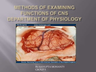
EXTIRPATION METHOD- The
extirpation method attempts to determine
the function of a given part of the brain by
removing or destroying it and observing
the resulting changes in the animal's
behavior A method for extirpation of the
pineal gland in albino rats and other
rodents (e. g., ground squirrels) is
proposed. Epiphysectomy is carried out by
resection of a fragment of the bone with the
underlying pineal gland. Using this
method, many animals can be operated
within a short period; the method is
reliable and simple, which recommends it
for chronobiological studies.
  
      . Pierre Flourens (1794-1867) was a professor of natural history at the College de France in Paris,
      who systematically destroyed parts of the brain and spinal cord in pigeons and observed the
      consequences of doing so. Flourens concluded that the cerebrum controls higher mental
      processes, parts of the midbrain control visual and auditory reflexes, the cerebellum controls
      coordination, and the medulla governs heartbeat, respiration, and other vital functions.
      Marshall Hall (1790-1857) Focused more on different parts of the brain and nervous system. He
      postulated that voluntary moveThe extirpation method attempts to determine the function of a
      given part of the brain by removing or destroying it and observing the resulting changes in the
      animal's behaviorment depends on the cerebrum, reflex movement on the spinal cord,
      involuntary movement on direct stimulation of the muscles, and respiratory movement on the
      medulla
Section of various parts of CNS where extirpation can be done-

1.SPINAL LEVEL-at the level of upper segments of spinal cord.
2.BULBAR LEVEL-between Medulla Oblongata and Mesencephalon.
3.MESENCEPHALIC LEVEL-section between mid brain and hind brain.
4.DIENCEPHALIC LEVEL-section above diencephalon.

Local Damage-
1.Mechanical-Pricking with a needle or scalpel.
2.ELECTRICAL-Inserting thin electrodes into the brain through which direct
current is passed and produces destruction of tissues.
3.FREEZING OR THERMAL COAGULATION
4.INTENSE X RAY OR ULTRASONIC VIBRATION-portions of brain tissue can
be damaged.nerve pathways can be damaged by vibrations of intensity that does
not effect the nerve cells.
5.PROTON RADIATION

                     -Non-invasive.
                     inserts electrodes.
                     does not destroy skin or bones.
                     apparatus is applied on some
                     portions of brain.
Stimulation-
               Electrical stimulation-applying a weak electrical
               stimulation to definite parts of CNS to produce
               different motor reactions.
               Used in neurosurgical operations on humans.
               Employed to examine the functions of brain stem and
               spinal cord.
               For this purpose,electrodes are implanted in different
               brain structures and attached to cranial bones.

               Non-invasive technique known as transcranial direct
               current stimulation (TDCS).
               TDCS involves stimulating specific regions of the brain
               with low-level electrical currents to enhance or reduce
               the activity of neurons. Over the last decade, the
               procedure has shown promise at improving brain
.              functioning in stroke victims as well as in people
               withParkinson's disease. But this is the first study to
               show that TDCS can help healthy individuals do better
               on math tests.
Chemical brain stimulation is the application of chemicals to brain tissue in order
to study aspects of neurochemistry , neuroanatomy andneurophysiology.
Intoduction of different chemicals stimulate different parts of CNS.
Uses the technique of electrophoresis.

A small micro pipette filled with solution is introduced into nerve centres.one small
electrode is inserted into mico pipette.another electrode is applied to the surface o
the body.when a weak DC is passed through the electrodes,the solution from the
pipette is introduced into the tissue.

 Electrophysiology is the study of the electrical
 properties of biological cells and tissues.
  It involves measurements of voltage change or electric
 current on a wide variety of scales from single ion
 channel proteins to whole organs like the heart.
 In neuroscience, it includes measurements of the
 electrical activity of neurons, and particularly action
 potential activity.
 Recordings of large-scale electric signals from the
 nervous system.
 Used in acute and chronic expts and neurosurgical
 operations.
Stereotactic surgery or stereotaxy is
  a minimally invasive form
  of surgical intervention which makes use
  of a three-dimensional coordinates system
  to locate small targets inside the body and
  to perform on them some action such
  as ablation (removal),biopsy, lesion,
  injection, stimulation,
  implantation, radiosurgery (SRS) etc.

its applications have been limited to brain surgery

   The Horsley–Clarke apparatus they
   developed was used for animal
   experimentation and implemented
   a Cartesian (three-orthogonal axis)
   system. Improved designs of their original
   device came into use in the 1930s for
   animal experimentation and are still in
   wide use today in all
   animal neuroscience laboratories
Electroencephalography-EEG refers to the recording of the brain's spontaneous
electrical activity over a short period of time, usually 20–40 minutes, as recorded
from multiple electrodes placed on the scalp.

In neurology, the main diagnostic application
of EEG is in the case of epilepsy, as epileptic
activity can create clear abnormalities on a
standard EEG study.[3]
 A secondary clinical use of EEG is in the
diagnosis of coma, encephalopathies,
and brain death.

 EEG used to be a first-line method for the
diagnosis of tumors, stroke and other focal
brain disorders, but this use has decreased
with the advent of anatomical imaging
techniques such as MRI and CT.
Wave patterns
delta waves.
Delta is the frequency range up to 4 Hz. It tends to be
the highest in amplitude and the slowest waves. It is
seen normally in adults in deep sleep. It is also seen
normally in babies. It may occur focally with subcortical
lesions and in general distribution with diffuse lesions,
metabolic encephalopathy hydrocephalus or deep
midline lesions. It is usually most prominent frontally in
adults (e.g. FIRDA - Frontal Intermittent Rhythmic
Delta) and posteriorly in children (e.g. OIRDA -
Occipital Intermittent Rhythmic Delta).
theta waves.
Theta is the frequency range from 4 Hz to 7 Hz. Theta is
seen normally in young children. It may be seen in
drowsiness or arousal in older children and adults; it
can also be seen in meditation.[17] Excess theta for age
represents abnormal activity. It can be seen as a focal
disturbance in focal subcortical lesions; it can be seen in
generalized distribution in diffuse disorder or metabolic
encephalopathy or deep midline disorders or some
instances of hydrocephalus. On the contrary this range
has been associated with reports of relaxed, meditative,
and creative states.
Alpha is the frequency range from 8 Hz to 12 Hz. Hans Berger named the first
rhythmic EEG activity he saw as the "alpha wave". This was the "posterior basic
rhythm" (also called the "posterior dominant rhythm" or the "posterior alpha
rhythm"), seen in the posterior regions of the head on both sides, higher in amplitude
on the dominant side. It emerges with closing of the eyes and with relaxation, and
attenuates with eye opening or mental exertion. The posterior basic rhythm is
actually slower than 8 Hz in young children (therefore technically in the theta range).
beta waves.
Beta is the frequency range from 12 Hz to about 30 Hz. It is seen usually on both
sides in symmetrical distribution and is most evident frontally. Beta activity is closely
linked to motor behavior and is generally attenuated during active
movements.[20] Low amplitude beta with multiple and varying frequencies is often
associated with active, busy or anxious thinking and active concentration. Rhythmic
beta with a dominant set of frequencies is associated with various pathologies and
drug effects, especially benzodiazepines. It may be absent or reduced in areas of
cortical damage. It is the dominant rhythm in patients who are alert or anxious or
who have their eyes open.
gamma waves.
Gamma is the frequency range approximately 30–100 Hz. Gamma rhythms are
thought to represent binding of different populations of neurons together into a
network for the purpose of carrying out a certain cognitive or motor function.[2]

Weitere ähnliche Inhalte

Was ist angesagt?

Cortical stimulation and mapping
Cortical stimulation and mappingCortical stimulation and mapping
Cortical stimulation and mappingPramod Krishnan
 
Brain implants By Neelima Sharma,M.Sc Biotechnology,Women Christian College,C...
Brain implants By Neelima Sharma,M.Sc Biotechnology,Women Christian College,C...Brain implants By Neelima Sharma,M.Sc Biotechnology,Women Christian College,C...
Brain implants By Neelima Sharma,M.Sc Biotechnology,Women Christian College,C...Neelima Sharma
 
EEG - Montages, Equipment and Basic Physics
EEG - Montages, Equipment and Basic PhysicsEEG - Montages, Equipment and Basic Physics
EEG - Montages, Equipment and Basic PhysicsRahul Kumar
 
Electroencephalogram (eeg)
Electroencephalogram (eeg)Electroencephalogram (eeg)
Electroencephalogram (eeg)divyabms
 
NEURODEGENERATIVE DISORDERS
NEURODEGENERATIVE DISORDERSNEURODEGENERATIVE DISORDERS
NEURODEGENERATIVE DISORDERSMariya Raju
 
Classification of Sensory receptor 2
Classification of Sensory receptor  2Classification of Sensory receptor  2
Classification of Sensory receptor 2Surjya Kumar Saikia
 
gene mapping, clonning of disease gene(1).pptx
gene mapping, clonning of disease gene(1).pptxgene mapping, clonning of disease gene(1).pptx
gene mapping, clonning of disease gene(1).pptxRajesh Yadav
 
Animal Models for Seizure and Epilepsy
Animal Models for Seizure and EpilepsyAnimal Models for Seizure and Epilepsy
Animal Models for Seizure and EpilepsyPranav Sopory
 
Normal EEG patterns, frequencies, as well as patterns that may simulate disease
Normal EEG patterns, frequencies, as well as patterns that may simulate diseaseNormal EEG patterns, frequencies, as well as patterns that may simulate disease
Normal EEG patterns, frequencies, as well as patterns that may simulate diseaseRahul Kumar
 
10b motor system voluntary control
10b motor system voluntary control10b motor system voluntary control
10b motor system voluntary controlPS Deb
 
Magnetoencephalography
MagnetoencephalographyMagnetoencephalography
MagnetoencephalographyNeurologyKota
 
Importance of si rna and microrna
Importance of si rna and microrna Importance of si rna and microrna
Importance of si rna and microrna Deepak Kumar
 
Cerebral cortex ppt
Cerebral cortex pptCerebral cortex ppt
Cerebral cortex pptfiza rizvi
 

Was ist angesagt? (20)

Cortical stimulation and mapping
Cortical stimulation and mappingCortical stimulation and mapping
Cortical stimulation and mapping
 
Electroencephal ogram (eeg)
Electroencephal ogram (eeg)Electroencephal ogram (eeg)
Electroencephal ogram (eeg)
 
Brain implants By Neelima Sharma,M.Sc Biotechnology,Women Christian College,C...
Brain implants By Neelima Sharma,M.Sc Biotechnology,Women Christian College,C...Brain implants By Neelima Sharma,M.Sc Biotechnology,Women Christian College,C...
Brain implants By Neelima Sharma,M.Sc Biotechnology,Women Christian College,C...
 
EEG - Montages, Equipment and Basic Physics
EEG - Montages, Equipment and Basic PhysicsEEG - Montages, Equipment and Basic Physics
EEG - Montages, Equipment and Basic Physics
 
Electroencephalogram (eeg)
Electroencephalogram (eeg)Electroencephalogram (eeg)
Electroencephalogram (eeg)
 
NEURODEGENERATIVE DISORDERS
NEURODEGENERATIVE DISORDERSNEURODEGENERATIVE DISORDERS
NEURODEGENERATIVE DISORDERS
 
Classification of Sensory receptor 2
Classification of Sensory receptor  2Classification of Sensory receptor  2
Classification of Sensory receptor 2
 
Electroencephalography
ElectroencephalographyElectroencephalography
Electroencephalography
 
gene mapping, clonning of disease gene(1).pptx
gene mapping, clonning of disease gene(1).pptxgene mapping, clonning of disease gene(1).pptx
gene mapping, clonning of disease gene(1).pptx
 
Animal Models for Seizure and Epilepsy
Animal Models for Seizure and EpilepsyAnimal Models for Seizure and Epilepsy
Animal Models for Seizure and Epilepsy
 
Normal EEG patterns, frequencies, as well as patterns that may simulate disease
Normal EEG patterns, frequencies, as well as patterns that may simulate diseaseNormal EEG patterns, frequencies, as well as patterns that may simulate disease
Normal EEG patterns, frequencies, as well as patterns that may simulate disease
 
10b motor system voluntary control
10b motor system voluntary control10b motor system voluntary control
10b motor system voluntary control
 
Magnetoencephalography
MagnetoencephalographyMagnetoencephalography
Magnetoencephalography
 
EEG dr archana
EEG dr archanaEEG dr archana
EEG dr archana
 
Electromyography (EMG)
Electromyography (EMG)Electromyography (EMG)
Electromyography (EMG)
 
Importance of si rna and microrna
Importance of si rna and microrna Importance of si rna and microrna
Importance of si rna and microrna
 
Cerebral cortex ppt
Cerebral cortex pptCerebral cortex ppt
Cerebral cortex ppt
 
Huntington disease
Huntington diseaseHuntington disease
Huntington disease
 
Epilepogenesis
EpilepogenesisEpilepogenesis
Epilepogenesis
 
Deep Brain Stimulation
Deep Brain StimulationDeep Brain Stimulation
Deep Brain Stimulation
 

Andere mochten auch

Case study electronic equipments in hospital ece-131
Case study electronic equipments in hospital  ece-131Case study electronic equipments in hospital  ece-131
Case study electronic equipments in hospital ece-131Pushkar N/A
 
Sterilization of ot & ot equipments pritam
Sterilization of ot & ot equipments  pritamSterilization of ot & ot equipments  pritam
Sterilization of ot & ot equipments pritamhavalprit
 
Electromyography
ElectromyographyElectromyography
ElectromyographyRajesh Goit
 
BLOOD:facts and fun
BLOOD:facts and funBLOOD:facts and fun
BLOOD:facts and funMehbob Mi
 

Andere mochten auch (8)

Emg presentation
Emg presentationEmg presentation
Emg presentation
 
Case study electronic equipments in hospital ece-131
Case study electronic equipments in hospital  ece-131Case study electronic equipments in hospital  ece-131
Case study electronic equipments in hospital ece-131
 
Sterilization of ot & ot equipments pritam
Sterilization of ot & ot equipments  pritamSterilization of ot & ot equipments  pritam
Sterilization of ot & ot equipments pritam
 
Emg fundamental
Emg fundamentalEmg fundamental
Emg fundamental
 
Electromyography
ElectromyographyElectromyography
Electromyography
 
BLOOD:facts and fun
BLOOD:facts and funBLOOD:facts and fun
BLOOD:facts and fun
 
Brain studies
Brain studiesBrain studies
Brain studies
 
BioPsych
BioPsychBioPsych
BioPsych
 

Ähnlich wie Methods of examining functions of CNS

Clinical teaching on electroencephelography
Clinical teaching on electroencephelographyClinical teaching on electroencephelography
Clinical teaching on electroencephelographyAquiflal KM
 
Intraoperative Neurophysiological Monitoring Brain
Intraoperative Neurophysiological Monitoring BrainIntraoperative Neurophysiological Monitoring Brain
Intraoperative Neurophysiological Monitoring BrainFarrukh Javeed
 
Body electric wireless power transfer for implant applications
Body electric wireless power transfer for implant applicationsBody electric wireless power transfer for implant applications
Body electric wireless power transfer for implant applicationsieeeprojectsbangalore
 
Neuromodulation in psychiatry.
Neuromodulation in psychiatry.Neuromodulation in psychiatry.
Neuromodulation in psychiatry.Vidhya Arunkumar
 
Brain-Computer-Interfaces
Brain-Computer-InterfacesBrain-Computer-Interfaces
Brain-Computer-InterfacesDominic Portain
 
Final prasoon
Final prasoonFinal prasoon
Final prasoonp_ganesh
 
Brain waves.ppt
Brain waves.pptBrain waves.ppt
Brain waves.pptShama
 
Simple information about nervous cell
Simple information about nervous cellSimple information about nervous cell
Simple information about nervous cellMohamed Ali Radwan
 
Electro convulsive therapy
Electro convulsive therapyElectro convulsive therapy
Electro convulsive therapysuswara
 
Anatomy and physiology of the nervous system
Anatomy and physiology of the nervous systemAnatomy and physiology of the nervous system
Anatomy and physiology of the nervous systemShaimaa Ibrahim
 
Cerebrum Essay
Cerebrum EssayCerebrum Essay
Cerebrum EssayDana Boo
 
POWER SPECTRAL ANALYSIS OF EEG AS A POTENTIAL MARKER IN THE DIAGNOSIS OF SPAS...
POWER SPECTRAL ANALYSIS OF EEG AS A POTENTIAL MARKER IN THE DIAGNOSIS OF SPAS...POWER SPECTRAL ANALYSIS OF EEG AS A POTENTIAL MARKER IN THE DIAGNOSIS OF SPAS...
POWER SPECTRAL ANALYSIS OF EEG AS A POTENTIAL MARKER IN THE DIAGNOSIS OF SPAS...ijbesjournal
 
Non pharmacological Treatments in Psychiatry
Non pharmacological Treatments in PsychiatryNon pharmacological Treatments in Psychiatry
Non pharmacological Treatments in PsychiatryDr Bhakti Murkey
 
Brain waves and biofeedback training
Brain waves and biofeedback trainingBrain waves and biofeedback training
Brain waves and biofeedback trainingvilla1451
 
Neuroimaging in psychiatry
Neuroimaging in psychiatryNeuroimaging in psychiatry
Neuroimaging in psychiatryDr.Pj Chakma
 

Ähnlich wie Methods of examining functions of CNS (20)

Clinical teaching on electroencephelography
Clinical teaching on electroencephelographyClinical teaching on electroencephelography
Clinical teaching on electroencephelography
 
Intraoperative Neurophysiological Monitoring Brain
Intraoperative Neurophysiological Monitoring BrainIntraoperative Neurophysiological Monitoring Brain
Intraoperative Neurophysiological Monitoring Brain
 
Body electric wireless power transfer for implant applications
Body electric wireless power transfer for implant applicationsBody electric wireless power transfer for implant applications
Body electric wireless power transfer for implant applications
 
Eeg
EegEeg
Eeg
 
kgavura 2 biology
kgavura 2 biologykgavura 2 biology
kgavura 2 biology
 
Neuromodulation in psychiatry.
Neuromodulation in psychiatry.Neuromodulation in psychiatry.
Neuromodulation in psychiatry.
 
Brain-Computer-Interfaces
Brain-Computer-InterfacesBrain-Computer-Interfaces
Brain-Computer-Interfaces
 
Final prasoon
Final prasoonFinal prasoon
Final prasoon
 
Brain waves.ppt
Brain waves.pptBrain waves.ppt
Brain waves.ppt
 
Reticular formation
Reticular formationReticular formation
Reticular formation
 
Simple information about nervous cell
Simple information about nervous cellSimple information about nervous cell
Simple information about nervous cell
 
Electro convulsive therapy
Electro convulsive therapyElectro convulsive therapy
Electro convulsive therapy
 
Anatomy and physiology of the nervous system
Anatomy and physiology of the nervous systemAnatomy and physiology of the nervous system
Anatomy and physiology of the nervous system
 
Cerebrum Essay
Cerebrum EssayCerebrum Essay
Cerebrum Essay
 
Eeg presentation
Eeg presentationEeg presentation
Eeg presentation
 
POWER SPECTRAL ANALYSIS OF EEG AS A POTENTIAL MARKER IN THE DIAGNOSIS OF SPAS...
POWER SPECTRAL ANALYSIS OF EEG AS A POTENTIAL MARKER IN THE DIAGNOSIS OF SPAS...POWER SPECTRAL ANALYSIS OF EEG AS A POTENTIAL MARKER IN THE DIAGNOSIS OF SPAS...
POWER SPECTRAL ANALYSIS OF EEG AS A POTENTIAL MARKER IN THE DIAGNOSIS OF SPAS...
 
Non pharmacological Treatments in Psychiatry
Non pharmacological Treatments in PsychiatryNon pharmacological Treatments in Psychiatry
Non pharmacological Treatments in Psychiatry
 
Brain waves and biofeedback training
Brain waves and biofeedback trainingBrain waves and biofeedback training
Brain waves and biofeedback training
 
Neuroimaging in psychiatry
Neuroimaging in psychiatryNeuroimaging in psychiatry
Neuroimaging in psychiatry
 
H177 Midterm Dezoysa
H177 Midterm DezoysaH177 Midterm Dezoysa
H177 Midterm Dezoysa
 

Mehr von Aayupta Mohanty

Maternal mortality in India
Maternal mortality in IndiaMaternal mortality in India
Maternal mortality in IndiaAayupta Mohanty
 
Medicinal electrophoresis(iontophoresis) by aayupta mohanty
Medicinal electrophoresis(iontophoresis) by aayupta mohantyMedicinal electrophoresis(iontophoresis) by aayupta mohanty
Medicinal electrophoresis(iontophoresis) by aayupta mohantyAayupta Mohanty
 
Neuromuscular diseases by aayupta mohanty
Neuromuscular diseases by  aayupta mohantyNeuromuscular diseases by  aayupta mohanty
Neuromuscular diseases by aayupta mohantyAayupta Mohanty
 
Religion as a social group
Religion as a social groupReligion as a social group
Religion as a social groupAayupta Mohanty
 
Computer as an exposure.risk prevention.
Computer as an exposure.risk prevention.Computer as an exposure.risk prevention.
Computer as an exposure.risk prevention.Aayupta Mohanty
 

Mehr von Aayupta Mohanty (7)

Porphyria
PorphyriaPorphyria
Porphyria
 
Maternal mortality in India
Maternal mortality in IndiaMaternal mortality in India
Maternal mortality in India
 
Indian culture
Indian cultureIndian culture
Indian culture
 
Medicinal electrophoresis(iontophoresis) by aayupta mohanty
Medicinal electrophoresis(iontophoresis) by aayupta mohantyMedicinal electrophoresis(iontophoresis) by aayupta mohanty
Medicinal electrophoresis(iontophoresis) by aayupta mohanty
 
Neuromuscular diseases by aayupta mohanty
Neuromuscular diseases by  aayupta mohantyNeuromuscular diseases by  aayupta mohanty
Neuromuscular diseases by aayupta mohanty
 
Religion as a social group
Religion as a social groupReligion as a social group
Religion as a social group
 
Computer as an exposure.risk prevention.
Computer as an exposure.risk prevention.Computer as an exposure.risk prevention.
Computer as an exposure.risk prevention.
 

Methods of examining functions of CNS

  • 2. EXTIRPATION METHOD- The extirpation method attempts to determine the function of a given part of the brain by removing or destroying it and observing the resulting changes in the animal's behavior A method for extirpation of the pineal gland in albino rats and other rodents (e. g., ground squirrels) is proposed. Epiphysectomy is carried out by resection of a fragment of the bone with the underlying pineal gland. Using this method, many animals can be operated within a short period; the method is reliable and simple, which recommends it for chronobiological studies.  . Pierre Flourens (1794-1867) was a professor of natural history at the College de France in Paris, who systematically destroyed parts of the brain and spinal cord in pigeons and observed the consequences of doing so. Flourens concluded that the cerebrum controls higher mental processes, parts of the midbrain control visual and auditory reflexes, the cerebellum controls coordination, and the medulla governs heartbeat, respiration, and other vital functions. Marshall Hall (1790-1857) Focused more on different parts of the brain and nervous system. He postulated that voluntary moveThe extirpation method attempts to determine the function of a given part of the brain by removing or destroying it and observing the resulting changes in the animal's behaviorment depends on the cerebrum, reflex movement on the spinal cord, involuntary movement on direct stimulation of the muscles, and respiratory movement on the medulla
  • 3. Section of various parts of CNS where extirpation can be done- 1.SPINAL LEVEL-at the level of upper segments of spinal cord. 2.BULBAR LEVEL-between Medulla Oblongata and Mesencephalon. 3.MESENCEPHALIC LEVEL-section between mid brain and hind brain. 4.DIENCEPHALIC LEVEL-section above diencephalon. Local Damage- 1.Mechanical-Pricking with a needle or scalpel. 2.ELECTRICAL-Inserting thin electrodes into the brain through which direct current is passed and produces destruction of tissues. 3.FREEZING OR THERMAL COAGULATION 4.INTENSE X RAY OR ULTRASONIC VIBRATION-portions of brain tissue can be damaged.nerve pathways can be damaged by vibrations of intensity that does not effect the nerve cells.
  • 4. 5.PROTON RADIATION -Non-invasive. inserts electrodes. does not destroy skin or bones. apparatus is applied on some portions of brain.
  • 5. Stimulation- Electrical stimulation-applying a weak electrical stimulation to definite parts of CNS to produce different motor reactions. Used in neurosurgical operations on humans. Employed to examine the functions of brain stem and spinal cord. For this purpose,electrodes are implanted in different brain structures and attached to cranial bones. Non-invasive technique known as transcranial direct current stimulation (TDCS). TDCS involves stimulating specific regions of the brain with low-level electrical currents to enhance or reduce the activity of neurons. Over the last decade, the procedure has shown promise at improving brain . functioning in stroke victims as well as in people withParkinson's disease. But this is the first study to show that TDCS can help healthy individuals do better on math tests.
  • 6. Chemical brain stimulation is the application of chemicals to brain tissue in order to study aspects of neurochemistry , neuroanatomy andneurophysiology. Intoduction of different chemicals stimulate different parts of CNS. Uses the technique of electrophoresis. A small micro pipette filled with solution is introduced into nerve centres.one small electrode is inserted into mico pipette.another electrode is applied to the surface o the body.when a weak DC is passed through the electrodes,the solution from the pipette is introduced into the tissue. Electrophysiology is the study of the electrical properties of biological cells and tissues. It involves measurements of voltage change or electric current on a wide variety of scales from single ion channel proteins to whole organs like the heart. In neuroscience, it includes measurements of the electrical activity of neurons, and particularly action potential activity. Recordings of large-scale electric signals from the nervous system. Used in acute and chronic expts and neurosurgical operations.
  • 7. Stereotactic surgery or stereotaxy is a minimally invasive form of surgical intervention which makes use of a three-dimensional coordinates system to locate small targets inside the body and to perform on them some action such as ablation (removal),biopsy, lesion, injection, stimulation, implantation, radiosurgery (SRS) etc. its applications have been limited to brain surgery The Horsley–Clarke apparatus they developed was used for animal experimentation and implemented a Cartesian (three-orthogonal axis) system. Improved designs of their original device came into use in the 1930s for animal experimentation and are still in wide use today in all animal neuroscience laboratories
  • 8. Electroencephalography-EEG refers to the recording of the brain's spontaneous electrical activity over a short period of time, usually 20–40 minutes, as recorded from multiple electrodes placed on the scalp. In neurology, the main diagnostic application of EEG is in the case of epilepsy, as epileptic activity can create clear abnormalities on a standard EEG study.[3] A secondary clinical use of EEG is in the diagnosis of coma, encephalopathies, and brain death. EEG used to be a first-line method for the diagnosis of tumors, stroke and other focal brain disorders, but this use has decreased with the advent of anatomical imaging techniques such as MRI and CT.
  • 9. Wave patterns delta waves. Delta is the frequency range up to 4 Hz. It tends to be the highest in amplitude and the slowest waves. It is seen normally in adults in deep sleep. It is also seen normally in babies. It may occur focally with subcortical lesions and in general distribution with diffuse lesions, metabolic encephalopathy hydrocephalus or deep midline lesions. It is usually most prominent frontally in adults (e.g. FIRDA - Frontal Intermittent Rhythmic Delta) and posteriorly in children (e.g. OIRDA - Occipital Intermittent Rhythmic Delta). theta waves. Theta is the frequency range from 4 Hz to 7 Hz. Theta is seen normally in young children. It may be seen in drowsiness or arousal in older children and adults; it can also be seen in meditation.[17] Excess theta for age represents abnormal activity. It can be seen as a focal disturbance in focal subcortical lesions; it can be seen in generalized distribution in diffuse disorder or metabolic encephalopathy or deep midline disorders or some instances of hydrocephalus. On the contrary this range has been associated with reports of relaxed, meditative, and creative states.
  • 10. Alpha is the frequency range from 8 Hz to 12 Hz. Hans Berger named the first rhythmic EEG activity he saw as the "alpha wave". This was the "posterior basic rhythm" (also called the "posterior dominant rhythm" or the "posterior alpha rhythm"), seen in the posterior regions of the head on both sides, higher in amplitude on the dominant side. It emerges with closing of the eyes and with relaxation, and attenuates with eye opening or mental exertion. The posterior basic rhythm is actually slower than 8 Hz in young children (therefore technically in the theta range). beta waves. Beta is the frequency range from 12 Hz to about 30 Hz. It is seen usually on both sides in symmetrical distribution and is most evident frontally. Beta activity is closely linked to motor behavior and is generally attenuated during active movements.[20] Low amplitude beta with multiple and varying frequencies is often associated with active, busy or anxious thinking and active concentration. Rhythmic beta with a dominant set of frequencies is associated with various pathologies and drug effects, especially benzodiazepines. It may be absent or reduced in areas of cortical damage. It is the dominant rhythm in patients who are alert or anxious or who have their eyes open. gamma waves. Gamma is the frequency range approximately 30–100 Hz. Gamma rhythms are thought to represent binding of different populations of neurons together into a network for the purpose of carrying out a certain cognitive or motor function.[2]