Michael Gutkin (Biology)
•Als PPTX, PDF herunterladen•
2 gefällt mir•370 views

Michael Gutkin, a 2010 graduating senior at Wagner College (B.S. in Biology), uses this Power Point slideshow to help make his thesis presentation.

Empfohlen
Weitere ähnliche Inhalte
Was ist angesagt?
Was ist angesagt? (20)
Ähnlich wie Michael Gutkin (Biology)
Ähnlich wie Michael Gutkin (Biology) (20)
Mehr von Wagner College
Mehr von Wagner College (20)
Kürzlich hochgeladen
Kürzlich hochgeladen (20)
Michael Gutkin (Biology)
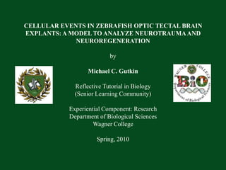
- 1. CELLULAR EVENTS IN ZEBRAFISH OPTIC TECTAL BRAIN EXPLANTS: A MODEL TO ANALYZE NEUROTRAUMA AND NEUROREGENERATION by Michael C. GutkinReflective Tutorial in Biology(Senior Learning Community) Experiential Component: ResearchDepartment of Biological SciencesWagner College Spring, 2010
- 2. Research Focuses in the Laboratory To study cellular events taking place in nervous tissue in cases of Traumatic Brain Injury (TBI). To use a model animal whose brain has known regenerative capacities. To use a model animal which inexpensive and easy to handle.
- 6. Light microscopic analysis showed formation of embryoid structures as early as 48 hours in culture which further differentiated with the extended time in culture. This picture is from a sample cultivated for 96 hours.
- 7. My Personal Research Goals Scanning electron microscopy Confocal laser scanning microscopy BrdU Cell Proliferation Immunohistochemistry
- 8. Scanning Electron MicroscopyTop Row – 12 Hours of cultivationBottom Row – 24 Hours of Cultivation
- 9. Scanning Electron Microscopy48 Hours of Cultivation
- 10. Scanning Electron Microscopy96 Hours of Cultivation
- 11. Zebrafish Brain Cytoarchitecture seen with Immunohistochemistry and Confocal Microscopy
- 12. BrdU Assay for Cell Proliferation6 & 48 Hours of Cultivation 6 h 48 h
- 13. BrdU Assay for Cell Proliferation96 Hours of Cultivation
- 14. BrdU Assay for Cell ProliferationPositive Control: CHO Cells
- 15. Conclusion Adult zebrafish brain demonstrates regenerative capacities in surviving organotypic culture. This regeneration is characterized in SEM by relocation of “stem-like cells” and the formation of embryoid bodies accompanied by neovascularization within spongiform degenerative regions. Vital cells can be detected by IHC as well as cell proliferation, but dividing cells are seen less than expected.
- 16. Acknowledgements Professor Christopher Corbo Dr. Alejandra del C. Alonso Dr. William L’Amoreaux Dr. ZoltanFulop Professor Linda Raths