

Empfohlen
Weitere ähnliche Inhalte
Was ist angesagt?
Was ist angesagt? (20)
Andere mochten auch
Andere mochten auch (20)
Ähnlich wie bacterial affections Equine
Ähnlich wie bacterial affections Equine (20)
bacterial affections Equine
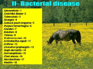
- 1. .Salmonellosis -1 .Clostridial disease -2 .Tuberculosis -3 .Strangles -4 .Guttural pouch empyema -5 .Purpura hemorrhagica -6 .Glanders -7 .Botulism -8 .Tetanus -9 .Rodococcus equi -10 .Actinobacillus equuli -11 .Anthrax -12 .Ulcerative lymphangitis -13 .Staph dermatitis -14 .Dermatophilosis -15 .Chest abscess -16 .Necrobacillosis -17 .Mastitis -18
- 2. Profuse dark diarrhea over wall of stable. Diarrhea contained blood and .shreds of intestinal mucosa Profuse diarrhea, often with blood and shreds of mucosa
- 3. Very limited area of distal jejunum and Extensive severe inflammation of proximal ileum affected with hemorrhagic jejunum which is not distended. inflammation Proximal distension )portion held( and normal large colon Well defined patches of compromised small intestinal )wall )arrow
- 4. Granulomatous lesion in wall of ileum Bilateral purulent hemorrhagic nasal discharge
- 5. Bilateral purulent nasal Bilateral mucopurulent )discharge )with blood nasal discharge Ruptured pharyngeal lymph node Massive pharyngeal and Bastered Strangles, pharyngeal submandibular swelling, discharging abscesses, lymphadenopathy weight loss, abducted elbow associated with dyspnea and ventral edema
- 6. Bastered Strangles, extensive Massive pulmonary pharyngeal and abscess with submandibular multiple lymphadenopathy compartments and thick fibrous reaction Endoscopic view of pharynx difference in discharge from guttural pouches indicating time difference to rupture of pharyngeal abscess Bastered Strangles, in guttural pouches. hypopyon with Discharge from left minimal corneal pouch is older than involvement right
- 7. Swelling ) non tympanic( below Endoscopic view,chondroids tendon of insertion of )inspissated and tumbled sternocephalicus m. purulent material. Generalized diverticulitis with loss of detailed structure such as blood vessels on mucosa Endoscopic view, caseated, purulent material on floor of medial compartment. Some loss of surface detail
- 8. Endoscopic view, purulent discharge from pharyngeal ostium of left pouch )arrow(. Endoscopic view, very liquid Marked dorsal pharyngeal contents, generalized .compression diverticulitis with loss of normally obvious detalid anatomical features such as blood vessels and nerves
- 9. Sharply Bilateral demarcated hemorrhagic edema of fore nasal limb at level discharge of elbow .))arrow Bilateral venous epistaxis Petechial hemorrhages in vaginal m.m
- 10. Chains of ulcerated lymphatic nodules Chains of ulcerated, discharging nodules Bilateral purulent following lymphatic hemorrhagic versals nasal discharge )scanty( with fetid odor
- 12. Opisthotonus and extensor rigidity of neck, limbs, trunk and tail Tail )hand pump( head elevation. Horse showed stiff and stilted gait Fixed, alert facial expression, erect ears, prolapsed third eye lid, nostrils drawn open and tense Prolapsed third eye lid and mouth enophthalmos in response to sudden noise and when )face is tapped with a finger
- 13. Miliary abscesses on Large numbers of abscesses the lung lesions in lung parenchyma Foal presented with severe diarrhea. Large numbers of abscesses in mesentery and lymph nodes of colon and cecum
- 14. Neonatal septicemia, Five day old foal with sign of generalized petechiation of neonatal septicemia, aqueous all body surfaces including flare )due to inflammatory debris .this pleural surface in aqueous humor ( , hyphema )blood in anterior chamber(. Muddy appearance of iris and miosis Multiple pyaemic abscesses encountered in a 10 day old foal with septicemia
- 15. Septicemic form, edematous swelling .of neck and throat
- 16. Caused by Corynebacterium ovis Chronic form, Corded lymphatic Lesion affecting the extensive vessels and lower hind limbs of fibrosis and purulent discharge foal above the hock. It distal edema of from ulcerated site is unusual to find hind leg )arrow(. Chronic lesions above the hock thickening and or on the front limbs exudate over distal parts of limb
- 17. Lesion restricted to saddle contact area. Very painful lesion with exudates Lesion had been present with little change for several years
- 18. Extensive hair Extensive deep loss leaving exudative hyperkeratotic dermatitis with linear scabs and cracking and skin denuded of thickening of skin hair on palmar pastern of white foot Loss of hair follows water run off pattern Extensive hair loss followed minor remaining hair is matted with exudate, grooming effort, leaving denuded some areas of hypopigmentation over skin with minimal scraping and no gluteal region apparent exudates
- 19. Hair matted in a tesselated Pale glistening surface with pattern purulent exudate found under matted hair Hair plucked from a case Paint brush effect with showing paint brush effect denudation of hair. No purulent exudate, skin surface very dry
- 20. Chest abscesses )pigeon breast / Wyoming strangles(Corynebacterium equi
- 21. Highly painful necrotic changes in skin of coronet .))arrow
- 22. Enlarged gland, mare lame, obvious engorgement of Gland was hot, painful, and )transthoracic vein )arrow had stringy milk without clots. Edema above gland Enlarged gland with fibrous induration
- 24. Obvious heavy encrustation Raised hair patches of very with minimal exudation early lesions )10 days post )infection Distribution of lesions corresponding with girth position. Several horses sharing saddlery developed an almost identical syndrome over 2 weeks. Lesion approximately 14 days post infection
- 25. Microsporum gypseum transmitted Hair loss leaves silvery by insect bite, note: location and grey glistening skin pattern of lesions correspond with which heals within 4-5 biting site of insect, hair loss not days. Border of lesion complete and scabs less easily poorly demarcated removed
- 26. Multiple nodules and crusted plaques over the shoulder
- 27. Lesion on third eye lid and/ or A mild, self limiting conjunctivitis with at the lacrimal puncta often marked epiphora was present in this case have an aggressive with the typically more severe lesions neoplastic appearance characteristic of the disease on the eye lids and within the naso lacrimal apparatus. Marked tear dermatitis is often present .down the sides of the face