1. Molecular Docking Studies of 9-Substituted
Adenine Derivatives As Selective
Phosphodiesterase Type-4 Inhibitors
Janagi, T.1, Velmurugan, D2. and Tamizh Muhil, P3.
monophosphate (cAMP) and cyclic guanosine monophosphate (cGMP),
Abstract by the respective PDE subtypes (Smith et al., 2006).
Abnormalities associated with inflammation comprise a large, Selective inhibitors of some PDE families are currently used in clinical
unrelated group of disorders, which underlie a variety of human practice for the treatment of cardiovascular disorders and erectile
diseases. The immune system is often involved with inflammatory dysfunction and other PDE inhibitors are under development for the
disorders, demonstrated in both allergic reactions and some treatment of CNS and inflammatory disorders (Schmitz et al., 2007).
myopathies. An example of the disorder associated with
inflammation includes Asthma, which is a chronic disease of airways Earliest described inhibitors of PDE4, such as rolipram, demonstrated
that is characterized by exacerbations of significant bronchospasm marked anti-inflammatory and bronchodilatory effects in vitro and in
and marked airway inflammation. Cyclic adenosine monophosphate vivo. Unfortunately, the clinical utility of these earlier compounds was
(cAMP) is thought to be associated with inflammatory cell activity: limited by their propensity to elicit various side effects such as nausea
high levels tend to decrease proliferation and cytokine secretion, and gastrointestinal distress. This has led to an extensive effort to
whereas low concentartions have the opposite effect. Since many identify novel PDE4 inhibitors that maintain the anti-inflammatory
phosphodiesterases (PDEs) degrade cAMP, inhibitors of this enzyme activity and bronchodilatory activity of rolipram but with a reduced
decrease inflammatory cell activity. Hence, the PDE enzymes are potential to produce side effects (Dastidar et al., 2009).
used as target for pharmacological inhibition. Inhibitors from the 9- 9-substituted adenine derivatives:
substituted adenine derivatives (6, 9-Disubstituted adenines & 2, 9-
Disubstituted N6 – Methyl adenines) have been studied for their Raboisson et al., described that 9-(2-fluorobenzyl-N6-methyladenine) as
inhibitory activity against PDE4 using docking softwares GoldTM a potent anticonvulsant and they found that several 9-substituted adenine
(CCDC software ltd, UK) and GlideTM (Schrödinger®, USA). derivatives elicited a concentration-dependent inhibiton of the TNF-α
Virtual Screening has been done for all these inhibitors and the release from mononuclear cells. They found the fact that this series of
ligands were chosen for induced fit docking based on their binding PDE4 inhibitors did not stimulate the in vivo gastric acid secretion in rats
affinity, glide energy and glide score. suggesting that they may produce fewer gastrointestinal side effects than
other PDE4 inhibitors (Raboisson et al., 2003). Hence the current study
Key words: 9-substituted adenine derivatives, 6, 9-Disubstituted deals with the in silico docking analysis of the ligands 9-substituted
adenines, 2, 9-Disubstituted N6 Methyl adenines, Phosphodiesterase adenine derivatives as inhibitors of PDE4 with target PDE4B using
type-4 inhibitors docking softwares and aims to evaluate the anti-inflammatory potential
of these ligands.
Introduction
Materials and Methods
Phosphodiesterases, also known as the cyclic nucleotide
phosphodiesterases (PDEs) comprise a group of enzymes, 11 in number Docking is a method which predicts the preferred orientation of one
(PDE1-PDE11) that degrade the phosphodiester bond in the second molecule to a second when bound to each other to form a stable complex
messenger molecules cAMP and cGMP (Huai et al., 2006). They in three-dimensional space. Docking is imminently important in many
regulate the localization, duration, and amplitude of cyclic nucleotide areas. Specifically, in cellular biology, the function of proteins is a result
signaling within sub cellular domains. PDEs are therefore important of its interaction (i.e. docking) with other proteins as well as other
regulators of signal transduction mediated by these second messenger molecular components (Jain, 2008). Therefore if we could predict how
molecules (Clayton et al., 2004; Omori and Kotera, 2007). In particular, proteins interact (dock) with other molecules we could possibly infer or
Phosphodiesterase type4 (PDE4) is a cAMP specific isoenzyme family inhibit function. The inhibiting function is of particular interest to drug
of PDEs that is predominantly expressed in many inflammaory cells and pharmaceutical companies (Warren et al., 2006).
(Schudt et al., 1995). GOLD is Genetic Optimization for Ligand Docking; it follows a genetic
Phosphodiesterase inhibitor: algorithm for calculating the solutions. It can be used for docking
flexible ligands into protein binding sites. Predicting how a small
A phosphodiesterase inhibitor is a drug which blocks one or more of the molecule will bind to a protein is difficult, and no program can guarantee
five subtypes of the enzyme phosphodiesterase, therefore preventing the success. The next best thing therefore is to measure as accurately as
inactivation of the intracellular second messengers, cyclic adenosine possible the reliability of the program, i.e. the chance that it will make a
2. successful prediction in a given instance. For that reason, GOLD has Table 2: 6, 9-Disubstituted Adenines
been tested on a large number of complexes extracted from the Protein
Data Bank.
Glide searches for favorable interactions between one or more typically
small ligand molecules and a larger receptor molecule usually a protein.
Each ligand must be a single molecule, while the receptor may include
Table 1: List Of Databases And Tools Used
Databases & Tools ' H FUSW Q
V L LR From the above basic structure about 22 compounds were
derived with varying R groups (Raboisson et al., 2002)
DrugBank l Target protein structure (PDE4B) was as shown in the table below:
selected from this database using the Compound Molecular R1 R2 R3
details provided for the drug Rolipram. Formula
1 C12H12N6 NH2 H Bn
PDB l Target protein structure PDE4B with 2 C12H11N5O OH H Bn
ligand component AMP was downloaded
3 C16H17N5 Bn
from this database.
4 C16H18N6 Bn
PDBSUM l To identify the active site residues from
5 C13H13N5O OMe H Bn
Ligplot of interactions with AMP.
6 C13H12ClN5 Me H 3-ClBn
Swiss PDB Viewer l To view the target protein structure 7 C15H15N5O2 Me H (MeOCO)(Ph)CH
complexed with AMP and to identify the 8 C15H16N6O Me H (MeNHCO)(Ph)CH
active site residues and export it as 9 C14H15N5O Me H (HOCH2)(Ph)CH
“.pdb” file. 10 C12H11N5 Me H Ph
11 C13H13N5 Me H Ph
Chemsketch l Ligand molecules were drawn and saved 12 C10H15N5 Me H n-Bu
as “.mol” files. 13 C11H15N5 Me H C-Pen
14 C14H15N5 Me H Ph(CH2)2
ArgusLab l To energy minimize the protein structure 15 C15H17N5 Me H Ph(CH2)3
and ligand structures (.mol files from 16 C13H12 ClN5 Me H 4-ClBn
chemsketch) and to export them as 17 C13H12 BrN5 Me H 2-BrBn
“.pdb” files, in order to use in docking. 18 C14H15N5O Me H 2-(MeO)Bn
19 C14H15N5O Me H 4-(MeO)Bn
Gold & Silver l For docking study and viewing the C14H13N5O PhCOCH2
20 Me H
results respectively. 21 C21H21N5 Me H (Ph)2CH(CH)2
22 C20H17N5O Me H 4-(PhCO)PhCH2
Glide l For docking study.
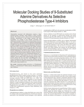
Fig 1: Interaction Of Co-crystal Ligand Docked Into
The Active Site Of Pde4 Receptor Fig 2: Interaction Of Compound 26 Docked Into
The Active Site Of Pde4 Receptor
3. TABLE 3: 2, 9-Disubstituted N6 Methyl Adenines Compound 23 NH CH 3 (N-H ...N)
2.682
N ASN 395:N
9-(2-fluoro F
N
benzyl)-N-methyl- F
N 44.68
2-trifluoromethyl- F N (O-H ...N)
9H-purin-6-amine 2.498
TYR 233:O
C14H11F4 N5 F
Compound 25 H3C
(N-H ...N)
From the above basic structure about 24 compounds were derived NH 2.629
with varying R groups (Raboisson et al., 2002) as shown in the table below: 9-benzyl-2- H3C GLN 443:N
N
isopropyl N6- 45.56
Compound Molecular R1 R2 methyladenine
N
H3C N
Formula N
(O-H ...N) 2.315
C16H19N5 TYR 233:O
23 C14H11F4 N5 CF3 2-FBn
24 C16H19N5 n-Pr Bn
25 C16H19N5 i-Pr Bn
26 C19H23N5 c-Hex Bn Compound 11 H3C (O-H ...N) 2.325
27 C17H21N5 t-Bu Bn 9-benzyl- N
TYR 233:O
N H
28 C21H19N5 PhCH=CH Bn N6-methyl - (N-H ...N)
adenine 2.482
29 C13H14N6 NH2 Bn ASN 395:N
C13H13N5 N N 45.74
30 C15H17N5O Me 2-(MeO)Bn
(N-H ...N)
31 C15H17N5O Me 4-(MeO)Bn N GLN 443:N 2.516
32 C16H19N5O Me 2-(MeO)Ph(CH2)2
(N-H ...N)
33 C12H19N5O2 Me CH3O(CH2)2O(CH2)2 GLN 443:N 2.506
34 C18H23N5 n-Pen Bn Compound 24 NH CH3 (O-H ...N)
35 Ph(CH2)2 2.745
C21H21N5 Bn TYR 233:O
9-benzyl- N
36 C22H23N5 Ph(CH2)3 Bn N6-methyl -2-n-
N
(N-H ...N) 46.13
37 C15H14F3N5O CF3 2-(MeO)Bn propyladenine N
N ASN 395:N 2.555
H3C
38 C17H21N5O n-Pr 2-(MeO)Bn C16H19N5
(N-H ...N)
39 C23H23 N5O n-Pr 4-(PhCO)PhCH2 GLN 443:N 2.389
40 C14H14FN5 Me 2-FBn Compound 38 NH2 (O-H ...N)
41 C13H12IN5 I Bn 2.625
9-(2-methoxy N
TYR 233:O
42 C14H14IN5O I 2-(MeO)Bn N
benzyl)- N6-
43 CH3C = C (N-H ...N)
C16H15N5 Bn methyl -2-n- N
GLN 443:N 2.708 47.47
H3C N
44 C17H17N5O CH3C = C 2-(MeO)Bn propyladenine
45 C17H19N5O CH3CH=CH 2-(MeO)Bn C17H21N5O (N-H ...N)
GLN 443:N 2.558
46 C15H17N5OS CH3S 2-(MeO)Bn O
CH3
Results Compound 8 H
N
CH3
(N-H …O) 2.583
N ASP 392:O
Table 4: Gold Fitness Score And Interactions Of Best Ligands N-{[6- (methyl N
N
amino)-9H-purin- N
(O-H ...N) 2.551 48.50
(Interactions : Hydrogen bonds viewed from Silver – listed for reference ligand 9-yl]methyl}-2- HN
TYR 233:O
O
AMP and all other ligands that have Gold score > AMP i.e. > 44.40) phenyl-acetamide
Bond (N-H ...O) 2.517
Compounds Structures Interactions C15H16N6O TYR 233:O
Distance Gold
(D-H ...A) Between Score Compound 34 H3C NH N O-H ...N) 2.461
Donor& TYR 233:O
Acceptor (Å) 9-benzyl- N6- N
methyl-2-n-pentyl-
N 50.09
Co-crystal (O-H …O) N (N-H ...N) 2.622
2.479 adenine GLN 443:N
Amp ASP 392:O C18H23N5
{2-[(2R, 3S, H3C
4R, 5R)-5- (O-H …O)
2.413 Compound 35 (N-H …N) 2.710
(6-amino ASP 392:O H3C NH N
octahydro-9H- 6
ASN 395:N
44.40 9-benzyl- N - N
purin-9-yl)-3,4- N
(N-H ...O) methyl-(2-phenyl (O-H ...N)
dihydroxytetrahy N
2.721 50.26
2.516 ethyl)- adenine TYR 233:O
drofuran-2-yl] ASN395:N
ethyl}phosphonic
acid C21H21N5 (N-H ...O)
(N-H ...O) 2.502
2.506 TYR 233:O
C11H24N5O6P ASN 395:N
4. H 3C H with Protein Data Bank identifier 1TB5) using the docking programs
Compound 21 N
N
GOLD and GLIDE. Most of the ligand compounds that were docked
N
N6-methyl-9- (3,3- seemed to have interaction with the active site residues like ASN 395,
N
diphenyl propyl)-
N (N-H …N) TYR 233, ASP 392, HIS 234 and GLN 443. Other than this, residue ILE
2.467 51.12
adenine ILE 410:N 410 also exhibited interaction.
C21H21N5 PDB complex or co-crystal AMP in docking analysis was found to have a
gold sore of 44.40 and a glide score of -7.217 and the glide energy was
Compound 26 (N-H ...N) 2.452 found to be -52.002. From Ligplot of interactions with ligand (Pdb
ASN 395:N complex) and from Gold and Glide docking analysis of AMP, the active
9-benzyl-2-
site residues were found to be ASP 392, ASN 395, HIS 234, TYR 233,
cyclohexyl- N6- (N-H ...N)
methyladenine 2.401 52.25 GLU 304, THR 345, ASP 275,GLN 443, MET 347 & ILE 410.
GLN 443:N
C19H23N5 Among the compounds that were docked, compound 26 (9-benzyl-2-
(O-H...N) 2.709
TYR 233:O cyclohexyl-N6-methyladenine) has given the highest score compared to
other compounds (including co-crystallized ligand) in both GLIDE and
GOLD docking analysis. The compound 26 obtained the highest score of
Table 5: Induced Fit Docking Results
52.25 in GOLD and also it exhibited the best GLIDE docking score of -
Compounds Glide Glide Energy Hydrogen bond Distance 7.833 and glide energy of -45.723 and showed strong interactions with
Score (Kcal/Mol) Interactions between the residues ASN 395,GLN 443,ASP 392 and HIS 234 in the active site,
DH…A Donor and having hydrogen bonds of length 2.939, 2.855, 2.710 and 3.266 Å
Acceptor respectively.
(Å)
Cocrystal -7.217 -52.002 (NH…O) ASN 395 3.113 Other than compound 26, compounds 24 (9-benzyl-N6-methyl-2-n
Ligand AMP (OH…O) ASP 392 2.639 propyladenine), 38 (9-(2-methoxybenzyl)-N 6 -methyl-2-n-
6
HIS 234(NH…O) 2.811 propyladenine) and 42 (2-iodo-9- (2-methoxybenzyl)-N -
TYR 233(OH...O) 2.921 methyladenine) also exhibit good interactions with the receptor. Their
(OH…O)GLU 304 2.758 scores were better than the PDB complex AMP, which we have seen
Compound 21 -5.835 -47.244 HIS 278(NH…N) 3.101 above, exhibited a gold sore of 44.40 and a glide score of -7.216.
HIS 234(NH…N) 2.898 Compound 24 obtained a glide score of -7.521(Gold score 46.13) and has
(NH…O) ASP 275) 3.199 show strong interactions with active site residues ASN 395, TYR 233 &
GLN443 with hydrogen bonds of length 2.849,3.101 & 3.007 Å
Compound 11 -7.394 -39.044 (NH…O) ASN 395 2.905
respectively.
GLN 443(NH…N) 3.073
(NH…O)GLU 304 2.597 6
Compound 38 (9-(2-methoxybenzyl)-N -methyl-2-n-propyladenine)
HIS 278(NH…N) 3.001
was observed to have a glide score of -7.467(Gold score 47.47)
Compound 38 -7.659 -44.258 (NH…O) ASN 395 2.870 comparatively better than AMP scores and has shown strong interactions
GLN 443(NH…N) 3.134 with residues ASN 395,GLN443 & TYR 233 with hydrogen bonds of
(NH…O)GLN 443) 3.132 length 2.913, 3.134 & 2.625 Å respectively.
Compound 24 -7.521 -43.049 (NH…O) ASN 395 2.849
GLN 443(NH…N) 3.007 Surprisingly Compound 42(Raboisson et al., 2002), an iodo derivative
(NH…O) TYR 233 3.101 exhibited a glide score -7.394 and has shown strong interactions with
ASN 395(NH…N) 2.967 residues ASN 395,GLN 443, TYR 233, ASP 392 & HIS 234 with
hydrogen bonds of length 2.873, 3.018, 3.113, 2.791 & 3.153 Å
Compound 26 -7.833 -45.723 (NH…O) ASN 395 2.939
GLN 443(NH…N) 2.855 respectively.
ASN 395(NH…N) 2.869
Thus from studying the interactions which the above mentioned
(NH…O) ASP 392 2.710
compounds exhibited and comparing their Gold and Glide scores with
HIS 234(NH…N) 3.266
that of PDB complex AMP, we conclude that those ligands i.e.
Compound 26 -7.394 -42.439 (NH…O) ASN 395 2.873 compounds 26,24,38 & 42 have better interactions with the active site of
GLN 443(NH…N) 3.018 the target protein PDE4 and may possess potential PDE4 inhibitory
TYR 233(OH…N) 3.113
activity.
(NH…O) ASP 392 2.791
HIS 234(NH…N) 3.088 The type of interaction, which the inhibitors exhibit, and the active site
[Various poses (<20) were obtained by docking co-crystal AMP & other residues with which they interact convey that they are good inhibitors of
ligand compounds and the collective interactions (of several poses) with PDE4 as they exhibit drug like activity. The results suggest that the
active site residues for each ligand were shown in table] compounds (9-substituted adenine derivatives) herewith proposed are
more than one molecule E.g. a protein and a cofactor (Here its PDE4B showing orientation close to active site and the compounds 26,24,38 &
and AMP). GLIDE can be run in rigid or flexible docking modes; the 42 may be used as a lead for designing future pharmaceuticals that may
later automatically generates conformation for each input ligand. be used as potential inhibitors of PDE4.
Discussion References
In the present study, we proposed and evaluated the interaction of 9- 1) Clayton RA, Dick CAJ, Mackenzie A, Nagasawa M, Galbraith D,
substituted adenine derivatives with PDE4-isoform B (Target protein Hastings SF and MacKenzie SJ (2004), The effect of selective
5. phosphodiesterase inhibitors, alone and in combination, on a murine 6) Schmitz T, Souil E, Herve R, Nicco C, Batteux F, Germain G,
model of allergic asthma, Respiratory Research, 5(1):4. Dominique C, Brion DE, Leroy MJ and Mehats C (2007), PDE4
inhibition prevents Preterm delivery induced by an intrauterine
2) Dastidar SG, Ray A, Shirumalla R, Rajagopal D, Chaudry S, Nanda inflammation, The Journal of Immunology, 178: 1115-1121.
K, Sharma P, Seth MK, Balachandran S, Gupta N and Palle V (2009),
Pharmacology of a Novel, Orally Active PDE4 Inhibitor, 7) Schudt C, Tenor H and Hatzelmann A (1995), PDE isoenzymes as
Pharmacology,83:275-286. targets for anti-asthma drugs, Eur Respir J, 8:1179-1183..
3) Huai Q, Sun Y, Wang H, Macdonald D, Aspiotis R, Robinson H, 8) Smith VB, Spina D, and Page CP (2006), Phosphodiesterase
Huang Z and Ke H (2006), Enantiomer discrimination illustrated by Inhibitors, British Journal of Pharmacology, 147: S252-S2575
high resolution crystal structures of type 4 phosphodiesterase, J Med
Chem., 49(6): 1867-1873. 9. Warren GL, Andrews CW, Capelli AM, Clarke B, Lalonde J,
Lambert MH, Lindvall M, Nevins N, Semus SF, Senger S, Tedesco
4) Omori K and Kotera J (2007), Overview of PDEs and their G, Wall ID, Woolven JM, Peishoff CE and Head MS (2006), A
regulation, Circ. Res., 100:309-327. critical assessment of docking programs and scoring functions, J
Med Chem, 49(20): 912-931.
5) Raboisson P, Lugnier C, Muller C, Reimund JM, Schultz D, Pinna G,
Bec AL, Basaran H, Desaubry L, Gaudiot F, Seloum M and
1,3 Department of Biochemistry, Biotechnology and
Bourguignon JJ (2003), Design, Synthesis and structure-activity
Bioinformatics, Avinashilingam University for women,
relationships of a series of 9-substituted adenine derivatives as
Coimbatore
selective phosphodiesterase type-4 inhibitors, European journal of
2 Center of Advanced Study in Crystallography and Biophysics,
medicinal chemistry,38:199-214
University of Madras, Guindy campus, Chennai
For Correspondence : janagi.thirumurthy@rediffmail.com