Correction to 2004 Article in Journal of Experimental Medicine
•
1 gefällt mir•232 views


Empfohlen
Weitere ähnliche Inhalte
Ähnlich wie Correction to 2004 Article in Journal of Experimental Medicine
Ähnlich wie Correction to 2004 Article in Journal of Experimental Medicine (20)
Mehr von Tamara Jorquiera
Mehr von Tamara Jorquiera (17)
Correction to 2004 Article in Journal of Experimental Medicine
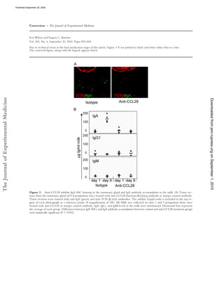
- 1. Published September 20, 2004 Correction • The Journal of Experimental Medicine Eric Wilson and Eugene C. Butcher Vol. 200, No. 6, September 20, 2004. Pages 805–809. Due to technical errors in the final production stages of this article, Figure 3 A was printed in black and white rather than in color. The corrected figure, along with the legend, appears below. The Journal of Experimental Medicine Downloaded from jem.rupress.org on September 1, 2010 Figure 3. Anti-CCL28 inhibits IgA ASC homing to the mammary gland and IgA antibody accumulation in the milk. (A) Tissue sec- tions from the mammary gland of 9-d postpartum mice treated with anti-CCL28 function-blocking antibody or isotype control antibody. Tissue sections were stained with anti-IgA (green) and anti–TCR- (red) antibodies. The subiliac lymph node is included in the top re- gion of each photograph as a reference point. A magnification of 200. (B) Milk was collected on days 1 and 9 postpartum from mice treated with anti-CCL28 or isotype control antibody. IgA, IgG1, and IgM levels in the milk were determined. Horizontal bars represent the average of each group. Differences between IgA ASCs and IgA antibody accumulation between control and anti-CCL28 treatment groups were statistically significant (P 0.001).
- 2. Published September 20, 2004 Brief Definitive Report CCL28 Controls Immunoglobulin (Ig)A Plasma Cell Accumulation in the Lactating Mammary Gland and IgA Antibody Transfer to the Neonate Eric Wilson and Eugene C. Butcher Department of Pathology, Stanford University School of Medicine, Stanford, CA 94305 Center for Molecular Biology and Medicine,Veterans Affairs Palo Alto Health Care System, Palo Alto, CA 94304 Abstract The accumulation of immunoglobulin (Ig)A antibody-secreting cells (ASCs) in the lactating mammary gland leads to secretion of antibodies into milk and their passive transfer to the suck- The Journal of Experimental Medicine Downloaded from jem.rupress.org on September 1, 2010 ling newborn. This transfer of IgA from mother to infant provides transient immune protection against a variety of gastrointestinal pathogens. Here we show that the mucosal epithelial che- mokine CCL28 is up-regulated in the mammary gland during lactation and that IgA ASCs from this tissue express CCR10 and migrate to CCL28. In vivo treatment with anti-CCL28 antibody blocks IgA ASC accumulation in the mammary gland, inhibiting IgA antibody secre- tion into milk and the subsequent appearance of antibody in the gastrointestinal tract of nursing neonates. We propose that CCL28 is a key regulator of IgA ASC accumulation in the mam- mary gland and thus controls the passive transfer of IgA antibodies from mother to infant. Key words: cell trafficking • common mucosal immune system • chemokine • passive immunity • milk Introduction The gut serves as a portal of entry for a myriad of patho- tract (9). The transfer of maternal antibodies to the nursing gens. Several mechanisms of protection have evolved to neonate provides transient immune protection to patho- protect the gut from microbes, including effector T cells gens previously encountered by the mother, and contrib- and antibody-secreting cells (ASCs). IgA ASCs are of par- utes to the dramatically reduced infant mortality levels in ticular importance because IgA antibodies are secreted children who are breast fed compared with those who are across the gut epithelium into the intestinal lumen where formula fed in developing countries (10). they can neutralize pathogens and toxins (1, 2). The gas- The participation of chemoattractants in the mammary trointestinal tract of the neonate is particularly vulnerable gland IgA response is suggested by early studies reporting to infection because the newborn is immunologically naive chemotactic activity for IgA ASCs in mouse colostrum (11). for the first several days of life, until effector T cells and It is now clear that chemoattractant cytokines (chemokines) ASCs are generated and disseminated throughout the body. play a vital role in lymphocyte trafficking, and participate as The adaptive immune system of the mother can provide key players in the multistep processes of lymphocyte re- passive protection to the suckling newborn through anti- cruitment from the blood into tissues (12). Recent studies bodies ingested in the mother’s milk (3, 4). During late have led to the hypothesis that the epithelial chemokines pregnancy and lactation, maternal IgA ASCs primed in the CCL25 and CCL28 mediate IgA ASC trafficking to gas- gut and respiratory tract, home to the mammary gland trointestinal and respiratory mucosal sites (13–15). The role (5–7), secreting antibody into the milk for passage to the of these or other chemokines in IgA ASC migration to the gastrointestinal tract of the nursing neonate (8). Secretory mammary gland has not been examined. In regard to mam- IgA is resistant to gastrointestinal enzymes, allowing the pas- mary gland homing, CCL28 is a particularly attractive can- sage of functional IgA through the infant’s gastrointestinal didate because most IgA ASCs in the body express the CCL28 receptor CCR10, migrate to CCL28 in vitro (13, 14), and CCL28 is found in milk (16). Address correspondence to Eric Wilson at his present address, Dept. of Microbiology and Molecular Biology, Brigham Young University, 773 In this report, we show that CCL28 is up-regulated in WIDB, Provo, UT 84602. Phone: (801) 422-4138; Fax: (801) 442-0519; the mammary gland during lactation, and demonstrate email: er4wilson@yahoo.com that antibodies to CCL28 inhibit the accumulation of IgA- 805 J. Exp. Med. © The Rockefeller University Press • 0022-1007/2004/09/805/5 $8.00 Volume 200, Number 6, September 20, 2004 805–809 http://www.jem.org/cgi/doi/10.1084/jem.20041069
- 3. Published September 20, 2004 producing cells in the mammary gland, providing direct ELISA. ELISA plates (Nunc) were coated with 2 g/ml of evidence that CCL28 can control local mucosal IgA ASC capture antibody diluted in PBS and coated overnight at 4 C. responses. Finally, we show that CCL28-mediated IgA Milk samples were diluted in blocking buffer and incubated in ASC accumulation is required for efficient transfer of ma- ELISA plates for 2 h at room temperature. Alkaline phosphatase– conjugated secondary antibodies were used as detection reagents. ternal IgA antibodies to the suckling neonate. Antibody concentrations were determined by constructing a standard curve of known values and calculating the microgram/ milliliter of antibody in milk or the microgram/milligram of anti- Materials and Methods body in feces. Milk from five or more mice was used to deter- PCR. Total RNA was collected from the mammary gland of mine antibody levels for each treatment group. 18 and 23 neo- BALB/c mice (the fourth abdominal mammary gland was used in nates were used to determine IgA levels in the feces of pups all experiments) at various stages of pregnancy and lactation using nursing on control- and anti-CCL28–treated mothers, respec- the RNeasy kit (QIAGEN). All PCR reactions were performed tively. Data are expressed as mean SEM. using an RNA PCR core kit (Applied Biosystems) according to Statistical Methods. Student’s t test was used to analyze the re- the manufacturer’s recommendations. The following primers were sults, and P 0.01 was considered significant. used: CCL28: sense ATGCAGCAAGCAGGGCTCACACTC, antisense ACGAGAGGCTTCGTGCCTGTGTGT; GAPHD: sense CCATGGAGAAGGCTGGGG, antisense CAAAGTTG- Results and Discussion TCATGGATGACC; CCR10: sense CCCGAAAGCCTCAC- CCL28 Is Up-regulated in the Mammary Gland during Lac- GCAGACTG, antisense GGAGCAGCCTCCGCAGGTCCC- Downloaded from jem.rupress.org on September 1, 2010 tation. Few lymphocytes are present in the mammary glands GGCGG; and CCR3: sense TCCACTGTACTCCCTGGTGT, of virgin mice and IgA ASCs are rare. IgA ASCs begin to ap- antisense GACTGCAGGAAAACTCTCCA. PCR product was pear late in pregnancy and increase dramatically in number run on a 1.5% agarose gel and visualized with ethidium bromide staining. soon after the start of lactation. By the third week of lactation, Chemotaxis and Cell Staining. Small intestine lamina propria the number of IgA ASCs has increased by several hundredfold lymphocytes and mammary gland lymphocytes were isolated by (6, 18). We determined if the level of CCL28 expression in collagenase digestion of the tissue (after removal of Peyer’s the mammary gland correlates with the accumulation of IgA patches and lymph nodes, respectively) as described previously ASCs. In contrast to constitutive mucosal expression reported (14). All tissues were collected from lactating mice 9 d postpar- for salivary gland and colon (19), we found that CCL28 ex- tum. Chemotaxis assays were performed and migrated lympho- pression in the mammary gland is tightly regulated and inti- cytes were enumerated using a bead-counting method as de- mately associated with the process of lactation. CCL28 mes- scribed previously (14). IgA ASCs were identified and defined as sage is not detected by semiquantitative RT-PCR in the described previously (14). CCL28–Ig binding was performed in mammary gland of virgin mice (Fig. 1). CCL28 message is the presence of 5 g normal goat IgG using a mCCL28-hIgG chimera detected with PE-conjugated donkey anti–human IgG slightly up-regulated during late pregnancy and early lactation, (Jackson ImmunoResearch Laboratories) as described previously correlating with the beginning of IgA ASC accumulation. (14). Negative controls were performed by inhibiting CCL28–Ig Approximately 48 h after the start of lactation, CCL28 ex- binding with 5 g polyclonal goat anti-mCCL28 (R&D Sys- pression rises dramatically and high levels of chemokine tems). The following rat anti–mouse antibodies were used for mRNA are maintained throughout lactation (Fig. 1). This re- staining: B220 (RA3-6B2), IgA (C10-3), and TCR- (H57-597; markable up-regulation of CCL28 correlates well with the all from BD Biosciences). Flow cytometry was performed on a time course of IgA ASC appearance and accumulation. FACSCalibur (BD Biosciences) using CELLQuest software. Mammary Gland IgA Cells Migrate to CCL28 and Express In Vivo Anti-CCL28 Blockade. Female BALB/c mice in their CCR10. Next, we asked whether IgA ASCs from the lac- first pregnancy were used in all experiments. In antibody-block- tating mammary gland can respond to CCL28 in in vitro ing experiments 100 g of monoclonal anti-CCL28 (R&D Sys- tems) or IgG2b isotype control antibody was injected i.p. on days 1, 3, 5, and 7 postpartum. Mouse milk was collected on days 1 and 9. Anesthetized mice were injected i.p. with 2 U oxytocin (Sigma-Aldrich) and milk was collected using a suction powered milking apparatus, similar to that described previously (17). Milk was then centrifuged at 14,000 RPM for 5 min at room tempera- ture, the fat was discarded, and the whey portion of the milk was stored at 20 C until use. Immunohistology. 8- m frozen sections were fixed in cold ac- etone for 10 min. After drying, slides were stained with FITC- labeled anti-IgA and PE-labeled anti–TCR- . Staining was visu- alized using confocal microscopy. IgA staining lymphocytes were counted by photographing random mammary gland sections and visually analyzing photographs for the number of stained cells/ field of view. Cell numbers were then scaled to reflect the num- ber of cells/mm2 of mammary gland tissue and data were ex- Figure 1. CCL28 expression in the mammary gland is up-regulated pressed as mean SEM. Multiple tissue sections from each of during lactation. RT-PCR was performed using primers specific for five mice were examined per treatment group. mouse CCL28 and GAPDH using mammary gland total RNA. 806 IgA Antibody-secreting Cell Homing to Lactating Breast
- 4. Published September 20, 2004 chemotaxis assays (Fig. 2 A). Mammary gland IgA ASCs resent a minor component of mammary ASCs. CCL28 has migrated approximately three times more efficiently to the been shown to bind two receptors, CCR3 and CCR10 CCR10 ligands CCL28 (mean migration: 36.2 5.4% (19), but mammary gland IgA ASCs did not migrate to the SEM) and CCL27 (not depicted), and less well to the to the CCR3 ligand eotaxin (not depicted). Moreover, IgA small intestinal chemokine CCL25 (mean migration: 12.1 ASCs sorted from the mammary glands of mice 9 d postpar- 3.2% SEM; P 0.01), which has been implicated in the tum showed strong expression of CCR10, but no expres- homing of CCR9-expressing IgA ASCs to the small intes- sion of CCR3 by RT-PCR (Fig. 2 C). We conclude that tine (Fig. 2 A; references 15, 20, and 21). In contrast, IgA mammary IgA ASCs, like IgA ASCs in the blood and other ASCs isolated from the small intestines migrated well to mucosal sites, express the CCL28 receptor CCR10 (22, 23). both CCL28 and CCL25 (Fig. 2 A). A CCL28–Ig fusion CCL28 Blockade Inhibits IgA ASC Accumulation to the protein bound specifically to the surface of most mammary Mammary Gland and IgA Accumulation in the Milk. To de- gland IgA ASCs (Fig. 2 B), confirming expression of termine whether CCL28 regulates IgA ASC accumulation CCL28 receptor by the majority of IgA-expressing lympho- in the mammary gland and secretory IgA levels in the milk, cytes. The robust migration of mammary gland IgA ASCs we treated mice with a function-blocking anti-CCL28 anti- to CCL28 but not CCL25 may indicate that mammary body and evaluated the number of IgA ASCs in the mam- gland IgA ASCs comprise a population of lymphocytes de- rived primarily from antigen responses in sites such as the respiratory tract and large intestine. Small intestine–derived Downloaded from jem.rupress.org on September 1, 2010 ASCs, which respond well to both chemokines, could rep- Figure 3. Anti-CCL28 inhibits IgA ASC homing to the mammary Figure 2. Mammary gland IgA ASCs migrate to CCL28, bind gland and IgA antibody accumulation in the milk. (A) Tissue sections CCL28–Ig chimera, and express CCR10. Lymphocytes were isolated from the mammary gland of 9-d postpartum mice treated with anti- from the mammary gland and small intestine of lactating mice. (A) Migra- CCL28 function-blocking antibody or isotype control antibody. Tissue tion of mammary gland and small intestine IgA ASCs to CCL25 (black sections were stained with anti-IgA (green) and anti–TCR- (red) anti- bars), CCL28 (hatched bars), and CXCL12 (white bars). **, differences bodies. The subiliac lymph node is included in the top region of each were statistically significant (P 0.01) between CCL28 and CCL25 mi- photograph as a reference point. A magnification of 200. (B) Milk was gration. Data are expressed as mean SEM. (B) CCL28–Ig binding. Left, collected on days 1 and 9 postpartum from mice treated with anti-CCL28 negative control; right, CCL28–Ig binding. (C) Total RNA was collected or isotype control antibody. IgA, IgG1, and IgM levels in the milk were from sorted mammary gland IgA ASCs. RT-PCR analysis shows expression determined. Horizontal bars represent the average of each group. Differ- of the chemokine receptor CCR10 but not CCR3 on mammary gland ences between IgA ASCs and IgA antibody accumulation between control IgA ASCs. and anti-CCL28 treatment groups were statistically significant (P 0.001). 807 Wilson and Butcher Brief Definitive Report
- 5. Published September 20, 2004 mary gland as well as the level of IgA antibody in the milk salivary gland epithelium (19), CCL28 is not highly ex- on day 9 postpartum. Immunostaining of mammary tissue pressed in the resting mammary gland, but instead is dra- sections revealed an almost complete inhibition of IgA ASC matically up-regulated postpartum in association with the accumulation in animals treated with anti-CCL28 (Fig. 3 A), onset of lactation. The up-regulation of CCL28 correlates with an average of 1.8 IgA ASCs/mm2 compared with 28.9 with an influx of large numbers of IgA-secreting cells. IgA ASCs/mm2 in isotype control mAb-treated mice (P Mammary gland ASCs, like IgA ASCs in other mucosal 0.001). Importantly, antibody blockade of CCL28 substan- sites and in blood, migrate efficiently to CCL28 in vitro tially inhibited IgA secretion into milk (Fig. 3 B). IgA anti- and express CCR10. In vivo, anti-CCL28 antibodies block body levels in milk collected 9 d postpartum from anti- the postpartum accumulation of IgA-secreting cells in the CCL28–treated animals were 20 g/ml, similar to IgA mammary gland, supporting the hypothesized role of CCL28 serum (not depicted) and milk levels on day 1 postpartum, at in the tissue recruitment of IgA plasma cells (14, 22, 26). which time IgA ASCs in the mammary gland are infrequent This blockade results in dramatically reduced levels of IgA and the majority of IgA antibody is derived from the blood antibody in milk and in the gastrointestinal tract of the (24). Conversely, IgA levels in the milk of control mice nursing infant. were 130 g/ml (P 0.001). Unlike IgA ASCs, most It has been proposed that IgA ASCs homing to diverse IgG and IgM secreting cells do not migrate to the chemo- mucosal surfaces creates a common mucosal immune sys- kine CCL28 in vitro (14) and do not accumulate, in appre- tem (27, 28). Our data indicates that the chemokine ciable numbers, in the mammary gland (18). The small CCL28 can play an integral role in such a common mu- Downloaded from jem.rupress.org on September 1, 2010 amount of IgG present in the milk is found at all stages of cosal immune system by linking homing mechanisms be- lactation and is likely serum derived (24, 25). Accordingly, tween the gut, respiratory tract, and the mammary gland. levels of IgG1 and IgM in the milk were low in lactating The “redirection” of IgA cells to the mammary gland is mice and remained unaffected by anti-CCL28 treatment controlled through the regulated expression of CCL28 (Fig. 3 B). during lactation, a process that enables the passive transfer CCL28 Blockade in the Mother Inhibits Passive Immune of maternal IgA antibody from the mother to the gut of the Transfer to the Neonate. Next, we asked whether CCL28 immunologically naive newborn. blockade in the mother significantly reduced maternal IgA levels in the gastrointestinal tract of the neonate. Mothers The authors thank B. Johnston, D.J. Campbell, and E. O’Hara for were treated with control antibody or anti-CCL28, and the advice and technical assistance. amount of IgA in the feces of nursing pups was determined E. Wilson is supported by a National Research Service Award (5F32HD042356). This work was also supported by National Institutes (Fig. 4). Newborn mice nursing on mothers treated with of Health (grants AI47822 and GM37734), by the FACS Core facility isotype control antibodies had high levels of IgA antibody of the Stanford Digestive Disease Center (under DK56339), and a in their stool (mean: 472.7 53.7 g antibody/mg feces). Merit Award from the Veterans Administration to E.C. Butcher. Conversely, newborn mice nursing on mothers treated The authors have no conflicting financial interests. with anti-CCL28 antibody had approximately sevenfold Submitted: 1 June 2004 lower levels of IgA in the stool (mean: 70.3 14.1 g an- Accepted: 27 July 2004 tibody/mg feces; P 0.001). In summary, we show that the mucosal epithelial che- mokine CCL28 regulates the accumulation of IgA ASCs in References the lactating mammary gland, and is required for transfer of 1. Williams, R.C., and R.J. Gibbons. 1972. Inhibition of bacte- maternal IgA antibodies to the suckling infant. In contrast rial adherence by secretory immunoglobulin A: a mechanism to its constitutive expression by intestinal, respiratory, and of antigen disposal. Science. 177:697–699. 2. Russell, M.W., M. Kilian, and M.E. Lamm. 1999. Biological activities of IgA. In Mucosal Immunology. P.L. Ogra, J. Mestecky, M.E. Lamm, W. Strober, J. Bienenstock, and J.R. McGhee, editors. Academic Press, San Diego. 225–240. 3. Holmgren, J., L.A. Hanson, B. Carlson, B.S. Lindblad, and J. Rahimtoola. 1976. Neutralizing antibodies against Escherichia coli and Vibrio cholerae enterotoxins in human milk from a de- veloping country. Scand. J. Immunol. 5:867–871. 4. Stoliar, O.A., R.P. Pelley, E. Kaniecki-Green, M.H. Kkaus, and C.C. Carpenter. 1976. Secretory IgA against enterotox- ins in breast-milk. Lancet. 1:1258–1261. 5. Roux, M.E., M. McWilliams, J.M. Phillips-Quagliata, P. Weisz-Carrington, and M.E. Lamm. 1977. Origin of IgA- secreting plasma cells in the mammary gland. J. Exp. Med. Figure 4. Anti-CCL28 blockade inhibits passive transfer of maternal IgA to the neonate. Feces were collected from 9-d-old neonates nursing 146:1311–1322. on mothers treated with anti-CCL28 or isotype control antibody, and 6. Tanneau, G.M., L. Hibrand-Saint Oyant, C.C. Chevaleyre, IgA levels were determined. **, statistically significant difference (P and H.P. Salmon. 1999. Differential recruitment of T- and 0.01). Data are expressed as mean SEM. IgA B-lymphocytes in the developing mammary gland in re- 808 IgA Antibody-secreting Cell Homing to Lactating Breast
- 6. Published September 20, 2004 lation to homing receptors and vascular addressins. J. His- Plasma cells and epithelial immunoglobulins in the mouse tochem. Cytochem. 47:1581–1592. mammary gland during pregnancy and lactation. J. Immunol. 7. Hanson, L.A., and E. Telemo. 1999. Immunobiology and 119:1306–1307. epidemiology of breastfeeding in relation to prevention of in- 19. Pan, J., E.J. Kunkel, U. Gosslar, N. Lazarus, P. Langdon, K. fections from a global perspective. In Mucosal Immunology. Broadwell, M.A. Vierra, M.C. Genovese, E.C. Butcher, and P.L. Ogra, J. Mestecky, M.E. Lamm, W. Strober, J. Bienen- D. Soler. 2000. A novel chemokine ligand for CCR10 and stock, and J.R. McGhee, editors. Academic Press, San Diego. CCR3 expressed by epithelial cells in mucosal tissues. J. Im- 1501–1510. munol. 165:2943–2949. 8. Goldblum, R.M., S. Ahlstedt, B. Carlsson, L.A. Hanson, U. 20. Zabel, B.A., W.W. Agace, J.J. Campbell, H.M. Heath, D. Par- Jodal, G. Lidin-Janson, and A. Sohl-Akerlund. 1975. Anti- ent, A.I. Roberts, E.C. Ebert, N. Kassam, S. Qin, M. Zovko, et body-forming cells in human colostrum after oral immunisa- al. 1999. Human G protein–coupled receptor GPR-9-6/CC tion. Nature. 257:797–798. chemokine receptor 9 is selectively expressed on intestinal 9. Lindh, E. 1975. Increased risistance of immunoglobulin A homing T lymphocytes, mucosal lymphocytes, and thymo- dimers to proteolytic degradation after binding of secretory cytes and is required for thymus-expressed chemokine-medi- component. J. Immunol. 114:284–286. ated chemotaxis. J. Exp. Med. 190:1241–1256. 10. Feachem, R.G., and M.A. Koblinski. 1984. Interventions for 21. Bowman, E.P., N.A. Kuklin, K.R. Youngman, N.H. Laz- the control of diarrhoeal diseases among young children: pro- arus, E.J. Kunkel, J. Pan, H.B. Greenberg, and E.C. Butcher. motion of breastfeeding. Bull. World Health Organ. 62:271– 2002. The intestinal chemokine thymus-expressed chemo- 291. kine (CCL25) attracts IgA antibody-secreting cells. J. Exp. 11. Czinn, S.J., and M.E. Lamm. 1986. Selective chemotaxis of Med. 195:269–275. Downloaded from jem.rupress.org on September 1, 2010 subsets of B lymphocytes from gut-associated lymphoid tissue 22. Kunkel, E.J., and E.C. Butcher. 2003. Plasma-cell homing. and its implications for the recruitment of mucosal plasma Nat. Rev. Immunol. 3:822–829. cells. J. Immunol. 136:3607–3611. 23. Nakayama, T., K. Hieshima, D. Izawa, Y. Tatsumi, A. Kan- 12. Butcher, E.C., and L.J. Picker. 1996. Lymphocyte homing amaru, and O. Yoshie. 2003. Cutting edge: profile of che- and homeostasis. Science. 272:60–66. mokine receptor expression on human plasma cells accounts 13. Kunkel, E.J., C.H. Kim, N.H. Lazarus, M.A. Vierra, D. Soler, for their efficient recruitment to target tissues. J. Immunol. E.P. Bowman, and E.C. Butcher. 2003. CCR10 expression is 170:1136–1140. a common feature of circulating and mucosal epithelial tissue 24. Halsey, J.F., C.S. Mitchell, and S.J. McKenzie. 1983. The or- IgA Ab-secreting cells. J. Clin. Invest. 111:1001–1010. igins of secretory IgA in milk: a shift during lactation from a 14. Lazarus, N.H., E.J. Kunkel, B. Johnston, E. Wilson, K.R. serum origin to local synthesis in the mammary gland. Ann. Youngman, and E.C. Butcher. 2003. A common mucosal che- N. Y. Acad. Sci. 409:452–460. mokine (mucosae-associated epithelial chemokine/CCL28) se- 25. Halsey, J.F., C. Mitchell, R. Meyer, and J.J. Cebra. 1982. lectively attracts IgA plasmablasts. J. Immunol. 170:3799–3805. Metabolism of immunoglobulin A in lactating mice: origins 15. Pabst, O., L. Ohl, M. Wendland, M.A. Wurbel, E. Krem- of immunoglobulin A in milk. Eur. J. Immunol. 12:107–112. mer, B. Malissen, and R. Forster. 2004. Chemokine receptor 26. Kunkel, E.J., and E.C. Butcher. 2002. Chemokines and the CCR9 contributes to the localization of plasma cells to the tissue-specific migration of lymphocytes. Immunity. 16:1–4. small intestine. J. Exp. Med. 199:411–416. 27. McDermott, M.R., and J. Bienenstock. 1979. Evidence for a 16. Hieshima, K., H. Ohtani, M. Shibano, D. Izawa, T. Na- common mucosal immunologic system. I. Migration of B kayama, Y. Kawasaki, F. Shiba, M. Shiota, F. Katou, T. immunoblasts into intestinal, respiratory, and genital tissues. Saito, and O. Yoshie. 2003. CCL28 has dual roles in mucosal J. Immunol. 122:1892–1898. immunity as a chemokine with broad-spectrum antimicrobial 28. Czerkinsky, C., S.J. Prince, S.M. Michalek, S. Jackson, activity. J. Immunol. 170:1452–1461. M.W. Russell, Z. Moldoveanu, J.R. McGhee, and J. Mestecky. 17. Nagasawa, H. 1979. A device for milk collection from mice. 1987. IgA antibody-producing cells in peripheral blood after Lab. Anim. Sci. 29:633–635. antigen ingestion: evidence for a common mucosal immune 18. Weisz-Carrington, P., M.E. Roux, and M.E. Lamm. 1977. system in humans. Proc. Natl. Acad. Sci. USA. 84:2449–2453. 809 Wilson and Butcher Brief Definitive Report