SlideShare a Scribd company logo
1 of 17
13
Gloria H. Su (ed.), Pancreatic Cancer: Methods and Protocols, Methods in Molecular Biology, vol. 980,
DOI 10.1007/978-1-62703-287-2_2, © Springer Science+Business Media, LLC 2013
Chapter 2
Tissue Microarrays: Construction and Use
Helen Remotti
Abstract
Tissue microarrays (TMAs) enable high-throughput tissue analysis by selecting a large number of
paraffin-embedded donor tissue block cores and transferring these tissue cores into a positionally encoded
array in the recipient TMA block. Once TMAs are constructed, a variety of analysis may be performed on
the arrays including histochemical, immunohistochemical, or immunofluorescent staining, and in situ
hybridization for DNA or RNA. TMAs offer a cost-effective method for performing parallel analysis of a
large number of tissue samples. In this chapter we outline the method of TMA construction with an
emphasis on providing useful information in the analysis of a variety of pancreatic neoplasms, including
pancreatic adenocarcinomas and pre-invasive lesions. The technique of TMA construction in this chapter
is restricted to the use of formalin-fixed paraffin-embedded tissue.
Key words: Tissue microarrays, Image analysis, Bioinformatics, Formalin-fixed paraffin-embedded
tissue, High-throughput tissue array protocol, Immunohistochemistry, In situ hybridization
The introduction of the multitumor (sausage) block as a novel
method for immunohistochemical antibody testing was initially
described by Battifora in 1986 with subsequent modification
creating the (checkerboard) block in 1990 (1, 2). Kononen and
colleagues refined the construction of the multicore tissue microar-
ray (TMA) and extended analysis to enable the parallel in situ
detection of DNA, RNA, and protein targets in a large number of
tumors (3, 4).
TMAs offer a cost-effective efficient resource for rapid
biomarker analysis for prognostic or therapeutic purposes. TMAs
facilitate high-throughput molecular profiling of cancer specimens
with minimal tissue requirements. There have been outstanding
reviews detailing methods used in constructing TMAs (5–12).
1. Introduction
14 H. Remotti
This review focuses on TMA construction-related issues that are of
particular relevance in the analysis of pancreatic neoplasms.
1. TMAs allow rapid and high-throughput discovery and valida-
tion of biomarkers. Hundreds of molecular targets can be
analyzed “in parallel” from consecutive TMA sections.
2. TMAs allow biomarker analysis in the context of tissue mor-
phology. TMAs allow localization of biomarkers to specific
cells within the tumor tissue including evaluation of the tumor
cells and interacting cells and stroma comprising the microen-
vironment of the tumor cells. TMAs also permit intracellular
compartmental localization of these biomarkers (i.e., cytoplas-
mic, membranous, and nuclear localization).
3. TMAs are cost effective and provide efficient use of reagents
and lab personnel. (Instead of performing immunohistochemi-
cal (IHC) staining on 100 slides, one can perform IHC on a
single TMA slide created with 100 tissue samples).
4. TMAs are useful for quality control standardization in IHC
and in situ hybridization (ISH). The same TMA can be uti-
lized for cross-validation or comparison studies between differ-
ent techniques.
5. TMAs can be used to evaluate sensitivity and specificity of anti-
bodies or ISH probes with respect to a large variety of tissue
types, and a wide spectrum of pathologic conditions and
neoplasms.
6. TMAs utilize only a portion of archival tissue, so limited tissue
is utilized efficiently.
7. TMAs constructed from formalin-fixed paraffin-embedded
(FFPE) tissue samples allow study of archival tissue samples
that cannot be analyzed using other high-throughput genomic
or proteomic methods.
1. One limitation of TMAs involves the issue of tissue heteroge-
neity, with a key concern of whether the TMA cores sampled
are representative of the tumor. In pathology, tumor sampling
is always an issue and the initial concern of TMAs was whether
they were representative of whole sections since they sample
far less tissue. Numerous studies have addressed concordance
of IHC staining in TMAs and whole sections. In general, for
many immunostains with relatively homogeneous staining, it
was observed that two to four tissue cores are representative of
whole sections with 95–97% concordance rates; furthermore,
five to six cores do not improve concordance rates (13). A sec-
ond concern involves biomarkers that show significant hetero-
geneity of expression within the tissue. Some biomarkers also
may show relative heterogeneity with regard to the topographic
regions of the tumor (central vs. peripheral regions). These
1.1. Benefits of TMAs
1.2. Limitations
of TMAs
152 Tissue Microarrays
topographic variations may reflect varying degrees of hypoxia
within the tumor, or differences in other components of the
microenvironment of the tumor cells.
Several studies have reported non-concordance in analysis
of TMA core samples compared to whole sections with respect
to IHC staining of markers that show heterogeneity of expres-
sion in the tumor including proliferation markers (Ki-67),
apoptosis markers (bcl-2, p53), and neoangiogenesis markers
(CD31, CD105) (14–16). Evaluation of biomarkers that show
heterogeneity in the tumor may require increased number of
cores sampling different areas of the tumor, as well as a larger
core diameter size to be representative of the tumor.
2. Early TMA validation studies demonstrated consistent and
representative protein expression by immunohistochemistry
(IHC) and DNA copy numbers by fluorescence in situ hybrid-
ization (FISH) assays (17–19). It has become increasingly clear
that nonuniformity of tissue fixation parameters has a consider-
able effect on the technical performance of a variety of in situ
assays of IHC, ISH, and FISH. Standardization of preanalytic
fixation parameters has become an area of intense focus on the
technical performance of these assays, particularly if the bio-
marker is utilized clinically as a predictive marker (20–26). We
have found immunohistochemical detection of labile phospho-
rylated proteins (e.g., pERK, pRB) may show discordant results
when comparing TMA studies derived from resection speci-
mens to studies performed on biopsies. This is largely the result
that labile proteins are more likely to be detected in core biop-
sies immediately fixed in 10% neutral buffered formalin, as
compared to resection specimens in which time to fixation may
vary greatly. In addition for RNA-ISH studies, since degrada-
tion of RNA occurs rapidly in pancreatic tissue before fixation,
it is imperative to develop a protocol in which representative
tissue to be used in the TMA is immediately fixed to optimally
preserve proteins and avoid RNA degradation.
Manual tissue arrayer-MTA-1 (Beecher instruments, available
through Estigen Tissue Science, http://www.estigen.com).
Tissue array punches (varying sizes: 0.6 mm, 1.0 mm, 1.5 mm,
or 2.0 mm, available through Estigen Tissue Science,
http://www.estigen.com).
Paraplast PlusTissue Embedding Media (McCormick
Scientific). Note: Paraffin kept at 60°C prior to use.
Oven (Fisher Scientific).
Magnifier on stand with attached light (Fisher Scientific).
2. Materials
16 H. Remotti
Stainless steel molds, extra-large (Labtek).
Tissue cassettes (Surgipath, Leica).
Flotation water bath.
Accu Edge blades.
Automated rotary microtome (Leica, Deerfield, IL).
Slide warmer (Fisher Scientific).
Pilot Pen ultrafine point (Register Office Supply, Baltimore, MD).
Although semiautomated and automated tissue microarrayers for
constructing TMAs are available, the manual microarrayer is used
in most laboratories. The first and largest commercial supplier of
arrayers was Beecher Instruments Inc. that manufactured a variety
of manual and robotic arrayers. The manual tissue arrayer MTA-I
and the automated tissue arrayer ATA-27 are available through
Estigen Tissue Science.
The first and most time-consuming step of constructing a TMA is
collecting and reviewing the appropriate starting material, consist-
ing of FFPE tissue blocks that accurately sample the disease state to
be analyzed. The material selected is dependent on the goals of the
study. An H&E section is cut from the donor paraffin blocks by
standard protocol to assess morphologic features of the tissue (see
Note 1). The H&E slide is annotated by the pathologist to indi-
cate the areas of interest for sampling (e.g., tumor and normal
areas). Each separate tissue diagnostic region can be given its own
number or letter so that it can be uniquely identified with case
number, block designation, and tissue diagnosis.
Construction of a TMA is primarily based on what particular
research question one is trying to answer.
Multi-tissue and multitumor arrays: These arrays contain tissues
from a variety of anatomic locations, sampling tumor and non-tumor
from these different sites. Small arrays can be used for quality control
for evaluating reagents/antibodies or documenting the specificity
of biomarkers in a spectrum of different tissues and tumors.
Specific tumor type arrays: These arrays may be constructed using
representative cases of a specific tumor type occurring in a single
tissue site (e.g., pancreatic adenocarcinoma). It is recommended
that control normal tissue of the primary tumor site also be sam-
pled. These arrays are helpful in studying the prevalence of a bio-
marker in a given tumor type and comparing different biomarkers
in different patients and comparing with their normal tissue.
3. Methods
3.1. Collection
and Selection
of Tissue Blocks
3.2. Design
and Organization
of TMA
3.2.1. Determining Type
of Array
172 Tissue Microarrays
Early progression arrays: These arrays analyze normal tissue, prein-
vasive lesions (e.g., pancreatic intraepithelial neoplasia (PanIN), or
intraductal papillary mucinous neoplasms (IPMN) that may include
a spectrum of low- and high-grade dysplastic lesions), in addition
to adenocarcinomas. IHC analysis of PanIN TMAs has been useful
in confirming the multistep model for pancreatic adenocarcinoma
with detection of “early, intermediate, and late” changes occurring
in pancreatic neoplasia (27).
Late progression arrays: These arrays may identify biomarkers dif-
ferentially expressed in the primary tumor, regional lymph node
metastases, or systemic metastases.
Tumor and microenvironment arrays: When studying the interac-
tions between tumor and stromal components, larger diameter
cores (1.5 or 2 mm) are recommended. Additional sampling of
stroma present at a distance from the tumor may also be useful.
Outcome-based arrays: One example of an outcome-based array
includes patients with pancreatic adenocarcinoma that received simi-
lar therapy and have been clinically followed with long-term out-
come data. These arrays may help identify predictive biomarkers that
identify specific tumor subtypes responsive to a particular therapy.
All tissue arrayers use two thin-walled needles with slightly differ-
ent core diameters, one to punch a hole in the recipient (composite
TMA) block and one to punch and transfer the core from the
donor block. The needles range in diameter from 0.6 to 2.0 mm.
When constructing a TMA with 1.5 mm cores, the recipient needle
(e.g., outer diameter 1.5 mm) punches a slightly larger hole than
the donor needle (e.g., inner diameter 1.5 mm), so the donor core
fits tightly into the recipient hole. Selection of the core size is based
on the (1) original tissue size in the donor block, (2) scope of the
study, and (3) number of blocks to be arrayed.
Usually four to five cores (0.6 or 1.0 mm) or alternatively two
to three cores (1.5–2.0 mm) are taken from two to three discrete
but representative regions. The increase in the number of cores
ensures minimal study case loss due to tissue core dropout, or tech-
nical difficulties. Larger sized cores also improve the chance of
sampling the entire lesion, or region of interest and adequate sur-
rounding tissue.
The number of cores required for representative sampling may
also depend on the degree of heterogeneity of a tumor. In addition,
if a tumor such as pancreatic ductal carcinoma is associated with a
desmoplastic stroma, a large component of the tumor may consist
of stroma and larger cores are recommended. For the majority of
TMA studies with pancreatic adenocarcinoma, we have preferred
the 1.5 or 2.0 mm core low-density composite TMAs. With more
homogeneous tumors (e.g., pancreatic neuroendocrine tumors),
sampling of smaller cores (0.6 or 1.0 mm) may be representative.
3.2.2. Determining Size
and Number of Cores
18 H. Remotti
1. The technologist matches the donor tissue blocks with the cor-
responding H&E slides in which areas of interest have been
previously marked by the pathologist with a marker (xylene-
free pen or Pilot pen).
2. The technologist creates a detailed map with core designations
for the tumor tissue (2–3 per case in general) and normal con-
trol tissue from the same case. The TMA layout is determined
and the corresponding TMA block summary is prepared to
record information about each tissue core in the array (Position
x, y: case #: Code: Tumor type, Organ tissue, Diagnosis/type,
etc.). For creation of pancreatic tumor TMAs in our labora-
tory, the number of sample cores per array ranges from 54 to
162 (see Table 1). If samples are performed in triplicate the
number of unique cases per TMA block ranges from 18 to 54
(see Figs. 1, 2, 3).
This protocol is tailored for the use of a manual arrayer MTA-1
(Beecher Instruments/Estigen Tissue Science).
1. The technologist matches the blocks to the annotated slides.
Blocks and slides are organized so that they match. Donor
blocks should be at least 1 mm thick. If the donor block is thin,
cores can be stacked (see Note 2). Slides, blocks, and TMA
map should be kept together.
TMA-coded samples should correspond to matching
blocks and slides (see Table 2). Prepare corresponding TMA
block summary to record all the information about each tissue
core in the array (Position x, y: case #: Code: Tumor type,
Organ tissue, Diagnosis/type, etc.). It may be helpful to pre-
pare a color-coded graphic map of the TMA (see Table 3).
3.2.3. Creation of TMA
Map
3.3. Technical
Construction/
Punching of TMA
Table 1
The number of cores on each tissue array block depends
on the size of the needle
Size of
the needle
Layout
(L×H)
Max # of
casesa
Max # of cores
(case triplicate)
Space between
each core Setup
0.6 mm 19×9 54b
162 1.0 mm 1.5 mm
1.0 mm 15×9 42c
126 1.0 mm 2 mm
1.5 mm 12×6 22d
66 1.0 mm 2.5 mm
2.0 mm 10×6 18e
54 1.0 mm 3 mm
a
Divide these numbers by two for cases that are comparing tumor/normal
b
54 cases plus 1 marker, 2 reference
c
42 cases plus 1 marker, 2 reference
d
22 cases plus 1 marker, 1 reference
e
18 cases plus 1 marker, 1 reference
192 Tissue Microarrays
Needle size: 0.6 mm
Max# of cores: 162 (L19 X H 9 = 162) * Max# of blocks: 54 (162 / 3 = 54) 3 cores per block*
* Blocks triplicate. 54 cases plus 1 marker, 2 references
Needle size: 1.0 mm
Max# of cores: 126 (L15 x H 9 = 126) * Max# of blocks: 42 (126 / 3 = 42) 3 cores per block *
* Blocks triplicate. 42 cases plus 1 marker, 2 references
Needle size: 1.5 mm
Max# of cores: 63 (L12 x H 6 = 63) * Max# of blocks: 21 (63 / 3 = 21) 3 cores per block *
* Blocks triplicate. 21 cases plus 1 marker, 2 references
Needle size: 2.0 mm
Max# of cores: 51 (L10 x H 6 = 51) * Max# of blocks: 17 (51 / 3 = 17) 3 cores per block *
* Blocks triplicate. 17 cases plus 1 marker, 2 references
H (9)
L (19)
Marker
Space for
reference (no
core)
.
.
.
.
.
.
.
.
.
.
.
. .
.
.
.
.
.
.
.
.
.
.
.
Empty
H (9)
L (15)
Marker
Empty
H (6)
L (12)
Marker
Empty
H (6)
L (10)
Marker
Empty
Space for
reference (no
core)
Space for
reference (no
core)
Space for
reference (no
core)
Fig. 1. Pictorial representation of TMAs with differing sized needle cores.
20 H. Remotti
2. Prepare the recipient paraffin block by pouring liquid paraffin
into a stainless steel base mold. A variety of paraffin can be
used. We use high-temperature Paraplast X-tra and extra large
molds. Cover with a slotted tissue cassette and allow to cool.
Remove the recipient block from the mold and check for any
bubbles or holes. Leave a margin of 3 mm around the array
(see Note 3).
Fig. 2. Workstation set up with manual tissue arrayer.
Fig. 3. Closer view of needle punches for recipient and donor blocks in the manual tissue
arrayer.
212 Tissue Microarrays
3. Place the recipient block in the block holder. Adjust the depth
stop by rotating the adjustment nut until the punch stops at
the desired depth within the paraffin block (typically 0.5–1 mm
above the base of the plastic tissue cassette).
4. Check the alignment of the donor (larger) and recipient
(smaller) punches of the MTA I arrayer. The circular imprints
of the punches on the paraffin block surface should be identi-
cally centered if they are correctly aligned. Adjust alignment of
punches if necessary.
Table 2
Initial spreadsheet with essential necessary for creating a
TMA array including ID number for donor block and tissue
diagnosis
Code Accession # Block # Diagnosis
IPMN5-001 SPXX-218608 A2 IPMN
IPMN5-002 SPXX-206635 A2 IPMN
IPMN5-003 SPXX-248434 A3 IPMN
IPMN5-004 SPXX-936324 A6 IPMN
IPMN5-005 SPXX-678733 A3 IPMN
IPMN5-006 SPXX-952667 B2 IPMN
IPMN5-007 SPXX-167946 A4 IPMN
IPMN5-008 SPXX-204091 A1 IPMN
IPMN5-009 SPXX-232040 A8 IPMN
IPMN5-010 SPXX-246582 A28 IPMN
IPMN5-011 SPXX-179234 A26 IPMN
MCN5-012 SPXX-270681 B14 MCN
MCN5-013 SPXX-157892 A1 MCN
IPMN5-014 SPXX-182621 A5 IPMN
IPMN5-015 SPXX-230490 A5 IPMN
IPMN5-016 SPXX-861556 A3 IPMN
IPMN5-017 SPXX-255629 A1 IPMN
PANCA5-018 SPXX-861523 A10 Adenocarcinoma
SA5-019 SPXX-451334 B8 Serous cystadenoma
AA5-020 SPXX-499721 B5 Acinar cystadenoma
AA5-021 SPXX-178690 B26 Acinar cystadenoma
22 H. Remotti
5. With the small punch mark a hole in the first position of the
array (intersection of the upper and left margins, position A1).
All other array positions will be in reference to this first spot.
Accordingly, set the X and Y micrometers of the MTA-1 to
zero. When the depth stop blocks the downward motion,
slowly release the tissue punch and eject the paraffin core.
6. Place the donor block bridge over the array block holder and
move the larger punch into the sampling position. Manually
hold the donor block in position on top of the donor block
bridge while positioning the area to be sampled directly
underneath the sample punch. Note: Superimposing the cor-
responding marked H&E slide over the tissue block will
assist in positioning the area to be sampled underneath the
tissue punch. For orientation purposes, we use a control lung
tissue as a standard marker for tissue in position A1 of all TMA
blocks, to facilitate orientation during microscopic evaluation
(see Note 4).
7. To retrieve the tissue core push downward on the sample
punch. Note: The depth stop will not block the punch motion
at the proper position for the donor block, so be careful to
prevent the punch from entering too deeply into the block
(see Note 5).
8. Remove the donor tissue block and bridge and push the punch
downward until its tip reaches the top of the hole in first hole
of the recipient array block. Use the large punch stylet to inject
the tissue core into the hole created by the smaller punch.
9. Adjust the micrometers to move the tissue punch to the next x-,
y-position. We use spacing of 1.5 and 2.0 mm between sample
centers when using 0.6 and 1.0 mm needles. For 1.5 and 2.0 mm
needles, a spacing of 2.5 mm and 3.0 mm is used.
10. Align the marked H&E slide and the corresponding block
(repeat steps 6–9, until TMA is completed).
11. The cores are gently pressed down with a spatula, to insure that
they are flushed with the surface of the block (see Note 6).
12. The recipient block is dusted clean or wiped with a kimwipe after
the placement of every punch. After several punches, the stylus/
punch complex may retain some paraffin. Move the stylus up and
down to dislodge the paraffin and wipe with a kimwipe.
13. The TMA block is placed in an adjusted metal mold with a
2 lb weight on it to prevent displacement of the cores when
the paraffin warms up. To allow annealing of cores with the
paraffin, the TMA block is placed in a slide warmer and gradu-
ally heated over 1 h (40°C for 15 min; 45°C for 15 min; 50°C
for 15 min; and 55°C for 15 min), followed at 60°C for 1 min
(see Note 7).
Table3
PictorialmapofTMAcoredataforeasyvisualization
Blocklabel:IPMN-5
Needlesize:1.5mm/numberofcases(cores):21(63)
123456789101112
1MarkerIPMN5-002IPMN5-004IPMN5-006IPMN5-008IPMN5-010MCN5-012IPMN5-014IPMN5-016PANCA5-018AA5-020AA5-021
2IPMN5-002IPMN5-004IPMN5-006IPMN5-008IPMN5-010MCN5-012IPMN5-014IPMN5-016PANCA5-018AA5-020AA5-021
3IPMN5-002IPMN5-004IPMN5-006IPMN5-008IPMN5-010MCN5-012IPMN5-014IPMN5-016PANCA5-018AA5-020AA5-021
4IPMN5-001IPMN5-003IPMN5-005IPMN5-007IPMN5-009IPMN5-011MCN5-013IPMN5-015IPMN5-017SA5-019
5IPMN5-001IPMN5-003IPMN5-005IPMN5-007IPMN5-009IPMN5-011MCN5-013IPMN5-015IPMN5-017SA5-019
6IPMN5-001IPMN5-003IPMN5-005IPMN5-007IPMN5-009IPMN5-011MCN5-013IPMN5-015IPMN5-017SA5-019
24 H. Remotti
14. Carefully transfer block in adjusted metal mold on cool plate
(−5°C) for 30 min. Weight should be kept on mold during
cooling.
1. The TMA block is trimmed on standard microtome (using new
blade). Sections should be no more than 5 m (see Note 8).
2. The TMA is placed on cool plate/ice water for standard micro-
tome sectioning.
3. In our laboratory we use standard tissue sectioning techniques
with a 34°C water bath to float off the sections onto positive-
charged sections or polylysine-coated slides. It is important to
maintain orientation of tissue on glass slides (see Note 9).
In the past, the tape transfer method (Instrumedics, Inc.)
was used due to the ease of transferring of tissue sections in the
desired orientation (3, 28). One disadvantage of the tape trans-
fer method involved increased background staining from the
adhesive residue that interfered with molecular assays, such as
ISH, FISH, and phosphorylation-specific IHC, particularly if
using automated methods.
4. In sectioning a TMA block, one H&E is stained for every 20
sections. For most studies we cut 40 sections at a time. Slides
are allowed to dry in vertical position in an open slide box for
48 h. Slides for IHC studies are optimally used within 1 or
2 weeks. For longer storage, unstained sections are stored at
−20°C, without baking the slides.
Histochemical, IHC, and ISH studies performed on TMAs can
follow similar protocols as conventional slides prepared with whole
tissue sections.
Although most TMAs are created from FFPE tissues, TMA meth-
ods may be modified to include sampling of frozen tissue or cell
lines.
Frozen TMAs are technically more difficult to construct and
require special handling. When constructing frozen TMAs special-
ized equipment is needed. Special donor tissue and recipient block
requirements with a common cutting media such as OCT and spe-
cial adhesives for section transfer for retained TMA core orienta-
tion are used (29).
One of the principal advantages of frozen TMAs includes bet-
ter RNA quality for applications in ISH assays. Frozen tissue
microarrays appear to provide excellent target material for the
study of DNA, RNA, and proteins by fixing each array slide in a
manner specific to the corresponding technique used (30). Another
advantage is that those procedures requiring fixation can be con-
ducted in samples fixed in an identical manner, since fixation is
performed for a limited time on the TMA slide.
3.4. Technical Cutting
of TMA
3.5. Biomarker
Analysis of TMA
3.6. TMA Construction
from Frozen Tissue
and Cell Line TMAs
252 Tissue Microarrays
The disadvantages include altered morphology with significant
loss of fine detail in frozen tissue compared to formalin-fixed tis-
sue. Newer commercially available methodologies for performing
molecular analysis including RNA-ISH on FFPE tissues provide
alternatives to frozen tissue TMAs (31).
Several protocols have been described for TMA preparation
from cell lines (32–34). One method involves growing cells, creat-
ing a cell pellet with subsequent formalin fixation to create an
FFPE block (5).
Analysis of TMA data can be assessed manually using an ordinal
grading system. If the number of markers and number of tissues
are relatively small, this method can be used; however it is time
consuming, semiquantitative, and requires an experienced
pathologist.
In order to optimally handle large-scale IHC analysis or to
objectively quantify IHC, ISH results, a variety of scanners are
available that will automate the acquisition of data generated with
the TMA technology. A variety of TMA analysis software programs
are available to standardize the analysis of biomarker data. These
programs may be able to analyze both chromogenic or
immunofluorescent markers to quantify biomarkers. The auto-
mated systems can use morphometric parameters to distinguish
tumor cells from background normal cells.
Data management and data analysis are therefore a major con-
cern to ensure both documentation and archiving data of the
experiment in addition to facilitating analysis in an efficient and
quantitative manner. Conventional spreadsheet-based data organi-
zation is feasible for relatively small projects. For larger projects,
analyzing thousands of samples with hundreds of biomarkers,
robust and scalable applications have to be utilized, which allow
data access and manipulation for researchers from different labora-
tories in a secure way.
1. A new recut H&E should be cut from the donor block, instead
of evaluating the H&E slide in the file. This will ensure that
the tissue has not been cut through and diagnostic tissue is still
present in the block.
2. If the thickness of the selected area of the donor block is too
thin (<1 mm), if possible another sample area or block should
be used, since the donor core will be exhausted and absent in
deeper sections of the TMA. Although it is recommended to
use paraffin blocks of similar thickness in order to maintain
3.7. Digital Image
Scanning and Analysis
4. Notes
26 H. Remotti
uniform sections with sampling of all cores throughout the
TMA, there is another alternative for utilizing thin tissue blocks.
This technique involves “stacking” or “packing” of the core
with two punches from the original donor block, one on top of
each other occupying the same hole in the recipient block.
3. We use a margin of 3.0 mm of paraffin around the entire TMA.
This prevents the recipient block from cracking during
construction.
4. The use of control tissue is recommended. We use a “marker”
lung tissue for the first core in the array. Other standard tissues
including normal pancreas and adenocarcinoma may be refer-
ences that can be utilized as a quality control for assessing uni-
formity of staining of multiple TMAs.
5. Although the depth of the recipient core can be set by the
depth stop, there is no depth stop that will set the depth of the
donor core. This depth varies when using different donor
blocks, since the tissue blocks are of varying thickness, which
determines the limit of the depth at which the cores are taken.
For an experienced technologist the depth of the donor core is
modified based on the thickness of the donor block. A separate
“Depth Stop Kit” can be purchased from Beecher that can be
used to obtain uniform lengths of donor cores; this kit may be
useful if all donor blocks are of similar thickness. If the size of
the donor core punched is too long, eject the core from the
stylus and place on a clean flat surface and trim with a clean
razor blade to the desired length. The core can then be placed
in the recipient hole with a small forceps.
6. Do not push the donor punch all the way into the receiver
hole; let it slightly protrude. It can be gently pressed with a
spatula to ensure that its position is level with the recipient
block surface.
7. It is important that the TMA block be remelted at a tempera-
ture slightly higher than the melting point of the wax used in
the recipient block for a short time (1 min) and then cooled.
This step is critical if sections of the TMA block are cut with a
standard microtome using water floatation.
8. It is important to face off the recipient block using a dedicated
rotary microtome prior to use. This ensures that the block face
is smooth and that all the arrays will be made in the identical
sectioning plane. This will minimize the amount of block
realignment that is necessary during sections and will help
272 Tissue Microarrays
optimize the number of complete sections that a TMA block
will provide.
9. It is important to maintain correct orientation of the slides in
both vertical and horizontal direction so that the sections are
oriented similarly on the slide and are easier to be read and
analyzed.
Acknowledgments
The author would like to acknowledge the assistance of Lin Yang,
Rina Wu, Ting Ting Wu, and Mariam Amrani, MD, in the prepara-
tion of this chapter.
References
1. Battifora H (1986) The multitumor (sausage)
tissue block: novel method for immunohis-
tochemical antibody testing. Lab Invest
55:244–248
2. Battifora H, Mehta P (1990) The checkerboard
tissue block. An improved multitissue control
block. Lab Invest 63:722–724
3. Kononen J, Bubendorf L, Kallioniemi A,
Barlund M, Schraml P, Leighton S, Torhorst J,
Mihatsch MJ, Sauter G, Kallioniemi OP (1998)
Tissue microarrays for high-throughput molec-
ular profiling of tumor specimens. Nat Med
4:844–847
4. Kallioniemi OP, Wagner U, Kononen J, Sauter
G (2001) Tissue microarray technology for
high-throughput molecular profiling of cancer.
Hum Mol Genet 10:657–662
5. Chandler I, Houlston R, Landberg G (2011) A
practical guide to constructing and using tissue
microarrays. Methods Mol Biol 675:363–373
6. Dhir R (2008) Tissue microarrays: an overview.
Methods Mol Biol 441:91–103
7. Eguiluz C, Viguera E, Millan L, Perez J (2006)
Multitissue array review: a chronological
description of tissue array techniques, applica-
tions and procedures. Pathol Res Pract 202:
561–568
8. Fedor HL, De Marzo AM (2005) Practical
methods for tissue microarray construction.
Methods Mol Med 103:89–101
9. Fowler CB, Man YG, Zhang S, O’Leary TJ,
Mason JT, Cunningham RE (2011) Tissue
microarrays: construction and uses. Methods
Mol Biol 724:23–35
10. Gately K, Kerr K, O’Byrne K (2011) Design,
construction, and analysis of cell line arrays and
tissue microarrays for gene expression analysis.
Methods Mol Biol 784:139–153
11. Hewitt SM (2004) Design, construction, and
use of tissue microarrays. Methods Mol Biol
264:61–72
12. Kajdacsy-Balla A, Geynisman JM, Macias V,
Setty S, Nanaji NM, Berman JJ, Dobbin K,
Melamed J, Kong X, Bosland M, Orenstein J,
Bayerl J, Becich MJ, Dhir R, Datta MW (2007)
Practical aspects of planning, building, and
interpreting tissue microarrays: the Cooperative
Prostate Cancer Tissue Resource experience.
J Mol Histol 38:113–121
13. Simon R, Mirlacher M, Sauter G (2004) Tissue
microarrays. Biotechniques 36:98–105
14. Chiesa-Vottero AG, Rybicki LA, Prayson RA
(2003) Comparison of proliferation indices in
glioblastoma multiforme by whole tissue sec-
tion vs tissue microarray. Am J Clin Pathol
120:902–908
15. Merseburger AS, Kuczyk MA, Serth J,
Bokemeyer C, Young DY, Sun L, Connelly
RR, McLeod DG, Mostofi FK, Srivastava SK,
Stenzl A, Moul JW, Sesterhenn IA (2003)
Limitations of tissue microarrays in the evalua-
tion of focal alterations of bcl-2 and p53 in
whole mount derived prostate tissues. Oncol
Rep 10:223–228
16. Charpin C, Dales JP, Garcia S, Carpentier S,
Djemli A, Andrac L, Lavaut MN, Allasia C,
Bonnier P (2004) Tumor neoangiogenesis by
CD31 and CD105 expression evaluation in
breast carcinoma tissue microarrays. Clin
Cancer Res 10:5815–5819
17. Bubendorf L, Kononen J, Koivisto P, Schraml
P, Moch H, Gasser TC, Willi N, Mihatsch MJ,
28 H. Remotti
Sauter G, Kallioniemi OP (1999) Survey of
gene amplifications during prostate cancer pro-
gression by high-throughout fluorescence in
situ hybridization on tissue microarrays. Cancer
Res 59:803–806
18. Rimm DL, Camp RL, Charette LA, Costa J,
Olsen DA, Reiss M (2001) Tissue microarray:
a new technology for amplification of tissue
resources. Cancer J 7:24–31
19. Schraml P, Kononen J, Bubendorf L, Moch H,
Bissig H, Nocito A, Mihatsch MJ, Kallioniemi
OP, Sauter G (1999) Tissue microarrays for
gene amplification surveys in many different
tumor types. Clin Cancer Res 5:1966–1975
20. Andersen CL, Hostetter G, Grigoryan A,
Sauter G, Kallioniemi A (2001) Improved pro-
cedure for fluorescence in situ hybridization on
tissue microarrays. Cytometry 45:83–86
21. Frantz GD, Pham TQ, Peale FV Jr, Hillan KJ
(2001) Detection of novel gene expression in
paraffin-embedded tissues by isotopic in situ
hybridization in tissue microarrays. J Pathol
195:87–96
22. De Marzo AM, Fedor HH, Gage WR, Rubin
MA (2002) Inadequate formalin fixation
decreases reliability of p27 immunohistochemi-
cal staining: probing optimal fixation time
using high-density tissue microarrays. Hum
Pathol 33:756–760
23. Chin SF, Daigo Y, Huang HE, Iyer NG,
Callagy G, Kranjac T, Gonzalez M, Sangan T,
Earl H, Caldas C (2003) A simple and reliable
pretreatment protocol facilitates fluorescent in
situ hybridisation on tissue microarrays of
paraffin wax embedded tumour samples. Mol
Pathol 56:275–279
24. Hammond ME, Hayes DF, Dowsett M, Allred
DC, Hagerty KL, Badve S, Fitzgibbons PL,
Francis G, Goldstein NS, Hayes M, Hicks DG,
Lester S, Love R, Mangu PB, McShane L,
Miller K, Osborne CK, Paik S, Perlmutter J,
Rhodes A, Sasano H, Schwartz JN, Sweep FC,
Taube S, Torlakovic EE, Valenstein P, Viale G,
Visscher D, Wheeler T, Williams RB, Wittliff
JL, Wolff AC (2010) American Society of
Clinical Oncology/College of American
Pathologists guideline recommendations for
immunohistochemical testing of estrogen and
progesterone receptors in breast cancer
(unabridged version). Arch Pathol Lab Med
134:e48–e72
25. Hammond ME, Hayes DF, Wolff AC (2011)
Clinical notice for American Society of Clinical
Oncology-College of American Pathologists
guideline recommendations on ER/PgR and
HER2 testing in breast cancer. J Clin Oncol
29:e458
26. Wolff AC, Hammond ME, Schwartz JN,
Hagerty KL, Allred DC, Cote RJ, Dowsett M,
Fitzgibbons PL, Hanna WM, Langer A,
McShane LM, Paik S, Pegram MD, Perez EA,
Press MF, Rhodes A, Sturgeon C, Taube SE,
Tubbs R, Vance GH, van de Vijver M, Wheeler
TM, Hayes DF (2007) American Society of
Clinical Oncology/College of American
Pathologists guideline recommendations for
human epidermal growth factor receptor 2
testing in breast cancer. Arch Pathol Lab Med
131:18–43
27. Maitra A, Adsay NV, Argani P, Iacobuzio-
Donahue C, De Marzo A, Cameron JL, Yeo CJ,
Hruban RH (2003) Multicomponent analysis
of the pancreatic adenocarcinoma progression
model using a pancreatic intraepithelial neopla-
sia tissue microarray. Mod Pathol 16:902–912
28. Sauter G, Simon R, Hillan K (2003) Tissue
microarrays in drug discovery. Nat Rev Drug
Discov 2:962–972
29. Schoenberg Fejzo M, Slamon DJ (2001)
Frozen tumor tissue microarray technology for
analysis of tumor RNA, DNA, and proteins.
Am J Pathol 159:1645–1650
30. Li H, Sun Y, Kong QY, Zhang KL, Wang XW,
Chen XY, Wang Q, Liu J (2003) Combination
of nucleic acid and protein isolation with tissue
array construction: using defined histologic
regions in single frozen tissue blocks for mul-
tiple research purposes. Int J Mol Med
12:299–304
31. Wang F, Flanagan J, Su N, Wang LC, Bui S,
Nielson A, Wu X, Vo HT, Ma XJ, Luo Y (2012)
RNAscope: a novel in situ RNA analysis plat-
form for formalin-fixed, paraffin-embedded
tissues. J Mol Diagn 14:22–29
32. Hoos A, Cordon-Cardo C (2001) Tissue
microarray profiling of cancer specimens and
cell lines: opportunities and limitations. Lab
Invest 81:1331–1338
33. Moskaluk CA, Stoler MH (2002) Agarose
mold embedding of cultured cells for tissue
microarrays. Diagn Mol Pathol 11:234–238
34. Abbott RT, Tripp S, Perkins SL, Elenitoba-
Johnson KS, Lim MS (2003) Analysis of the
PI-3-Kinase-PTEN-AKT pathway in human
lymphoma and leukemia using a cell line
microarray. Mod Pathol 16:607–612
http://www.springer.com/978-1-62703-286-5

More Related Content

What's hot

2015 City of Hope Summer Research
2015 City of Hope Summer Research 2015 City of Hope Summer Research
2015 City of Hope Summer Research
Joy Cai
 
Anti-cancer stem cell drug starts clinical trials
Anti-cancer stem cell drug starts clinical trialsAnti-cancer stem cell drug starts clinical trials
Anti-cancer stem cell drug starts clinical trials
randazzov
 

What's hot (20)

Single cell pcr
Single cell pcrSingle cell pcr
Single cell pcr
 
SCT60103 Group 2 Presentation
SCT60103 Group 2 PresentationSCT60103 Group 2 Presentation
SCT60103 Group 2 Presentation
 
SCT60103 - Chemotherapy
SCT60103 - Chemotherapy SCT60103 - Chemotherapy
SCT60103 - Chemotherapy
 
2015 City of Hope Summer Research
2015 City of Hope Summer Research 2015 City of Hope Summer Research
2015 City of Hope Summer Research
 
TriStar CSC
TriStar CSCTriStar CSC
TriStar CSC
 
PaperRozenchan 2015
PaperRozenchan 2015PaperRozenchan 2015
PaperRozenchan 2015
 
Cancer stem cell theory and evidence from colorecatal
Cancer stem cell theory and evidence from colorecatalCancer stem cell theory and evidence from colorecatal
Cancer stem cell theory and evidence from colorecatal
 
SCT60103 Group 2 Presentation
SCT60103 Group 2 PresentationSCT60103 Group 2 Presentation
SCT60103 Group 2 Presentation
 
A physical sciences network characterization of non-tumorigenic and metastati...
A physical sciences network characterization of non-tumorigenic and metastati...A physical sciences network characterization of non-tumorigenic and metastati...
A physical sciences network characterization of non-tumorigenic and metastati...
 
Cellular level cancer therapy
Cellular level  cancer therapyCellular level  cancer therapy
Cellular level cancer therapy
 
An Overview on Gene Expression Analysis
An Overview on Gene Expression AnalysisAn Overview on Gene Expression Analysis
An Overview on Gene Expression Analysis
 
SCT60103 Group 4 Assignment
SCT60103 Group 4 AssignmentSCT60103 Group 4 Assignment
SCT60103 Group 4 Assignment
 
Cancer therapy at protein level
Cancer therapy at protein levelCancer therapy at protein level
Cancer therapy at protein level
 
Circulating tumor cells in crc
Circulating tumor cells in crcCirculating tumor cells in crc
Circulating tumor cells in crc
 
Nanodroplet processing platform for deep and quantitative proteome profiling ...
Nanodroplet processing platform for deep and quantitative proteome profiling ...Nanodroplet processing platform for deep and quantitative proteome profiling ...
Nanodroplet processing platform for deep and quantitative proteome profiling ...
 
Genes and Tissue Culture Assignment Presentation (Group 3)
Genes and Tissue Culture Assignment Presentation (Group 3)Genes and Tissue Culture Assignment Presentation (Group 3)
Genes and Tissue Culture Assignment Presentation (Group 3)
 
Gtc presentation
Gtc presentationGtc presentation
Gtc presentation
 
Anti-cancer stem cell drug starts clinical trials
Anti-cancer stem cell drug starts clinical trialsAnti-cancer stem cell drug starts clinical trials
Anti-cancer stem cell drug starts clinical trials
 
urology 2013
urology 2013urology 2013
urology 2013
 
Minimal Criteria for Defining MSC's. The ISCT Position Statement
Minimal Criteria for Defining MSC's. The ISCT Position StatementMinimal Criteria for Defining MSC's. The ISCT Position Statement
Minimal Criteria for Defining MSC's. The ISCT Position Statement
 

Viewers also liked

Designing public procurement policy in developing countries
Designing public procurement policy in developing countriesDesigning public procurement policy in developing countries
Designing public procurement policy in developing countries
Springer
 
Linear algebra
Linear algebraLinear algebra
Linear algebra
Springer
 
Atlas of advanced endoaortic surgery
Atlas of advanced endoaortic surgeryAtlas of advanced endoaortic surgery
Atlas of advanced endoaortic surgery
Springer
 
11. menaxhimi me_kapitalin_punues
11. menaxhimi me_kapitalin_punues11. menaxhimi me_kapitalin_punues
11. menaxhimi me_kapitalin_punues
Valdet Shala
 

Viewers also liked (12)

Designing public procurement policy in developing countries
Designing public procurement policy in developing countriesDesigning public procurement policy in developing countries
Designing public procurement policy in developing countries
 
Linear algebra
Linear algebraLinear algebra
Linear algebra
 
Informatica(1)
Informatica(1)Informatica(1)
Informatica(1)
 
Ejercicio tabla dinamicas
Ejercicio tabla dinamicasEjercicio tabla dinamicas
Ejercicio tabla dinamicas
 
Presentación (leaneth marquez) junio 2015
Presentación (leaneth marquez) junio 2015Presentación (leaneth marquez) junio 2015
Presentación (leaneth marquez) junio 2015
 
Atlas of advanced endoaortic surgery
Atlas of advanced endoaortic surgeryAtlas of advanced endoaortic surgery
Atlas of advanced endoaortic surgery
 
La descentralización es el camino hacia el progreso del Perú
La descentralización es el camino hacia el progreso del PerúLa descentralización es el camino hacia el progreso del Perú
La descentralización es el camino hacia el progreso del Perú
 
Voxxed Days Thessaloniki 2016 - JDK 9 : Big Changes To Make Java Smaller
Voxxed Days Thessaloniki 2016 - JDK 9 : Big Changes To Make Java Smaller Voxxed Days Thessaloniki 2016 - JDK 9 : Big Changes To Make Java Smaller
Voxxed Days Thessaloniki 2016 - JDK 9 : Big Changes To Make Java Smaller
 
Measuring Supply Chain Resiliency
Measuring Supply Chain Resiliency Measuring Supply Chain Resiliency
Measuring Supply Chain Resiliency
 
Reutilizacion caso plantas musaceas venezuela
Reutilizacion  caso plantas musaceas venezuelaReutilizacion  caso plantas musaceas venezuela
Reutilizacion caso plantas musaceas venezuela
 
11. menaxhimi me_kapitalin_punues
11. menaxhimi me_kapitalin_punues11. menaxhimi me_kapitalin_punues
11. menaxhimi me_kapitalin_punues
 
International Marketing Research:Complete Aspect.
International Marketing Research:Complete Aspect.International Marketing Research:Complete Aspect.
International Marketing Research:Complete Aspect.
 

Similar to Pancreatic cancer

Majumder_B_et_al_Nature_Communications_2015
Majumder_B_et_al_Nature_Communications_2015Majumder_B_et_al_Nature_Communications_2015
Majumder_B_et_al_Nature_Communications_2015
Michelle Stevens
 
Characterization of microRNA expression profiles in normal human tissues
Characterization of microRNA expression profiles in normal human tissuesCharacterization of microRNA expression profiles in normal human tissues
Characterization of microRNA expression profiles in normal human tissues
Yu Liang
 
Identification of Rare and Novel Alleles in FFPE Tumor Samples | ESHG 2015 Po...
Identification of Rare and Novel Alleles in FFPE Tumor Samples | ESHG 2015 Po...Identification of Rare and Novel Alleles in FFPE Tumor Samples | ESHG 2015 Po...
Identification of Rare and Novel Alleles in FFPE Tumor Samples | ESHG 2015 Po...
Thermo Fisher Scientific
 
Pluripotent stem cells An in vitro model for nanotoxicity
Pluripotent stem cells An in vitro model for nanotoxicityPluripotent stem cells An in vitro model for nanotoxicity
Pluripotent stem cells An in vitro model for nanotoxicity
Dr. Harish Handral
 
Compartment specific micro rna expression profiles (poster) poster
Compartment specific micro rna expression profiles (poster) posterCompartment specific micro rna expression profiles (poster) poster
Compartment specific micro rna expression profiles (poster) poster
Jackie Lau
 
An expression meta-analysis of predicted microRNA targets identifies a diagno...
An expression meta-analysis of predicted microRNA targets identifies a diagno...An expression meta-analysis of predicted microRNA targets identifies a diagno...
An expression meta-analysis of predicted microRNA targets identifies a diagno...
Yu Liang
 

Similar to Pancreatic cancer (20)

The Acoustic Technology for Ctcs Isolation in Blood: Low-Cost Devices_Crimson...
The Acoustic Technology for Ctcs Isolation in Blood: Low-Cost Devices_Crimson...The Acoustic Technology for Ctcs Isolation in Blood: Low-Cost Devices_Crimson...
The Acoustic Technology for Ctcs Isolation in Blood: Low-Cost Devices_Crimson...
 
A bioengineering method for modeling alveolar Rhabdomyosarcoma and assessing ...
A bioengineering method for modeling alveolar Rhabdomyosarcoma and assessing ...A bioengineering method for modeling alveolar Rhabdomyosarcoma and assessing ...
A bioengineering method for modeling alveolar Rhabdomyosarcoma and assessing ...
 
Majumder_B_et_al_Nature_Communications_2015
Majumder_B_et_al_Nature_Communications_2015Majumder_B_et_al_Nature_Communications_2015
Majumder_B_et_al_Nature_Communications_2015
 
Cancer Immunogenomics
Cancer ImmunogenomicsCancer Immunogenomics
Cancer Immunogenomics
 
GLIIFCA 22 final (1)
GLIIFCA 22 final (1)GLIIFCA 22 final (1)
GLIIFCA 22 final (1)
 
Mutagenicity, Carcinogenicity, Genotoxicity Tests
Mutagenicity, Carcinogenicity, Genotoxicity TestsMutagenicity, Carcinogenicity, Genotoxicity Tests
Mutagenicity, Carcinogenicity, Genotoxicity Tests
 
Characterization of microRNA expression profiles in normal human tissues
Characterization of microRNA expression profiles in normal human tissuesCharacterization of microRNA expression profiles in normal human tissues
Characterization of microRNA expression profiles in normal human tissues
 
Identification of Rare and Novel Alleles in FFPE Tumor Samples | ESHG 2015 Po...
Identification of Rare and Novel Alleles in FFPE Tumor Samples | ESHG 2015 Po...Identification of Rare and Novel Alleles in FFPE Tumor Samples | ESHG 2015 Po...
Identification of Rare and Novel Alleles in FFPE Tumor Samples | ESHG 2015 Po...
 
Topology
TopologyTopology
Topology
 
Importance of Clinical and Pathological Characteristics of Meningiomas and Th...
Importance of Clinical and Pathological Characteristics of Meningiomas and Th...Importance of Clinical and Pathological Characteristics of Meningiomas and Th...
Importance of Clinical and Pathological Characteristics of Meningiomas and Th...
 
Pluripotent stem cells An in vitro model for nanotoxicity
Pluripotent stem cells An in vitro model for nanotoxicityPluripotent stem cells An in vitro model for nanotoxicity
Pluripotent stem cells An in vitro model for nanotoxicity
 
Compartment specific micro rna expression profiles (poster) poster
Compartment specific micro rna expression profiles (poster) posterCompartment specific micro rna expression profiles (poster) poster
Compartment specific micro rna expression profiles (poster) poster
 
Beller BHTP 7 17 09
Beller BHTP 7 17 09Beller BHTP 7 17 09
Beller BHTP 7 17 09
 
CAR- T Cell
CAR- T CellCAR- T Cell
CAR- T Cell
 
Cancer cell lines
Cancer cell linesCancer cell lines
Cancer cell lines
 
Characterization strategies for primary cell culture term paper
Characterization strategies for primary cell culture term paperCharacterization strategies for primary cell culture term paper
Characterization strategies for primary cell culture term paper
 
Cancer in vein 2 11-2018
Cancer in vein 2 11-2018Cancer in vein 2 11-2018
Cancer in vein 2 11-2018
 
Collision Tumour-IRB -Final.pptx
Collision Tumour-IRB -Final.pptxCollision Tumour-IRB -Final.pptx
Collision Tumour-IRB -Final.pptx
 
bioph Abstract
bioph Abstractbioph Abstract
bioph Abstract
 
An expression meta-analysis of predicted microRNA targets identifies a diagno...
An expression meta-analysis of predicted microRNA targets identifies a diagno...An expression meta-analysis of predicted microRNA targets identifies a diagno...
An expression meta-analysis of predicted microRNA targets identifies a diagno...
 

More from Springer

The chemistry of the actinide and transactinide elements (set vol.1 6)
The chemistry of the actinide and transactinide elements (set vol.1 6)The chemistry of the actinide and transactinide elements (set vol.1 6)
The chemistry of the actinide and transactinide elements (set vol.1 6)
Springer
 
Transition metal catalyzed enantioselective allylic substitution in organic s...
Transition metal catalyzed enantioselective allylic substitution in organic s...Transition metal catalyzed enantioselective allylic substitution in organic s...
Transition metal catalyzed enantioselective allylic substitution in organic s...
Springer
 
Total synthesis of natural products
Total synthesis of natural productsTotal synthesis of natural products
Total synthesis of natural products
Springer
 
Solid state nmr
Solid state nmrSolid state nmr
Solid state nmr
Springer
 
Mass spectrometry
Mass spectrometryMass spectrometry
Mass spectrometry
Springer
 
Higher oxidation state organopalladium and platinum
Higher oxidation state organopalladium and platinumHigher oxidation state organopalladium and platinum
Higher oxidation state organopalladium and platinum
Springer
 
Principles and applications of esr spectroscopy
Principles and applications of esr spectroscopyPrinciples and applications of esr spectroscopy
Principles and applications of esr spectroscopy
Springer
 
Inorganic 3 d structures
Inorganic 3 d structuresInorganic 3 d structures
Inorganic 3 d structures
Springer
 
Field flow fractionation in biopolymer analysis
Field flow fractionation in biopolymer analysisField flow fractionation in biopolymer analysis
Field flow fractionation in biopolymer analysis
Springer
 
Thermodynamics of crystalline states
Thermodynamics of crystalline statesThermodynamics of crystalline states
Thermodynamics of crystalline states
Springer
 
Theory of electroelasticity
Theory of electroelasticityTheory of electroelasticity
Theory of electroelasticity
Springer
 
Tensor algebra and tensor analysis for engineers
Tensor algebra and tensor analysis for engineersTensor algebra and tensor analysis for engineers
Tensor algebra and tensor analysis for engineers
Springer
 
Springer handbook of nanomaterials
Springer handbook of nanomaterialsSpringer handbook of nanomaterials
Springer handbook of nanomaterials
Springer
 
Shock wave compression of condensed matter
Shock wave compression of condensed matterShock wave compression of condensed matter
Shock wave compression of condensed matter
Springer
 
Polarization bremsstrahlung on atoms, plasmas, nanostructures and solids
Polarization bremsstrahlung on atoms, plasmas, nanostructures and solidsPolarization bremsstrahlung on atoms, plasmas, nanostructures and solids
Polarization bremsstrahlung on atoms, plasmas, nanostructures and solids
Springer
 
Nanostructured materials for magnetoelectronics
Nanostructured materials for magnetoelectronicsNanostructured materials for magnetoelectronics
Nanostructured materials for magnetoelectronics
Springer
 
Nanobioelectrochemistry
NanobioelectrochemistryNanobioelectrochemistry
Nanobioelectrochemistry
Springer
 
Modern theory of magnetism in metals and alloys
Modern theory of magnetism in metals and alloysModern theory of magnetism in metals and alloys
Modern theory of magnetism in metals and alloys
Springer
 
Mechanical behaviour of materials
Mechanical behaviour of materialsMechanical behaviour of materials
Mechanical behaviour of materials
Springer
 

More from Springer (20)

The chemistry of the actinide and transactinide elements (set vol.1 6)
The chemistry of the actinide and transactinide elements (set vol.1 6)The chemistry of the actinide and transactinide elements (set vol.1 6)
The chemistry of the actinide and transactinide elements (set vol.1 6)
 
Transition metal catalyzed enantioselective allylic substitution in organic s...
Transition metal catalyzed enantioselective allylic substitution in organic s...Transition metal catalyzed enantioselective allylic substitution in organic s...
Transition metal catalyzed enantioselective allylic substitution in organic s...
 
Total synthesis of natural products
Total synthesis of natural productsTotal synthesis of natural products
Total synthesis of natural products
 
Solid state nmr
Solid state nmrSolid state nmr
Solid state nmr
 
Mass spectrometry
Mass spectrometryMass spectrometry
Mass spectrometry
 
Higher oxidation state organopalladium and platinum
Higher oxidation state organopalladium and platinumHigher oxidation state organopalladium and platinum
Higher oxidation state organopalladium and platinum
 
Principles and applications of esr spectroscopy
Principles and applications of esr spectroscopyPrinciples and applications of esr spectroscopy
Principles and applications of esr spectroscopy
 
Inorganic 3 d structures
Inorganic 3 d structuresInorganic 3 d structures
Inorganic 3 d structures
 
Field flow fractionation in biopolymer analysis
Field flow fractionation in biopolymer analysisField flow fractionation in biopolymer analysis
Field flow fractionation in biopolymer analysis
 
Thermodynamics of crystalline states
Thermodynamics of crystalline statesThermodynamics of crystalline states
Thermodynamics of crystalline states
 
Theory of electroelasticity
Theory of electroelasticityTheory of electroelasticity
Theory of electroelasticity
 
Tensor algebra and tensor analysis for engineers
Tensor algebra and tensor analysis for engineersTensor algebra and tensor analysis for engineers
Tensor algebra and tensor analysis for engineers
 
Springer handbook of nanomaterials
Springer handbook of nanomaterialsSpringer handbook of nanomaterials
Springer handbook of nanomaterials
 
Shock wave compression of condensed matter
Shock wave compression of condensed matterShock wave compression of condensed matter
Shock wave compression of condensed matter
 
Polarization bremsstrahlung on atoms, plasmas, nanostructures and solids
Polarization bremsstrahlung on atoms, plasmas, nanostructures and solidsPolarization bremsstrahlung on atoms, plasmas, nanostructures and solids
Polarization bremsstrahlung on atoms, plasmas, nanostructures and solids
 
Nanostructured materials for magnetoelectronics
Nanostructured materials for magnetoelectronicsNanostructured materials for magnetoelectronics
Nanostructured materials for magnetoelectronics
 
Nanobioelectrochemistry
NanobioelectrochemistryNanobioelectrochemistry
Nanobioelectrochemistry
 
Modern theory of magnetism in metals and alloys
Modern theory of magnetism in metals and alloysModern theory of magnetism in metals and alloys
Modern theory of magnetism in metals and alloys
 
Mechanical behaviour of materials
Mechanical behaviour of materialsMechanical behaviour of materials
Mechanical behaviour of materials
 
Magnonics
MagnonicsMagnonics
Magnonics
 

Recently uploaded

Histor y of HAM Radio presentation slide
Histor y of HAM Radio presentation slideHistor y of HAM Radio presentation slide
Histor y of HAM Radio presentation slide
vu2urc
 

Recently uploaded (20)

Developing An App To Navigate The Roads of Brazil
Developing An App To Navigate The Roads of BrazilDeveloping An App To Navigate The Roads of Brazil
Developing An App To Navigate The Roads of Brazil
 
Scaling API-first – The story of a global engineering organization
Scaling API-first – The story of a global engineering organizationScaling API-first – The story of a global engineering organization
Scaling API-first – The story of a global engineering organization
 
Tech Trends Report 2024 Future Today Institute.pdf
Tech Trends Report 2024 Future Today Institute.pdfTech Trends Report 2024 Future Today Institute.pdf
Tech Trends Report 2024 Future Today Institute.pdf
 
Histor y of HAM Radio presentation slide
Histor y of HAM Radio presentation slideHistor y of HAM Radio presentation slide
Histor y of HAM Radio presentation slide
 
[2024]Digital Global Overview Report 2024 Meltwater.pdf
[2024]Digital Global Overview Report 2024 Meltwater.pdf[2024]Digital Global Overview Report 2024 Meltwater.pdf
[2024]Digital Global Overview Report 2024 Meltwater.pdf
 
ProductAnonymous-April2024-WinProductDiscovery-MelissaKlemke
ProductAnonymous-April2024-WinProductDiscovery-MelissaKlemkeProductAnonymous-April2024-WinProductDiscovery-MelissaKlemke
ProductAnonymous-April2024-WinProductDiscovery-MelissaKlemke
 
Mastering MySQL Database Architecture: Deep Dive into MySQL Shell and MySQL R...
Mastering MySQL Database Architecture: Deep Dive into MySQL Shell and MySQL R...Mastering MySQL Database Architecture: Deep Dive into MySQL Shell and MySQL R...
Mastering MySQL Database Architecture: Deep Dive into MySQL Shell and MySQL R...
 
Handwritten Text Recognition for manuscripts and early printed texts
Handwritten Text Recognition for manuscripts and early printed textsHandwritten Text Recognition for manuscripts and early printed texts
Handwritten Text Recognition for manuscripts and early printed texts
 
From Event to Action: Accelerate Your Decision Making with Real-Time Automation
From Event to Action: Accelerate Your Decision Making with Real-Time AutomationFrom Event to Action: Accelerate Your Decision Making with Real-Time Automation
From Event to Action: Accelerate Your Decision Making with Real-Time Automation
 
🐬 The future of MySQL is Postgres 🐘
🐬  The future of MySQL is Postgres   🐘🐬  The future of MySQL is Postgres   🐘
🐬 The future of MySQL is Postgres 🐘
 
AWS Community Day CPH - Three problems of Terraform
AWS Community Day CPH - Three problems of TerraformAWS Community Day CPH - Three problems of Terraform
AWS Community Day CPH - Three problems of Terraform
 
Connector Corner: Accelerate revenue generation using UiPath API-centric busi...
Connector Corner: Accelerate revenue generation using UiPath API-centric busi...Connector Corner: Accelerate revenue generation using UiPath API-centric busi...
Connector Corner: Accelerate revenue generation using UiPath API-centric busi...
 
Tata AIG General Insurance Company - Insurer Innovation Award 2024
Tata AIG General Insurance Company - Insurer Innovation Award 2024Tata AIG General Insurance Company - Insurer Innovation Award 2024
Tata AIG General Insurance Company - Insurer Innovation Award 2024
 
Apidays Singapore 2024 - Building Digital Trust in a Digital Economy by Veron...
Apidays Singapore 2024 - Building Digital Trust in a Digital Economy by Veron...Apidays Singapore 2024 - Building Digital Trust in a Digital Economy by Veron...
Apidays Singapore 2024 - Building Digital Trust in a Digital Economy by Veron...
 
Finology Group – Insurtech Innovation Award 2024
Finology Group – Insurtech Innovation Award 2024Finology Group – Insurtech Innovation Award 2024
Finology Group – Insurtech Innovation Award 2024
 
Data Cloud, More than a CDP by Matt Robison
Data Cloud, More than a CDP by Matt RobisonData Cloud, More than a CDP by Matt Robison
Data Cloud, More than a CDP by Matt Robison
 
Real Time Object Detection Using Open CV
Real Time Object Detection Using Open CVReal Time Object Detection Using Open CV
Real Time Object Detection Using Open CV
 
The 7 Things I Know About Cyber Security After 25 Years | April 2024
The 7 Things I Know About Cyber Security After 25 Years | April 2024The 7 Things I Know About Cyber Security After 25 Years | April 2024
The 7 Things I Know About Cyber Security After 25 Years | April 2024
 
Boost Fertility New Invention Ups Success Rates.pdf
Boost Fertility New Invention Ups Success Rates.pdfBoost Fertility New Invention Ups Success Rates.pdf
Boost Fertility New Invention Ups Success Rates.pdf
 
Strategies for Landing an Oracle DBA Job as a Fresher
Strategies for Landing an Oracle DBA Job as a FresherStrategies for Landing an Oracle DBA Job as a Fresher
Strategies for Landing an Oracle DBA Job as a Fresher
 

Pancreatic cancer

  • 1. 13 Gloria H. Su (ed.), Pancreatic Cancer: Methods and Protocols, Methods in Molecular Biology, vol. 980, DOI 10.1007/978-1-62703-287-2_2, © Springer Science+Business Media, LLC 2013 Chapter 2 Tissue Microarrays: Construction and Use Helen Remotti Abstract Tissue microarrays (TMAs) enable high-throughput tissue analysis by selecting a large number of paraffin-embedded donor tissue block cores and transferring these tissue cores into a positionally encoded array in the recipient TMA block. Once TMAs are constructed, a variety of analysis may be performed on the arrays including histochemical, immunohistochemical, or immunofluorescent staining, and in situ hybridization for DNA or RNA. TMAs offer a cost-effective method for performing parallel analysis of a large number of tissue samples. In this chapter we outline the method of TMA construction with an emphasis on providing useful information in the analysis of a variety of pancreatic neoplasms, including pancreatic adenocarcinomas and pre-invasive lesions. The technique of TMA construction in this chapter is restricted to the use of formalin-fixed paraffin-embedded tissue. Key words: Tissue microarrays, Image analysis, Bioinformatics, Formalin-fixed paraffin-embedded tissue, High-throughput tissue array protocol, Immunohistochemistry, In situ hybridization The introduction of the multitumor (sausage) block as a novel method for immunohistochemical antibody testing was initially described by Battifora in 1986 with subsequent modification creating the (checkerboard) block in 1990 (1, 2). Kononen and colleagues refined the construction of the multicore tissue microar- ray (TMA) and extended analysis to enable the parallel in situ detection of DNA, RNA, and protein targets in a large number of tumors (3, 4). TMAs offer a cost-effective efficient resource for rapid biomarker analysis for prognostic or therapeutic purposes. TMAs facilitate high-throughput molecular profiling of cancer specimens with minimal tissue requirements. There have been outstanding reviews detailing methods used in constructing TMAs (5–12). 1. Introduction
  • 2. 14 H. Remotti This review focuses on TMA construction-related issues that are of particular relevance in the analysis of pancreatic neoplasms. 1. TMAs allow rapid and high-throughput discovery and valida- tion of biomarkers. Hundreds of molecular targets can be analyzed “in parallel” from consecutive TMA sections. 2. TMAs allow biomarker analysis in the context of tissue mor- phology. TMAs allow localization of biomarkers to specific cells within the tumor tissue including evaluation of the tumor cells and interacting cells and stroma comprising the microen- vironment of the tumor cells. TMAs also permit intracellular compartmental localization of these biomarkers (i.e., cytoplas- mic, membranous, and nuclear localization). 3. TMAs are cost effective and provide efficient use of reagents and lab personnel. (Instead of performing immunohistochemi- cal (IHC) staining on 100 slides, one can perform IHC on a single TMA slide created with 100 tissue samples). 4. TMAs are useful for quality control standardization in IHC and in situ hybridization (ISH). The same TMA can be uti- lized for cross-validation or comparison studies between differ- ent techniques. 5. TMAs can be used to evaluate sensitivity and specificity of anti- bodies or ISH probes with respect to a large variety of tissue types, and a wide spectrum of pathologic conditions and neoplasms. 6. TMAs utilize only a portion of archival tissue, so limited tissue is utilized efficiently. 7. TMAs constructed from formalin-fixed paraffin-embedded (FFPE) tissue samples allow study of archival tissue samples that cannot be analyzed using other high-throughput genomic or proteomic methods. 1. One limitation of TMAs involves the issue of tissue heteroge- neity, with a key concern of whether the TMA cores sampled are representative of the tumor. In pathology, tumor sampling is always an issue and the initial concern of TMAs was whether they were representative of whole sections since they sample far less tissue. Numerous studies have addressed concordance of IHC staining in TMAs and whole sections. In general, for many immunostains with relatively homogeneous staining, it was observed that two to four tissue cores are representative of whole sections with 95–97% concordance rates; furthermore, five to six cores do not improve concordance rates (13). A sec- ond concern involves biomarkers that show significant hetero- geneity of expression within the tissue. Some biomarkers also may show relative heterogeneity with regard to the topographic regions of the tumor (central vs. peripheral regions). These 1.1. Benefits of TMAs 1.2. Limitations of TMAs
  • 3. 152 Tissue Microarrays topographic variations may reflect varying degrees of hypoxia within the tumor, or differences in other components of the microenvironment of the tumor cells. Several studies have reported non-concordance in analysis of TMA core samples compared to whole sections with respect to IHC staining of markers that show heterogeneity of expres- sion in the tumor including proliferation markers (Ki-67), apoptosis markers (bcl-2, p53), and neoangiogenesis markers (CD31, CD105) (14–16). Evaluation of biomarkers that show heterogeneity in the tumor may require increased number of cores sampling different areas of the tumor, as well as a larger core diameter size to be representative of the tumor. 2. Early TMA validation studies demonstrated consistent and representative protein expression by immunohistochemistry (IHC) and DNA copy numbers by fluorescence in situ hybrid- ization (FISH) assays (17–19). It has become increasingly clear that nonuniformity of tissue fixation parameters has a consider- able effect on the technical performance of a variety of in situ assays of IHC, ISH, and FISH. Standardization of preanalytic fixation parameters has become an area of intense focus on the technical performance of these assays, particularly if the bio- marker is utilized clinically as a predictive marker (20–26). We have found immunohistochemical detection of labile phospho- rylated proteins (e.g., pERK, pRB) may show discordant results when comparing TMA studies derived from resection speci- mens to studies performed on biopsies. This is largely the result that labile proteins are more likely to be detected in core biop- sies immediately fixed in 10% neutral buffered formalin, as compared to resection specimens in which time to fixation may vary greatly. In addition for RNA-ISH studies, since degrada- tion of RNA occurs rapidly in pancreatic tissue before fixation, it is imperative to develop a protocol in which representative tissue to be used in the TMA is immediately fixed to optimally preserve proteins and avoid RNA degradation. Manual tissue arrayer-MTA-1 (Beecher instruments, available through Estigen Tissue Science, http://www.estigen.com). Tissue array punches (varying sizes: 0.6 mm, 1.0 mm, 1.5 mm, or 2.0 mm, available through Estigen Tissue Science, http://www.estigen.com). Paraplast PlusTissue Embedding Media (McCormick Scientific). Note: Paraffin kept at 60°C prior to use. Oven (Fisher Scientific). Magnifier on stand with attached light (Fisher Scientific). 2. Materials
  • 4. 16 H. Remotti Stainless steel molds, extra-large (Labtek). Tissue cassettes (Surgipath, Leica). Flotation water bath. Accu Edge blades. Automated rotary microtome (Leica, Deerfield, IL). Slide warmer (Fisher Scientific). Pilot Pen ultrafine point (Register Office Supply, Baltimore, MD). Although semiautomated and automated tissue microarrayers for constructing TMAs are available, the manual microarrayer is used in most laboratories. The first and largest commercial supplier of arrayers was Beecher Instruments Inc. that manufactured a variety of manual and robotic arrayers. The manual tissue arrayer MTA-I and the automated tissue arrayer ATA-27 are available through Estigen Tissue Science. The first and most time-consuming step of constructing a TMA is collecting and reviewing the appropriate starting material, consist- ing of FFPE tissue blocks that accurately sample the disease state to be analyzed. The material selected is dependent on the goals of the study. An H&E section is cut from the donor paraffin blocks by standard protocol to assess morphologic features of the tissue (see Note 1). The H&E slide is annotated by the pathologist to indi- cate the areas of interest for sampling (e.g., tumor and normal areas). Each separate tissue diagnostic region can be given its own number or letter so that it can be uniquely identified with case number, block designation, and tissue diagnosis. Construction of a TMA is primarily based on what particular research question one is trying to answer. Multi-tissue and multitumor arrays: These arrays contain tissues from a variety of anatomic locations, sampling tumor and non-tumor from these different sites. Small arrays can be used for quality control for evaluating reagents/antibodies or documenting the specificity of biomarkers in a spectrum of different tissues and tumors. Specific tumor type arrays: These arrays may be constructed using representative cases of a specific tumor type occurring in a single tissue site (e.g., pancreatic adenocarcinoma). It is recommended that control normal tissue of the primary tumor site also be sam- pled. These arrays are helpful in studying the prevalence of a bio- marker in a given tumor type and comparing different biomarkers in different patients and comparing with their normal tissue. 3. Methods 3.1. Collection and Selection of Tissue Blocks 3.2. Design and Organization of TMA 3.2.1. Determining Type of Array
  • 5. 172 Tissue Microarrays Early progression arrays: These arrays analyze normal tissue, prein- vasive lesions (e.g., pancreatic intraepithelial neoplasia (PanIN), or intraductal papillary mucinous neoplasms (IPMN) that may include a spectrum of low- and high-grade dysplastic lesions), in addition to adenocarcinomas. IHC analysis of PanIN TMAs has been useful in confirming the multistep model for pancreatic adenocarcinoma with detection of “early, intermediate, and late” changes occurring in pancreatic neoplasia (27). Late progression arrays: These arrays may identify biomarkers dif- ferentially expressed in the primary tumor, regional lymph node metastases, or systemic metastases. Tumor and microenvironment arrays: When studying the interac- tions between tumor and stromal components, larger diameter cores (1.5 or 2 mm) are recommended. Additional sampling of stroma present at a distance from the tumor may also be useful. Outcome-based arrays: One example of an outcome-based array includes patients with pancreatic adenocarcinoma that received simi- lar therapy and have been clinically followed with long-term out- come data. These arrays may help identify predictive biomarkers that identify specific tumor subtypes responsive to a particular therapy. All tissue arrayers use two thin-walled needles with slightly differ- ent core diameters, one to punch a hole in the recipient (composite TMA) block and one to punch and transfer the core from the donor block. The needles range in diameter from 0.6 to 2.0 mm. When constructing a TMA with 1.5 mm cores, the recipient needle (e.g., outer diameter 1.5 mm) punches a slightly larger hole than the donor needle (e.g., inner diameter 1.5 mm), so the donor core fits tightly into the recipient hole. Selection of the core size is based on the (1) original tissue size in the donor block, (2) scope of the study, and (3) number of blocks to be arrayed. Usually four to five cores (0.6 or 1.0 mm) or alternatively two to three cores (1.5–2.0 mm) are taken from two to three discrete but representative regions. The increase in the number of cores ensures minimal study case loss due to tissue core dropout, or tech- nical difficulties. Larger sized cores also improve the chance of sampling the entire lesion, or region of interest and adequate sur- rounding tissue. The number of cores required for representative sampling may also depend on the degree of heterogeneity of a tumor. In addition, if a tumor such as pancreatic ductal carcinoma is associated with a desmoplastic stroma, a large component of the tumor may consist of stroma and larger cores are recommended. For the majority of TMA studies with pancreatic adenocarcinoma, we have preferred the 1.5 or 2.0 mm core low-density composite TMAs. With more homogeneous tumors (e.g., pancreatic neuroendocrine tumors), sampling of smaller cores (0.6 or 1.0 mm) may be representative. 3.2.2. Determining Size and Number of Cores
  • 6. 18 H. Remotti 1. The technologist matches the donor tissue blocks with the cor- responding H&E slides in which areas of interest have been previously marked by the pathologist with a marker (xylene- free pen or Pilot pen). 2. The technologist creates a detailed map with core designations for the tumor tissue (2–3 per case in general) and normal con- trol tissue from the same case. The TMA layout is determined and the corresponding TMA block summary is prepared to record information about each tissue core in the array (Position x, y: case #: Code: Tumor type, Organ tissue, Diagnosis/type, etc.). For creation of pancreatic tumor TMAs in our labora- tory, the number of sample cores per array ranges from 54 to 162 (see Table 1). If samples are performed in triplicate the number of unique cases per TMA block ranges from 18 to 54 (see Figs. 1, 2, 3). This protocol is tailored for the use of a manual arrayer MTA-1 (Beecher Instruments/Estigen Tissue Science). 1. The technologist matches the blocks to the annotated slides. Blocks and slides are organized so that they match. Donor blocks should be at least 1 mm thick. If the donor block is thin, cores can be stacked (see Note 2). Slides, blocks, and TMA map should be kept together. TMA-coded samples should correspond to matching blocks and slides (see Table 2). Prepare corresponding TMA block summary to record all the information about each tissue core in the array (Position x, y: case #: Code: Tumor type, Organ tissue, Diagnosis/type, etc.). It may be helpful to pre- pare a color-coded graphic map of the TMA (see Table 3). 3.2.3. Creation of TMA Map 3.3. Technical Construction/ Punching of TMA Table 1 The number of cores on each tissue array block depends on the size of the needle Size of the needle Layout (L×H) Max # of casesa Max # of cores (case triplicate) Space between each core Setup 0.6 mm 19×9 54b 162 1.0 mm 1.5 mm 1.0 mm 15×9 42c 126 1.0 mm 2 mm 1.5 mm 12×6 22d 66 1.0 mm 2.5 mm 2.0 mm 10×6 18e 54 1.0 mm 3 mm a Divide these numbers by two for cases that are comparing tumor/normal b 54 cases plus 1 marker, 2 reference c 42 cases plus 1 marker, 2 reference d 22 cases plus 1 marker, 1 reference e 18 cases plus 1 marker, 1 reference
  • 7. 192 Tissue Microarrays
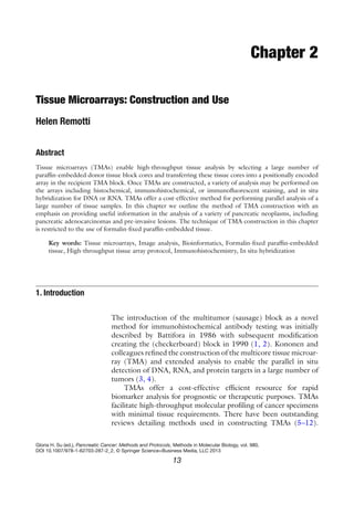
 Needle size: 0.6 mm Max# of cores: 162 (L19 X H 9 = 162) * Max# of blocks: 54 (162 / 3 = 54) 3 cores per block* * Blocks triplicate. 54 cases plus 1 marker, 2 references Needle size: 1.0 mm Max# of cores: 126 (L15 x H 9 = 126) * Max# of blocks: 42 (126 / 3 = 42) 3 cores per block * * Blocks triplicate. 42 cases plus 1 marker, 2 references Needle size: 1.5 mm Max# of cores: 63 (L12 x H 6 = 63) * Max# of blocks: 21 (63 / 3 = 21) 3 cores per block * * Blocks triplicate. 21 cases plus 1 marker, 2 references Needle size: 2.0 mm Max# of cores: 51 (L10 x H 6 = 51) * Max# of blocks: 17 (51 / 3 = 17) 3 cores per block * * Blocks triplicate. 17 cases plus 1 marker, 2 references H (9) L (19) Marker Space for reference (no core) . . . . . . . . . . . . . . . . . . . . . . . . Empty H (9) L (15) Marker Empty H (6) L (12) Marker Empty H (6) L (10) Marker Empty Space for reference (no core) Space for reference (no core) Space for reference (no core) Fig. 1. Pictorial representation of TMAs with differing sized needle cores.
  • 8. 20 H. Remotti 2. Prepare the recipient paraffin block by pouring liquid paraffin into a stainless steel base mold. A variety of paraffin can be used. We use high-temperature Paraplast X-tra and extra large molds. Cover with a slotted tissue cassette and allow to cool. Remove the recipient block from the mold and check for any bubbles or holes. Leave a margin of 3 mm around the array (see Note 3). Fig. 2. Workstation set up with manual tissue arrayer. Fig. 3. Closer view of needle punches for recipient and donor blocks in the manual tissue arrayer.
  • 9. 212 Tissue Microarrays 3. Place the recipient block in the block holder. Adjust the depth stop by rotating the adjustment nut until the punch stops at the desired depth within the paraffin block (typically 0.5–1 mm above the base of the plastic tissue cassette). 4. Check the alignment of the donor (larger) and recipient (smaller) punches of the MTA I arrayer. The circular imprints of the punches on the paraffin block surface should be identi- cally centered if they are correctly aligned. Adjust alignment of punches if necessary. Table 2 Initial spreadsheet with essential necessary for creating a TMA array including ID number for donor block and tissue diagnosis Code Accession # Block # Diagnosis IPMN5-001 SPXX-218608 A2 IPMN IPMN5-002 SPXX-206635 A2 IPMN IPMN5-003 SPXX-248434 A3 IPMN IPMN5-004 SPXX-936324 A6 IPMN IPMN5-005 SPXX-678733 A3 IPMN IPMN5-006 SPXX-952667 B2 IPMN IPMN5-007 SPXX-167946 A4 IPMN IPMN5-008 SPXX-204091 A1 IPMN IPMN5-009 SPXX-232040 A8 IPMN IPMN5-010 SPXX-246582 A28 IPMN IPMN5-011 SPXX-179234 A26 IPMN MCN5-012 SPXX-270681 B14 MCN MCN5-013 SPXX-157892 A1 MCN IPMN5-014 SPXX-182621 A5 IPMN IPMN5-015 SPXX-230490 A5 IPMN IPMN5-016 SPXX-861556 A3 IPMN IPMN5-017 SPXX-255629 A1 IPMN PANCA5-018 SPXX-861523 A10 Adenocarcinoma SA5-019 SPXX-451334 B8 Serous cystadenoma AA5-020 SPXX-499721 B5 Acinar cystadenoma AA5-021 SPXX-178690 B26 Acinar cystadenoma
  • 10. 22 H. Remotti 5. With the small punch mark a hole in the first position of the array (intersection of the upper and left margins, position A1). All other array positions will be in reference to this first spot. Accordingly, set the X and Y micrometers of the MTA-1 to zero. When the depth stop blocks the downward motion, slowly release the tissue punch and eject the paraffin core. 6. Place the donor block bridge over the array block holder and move the larger punch into the sampling position. Manually hold the donor block in position on top of the donor block bridge while positioning the area to be sampled directly underneath the sample punch. Note: Superimposing the cor- responding marked H&E slide over the tissue block will assist in positioning the area to be sampled underneath the tissue punch. For orientation purposes, we use a control lung tissue as a standard marker for tissue in position A1 of all TMA blocks, to facilitate orientation during microscopic evaluation (see Note 4). 7. To retrieve the tissue core push downward on the sample punch. Note: The depth stop will not block the punch motion at the proper position for the donor block, so be careful to prevent the punch from entering too deeply into the block (see Note 5). 8. Remove the donor tissue block and bridge and push the punch downward until its tip reaches the top of the hole in first hole of the recipient array block. Use the large punch stylet to inject the tissue core into the hole created by the smaller punch. 9. Adjust the micrometers to move the tissue punch to the next x-, y-position. We use spacing of 1.5 and 2.0 mm between sample centers when using 0.6 and 1.0 mm needles. For 1.5 and 2.0 mm needles, a spacing of 2.5 mm and 3.0 mm is used. 10. Align the marked H&E slide and the corresponding block (repeat steps 6–9, until TMA is completed). 11. The cores are gently pressed down with a spatula, to insure that they are flushed with the surface of the block (see Note 6). 12. The recipient block is dusted clean or wiped with a kimwipe after the placement of every punch. After several punches, the stylus/ punch complex may retain some paraffin. Move the stylus up and down to dislodge the paraffin and wipe with a kimwipe. 13. The TMA block is placed in an adjusted metal mold with a 2 lb weight on it to prevent displacement of the cores when the paraffin warms up. To allow annealing of cores with the paraffin, the TMA block is placed in a slide warmer and gradu- ally heated over 1 h (40°C for 15 min; 45°C for 15 min; 50°C for 15 min; and 55°C for 15 min), followed at 60°C for 1 min (see Note 7).
  • 12. 24 H. Remotti 14. Carefully transfer block in adjusted metal mold on cool plate (−5°C) for 30 min. Weight should be kept on mold during cooling. 1. The TMA block is trimmed on standard microtome (using new blade). Sections should be no more than 5 m (see Note 8). 2. The TMA is placed on cool plate/ice water for standard micro- tome sectioning. 3. In our laboratory we use standard tissue sectioning techniques with a 34°C water bath to float off the sections onto positive- charged sections or polylysine-coated slides. It is important to maintain orientation of tissue on glass slides (see Note 9). In the past, the tape transfer method (Instrumedics, Inc.) was used due to the ease of transferring of tissue sections in the desired orientation (3, 28). One disadvantage of the tape trans- fer method involved increased background staining from the adhesive residue that interfered with molecular assays, such as ISH, FISH, and phosphorylation-specific IHC, particularly if using automated methods. 4. In sectioning a TMA block, one H&E is stained for every 20 sections. For most studies we cut 40 sections at a time. Slides are allowed to dry in vertical position in an open slide box for 48 h. Slides for IHC studies are optimally used within 1 or 2 weeks. For longer storage, unstained sections are stored at −20°C, without baking the slides. Histochemical, IHC, and ISH studies performed on TMAs can follow similar protocols as conventional slides prepared with whole tissue sections. Although most TMAs are created from FFPE tissues, TMA meth- ods may be modified to include sampling of frozen tissue or cell lines. Frozen TMAs are technically more difficult to construct and require special handling. When constructing frozen TMAs special- ized equipment is needed. Special donor tissue and recipient block requirements with a common cutting media such as OCT and spe- cial adhesives for section transfer for retained TMA core orienta- tion are used (29). One of the principal advantages of frozen TMAs includes bet- ter RNA quality for applications in ISH assays. Frozen tissue microarrays appear to provide excellent target material for the study of DNA, RNA, and proteins by fixing each array slide in a manner specific to the corresponding technique used (30). Another advantage is that those procedures requiring fixation can be con- ducted in samples fixed in an identical manner, since fixation is performed for a limited time on the TMA slide. 3.4. Technical Cutting of TMA 3.5. Biomarker Analysis of TMA 3.6. TMA Construction from Frozen Tissue and Cell Line TMAs
  • 13. 252 Tissue Microarrays The disadvantages include altered morphology with significant loss of fine detail in frozen tissue compared to formalin-fixed tis- sue. Newer commercially available methodologies for performing molecular analysis including RNA-ISH on FFPE tissues provide alternatives to frozen tissue TMAs (31). Several protocols have been described for TMA preparation from cell lines (32–34). One method involves growing cells, creat- ing a cell pellet with subsequent formalin fixation to create an FFPE block (5). Analysis of TMA data can be assessed manually using an ordinal grading system. If the number of markers and number of tissues are relatively small, this method can be used; however it is time consuming, semiquantitative, and requires an experienced pathologist. In order to optimally handle large-scale IHC analysis or to objectively quantify IHC, ISH results, a variety of scanners are available that will automate the acquisition of data generated with the TMA technology. A variety of TMA analysis software programs are available to standardize the analysis of biomarker data. These programs may be able to analyze both chromogenic or immunofluorescent markers to quantify biomarkers. The auto- mated systems can use morphometric parameters to distinguish tumor cells from background normal cells. Data management and data analysis are therefore a major con- cern to ensure both documentation and archiving data of the experiment in addition to facilitating analysis in an efficient and quantitative manner. Conventional spreadsheet-based data organi- zation is feasible for relatively small projects. For larger projects, analyzing thousands of samples with hundreds of biomarkers, robust and scalable applications have to be utilized, which allow data access and manipulation for researchers from different labora- tories in a secure way. 1. A new recut H&E should be cut from the donor block, instead of evaluating the H&E slide in the file. This will ensure that the tissue has not been cut through and diagnostic tissue is still present in the block. 2. If the thickness of the selected area of the donor block is too thin (<1 mm), if possible another sample area or block should be used, since the donor core will be exhausted and absent in deeper sections of the TMA. Although it is recommended to use paraffin blocks of similar thickness in order to maintain 3.7. Digital Image Scanning and Analysis 4. Notes
  • 14. 26 H. Remotti uniform sections with sampling of all cores throughout the TMA, there is another alternative for utilizing thin tissue blocks. This technique involves “stacking” or “packing” of the core with two punches from the original donor block, one on top of each other occupying the same hole in the recipient block. 3. We use a margin of 3.0 mm of paraffin around the entire TMA. This prevents the recipient block from cracking during construction. 4. The use of control tissue is recommended. We use a “marker” lung tissue for the first core in the array. Other standard tissues including normal pancreas and adenocarcinoma may be refer- ences that can be utilized as a quality control for assessing uni- formity of staining of multiple TMAs. 5. Although the depth of the recipient core can be set by the depth stop, there is no depth stop that will set the depth of the donor core. This depth varies when using different donor blocks, since the tissue blocks are of varying thickness, which determines the limit of the depth at which the cores are taken. For an experienced technologist the depth of the donor core is modified based on the thickness of the donor block. A separate “Depth Stop Kit” can be purchased from Beecher that can be used to obtain uniform lengths of donor cores; this kit may be useful if all donor blocks are of similar thickness. If the size of the donor core punched is too long, eject the core from the stylus and place on a clean flat surface and trim with a clean razor blade to the desired length. The core can then be placed in the recipient hole with a small forceps. 6. Do not push the donor punch all the way into the receiver hole; let it slightly protrude. It can be gently pressed with a spatula to ensure that its position is level with the recipient block surface. 7. It is important that the TMA block be remelted at a tempera- ture slightly higher than the melting point of the wax used in the recipient block for a short time (1 min) and then cooled. This step is critical if sections of the TMA block are cut with a standard microtome using water floatation. 8. It is important to face off the recipient block using a dedicated rotary microtome prior to use. This ensures that the block face is smooth and that all the arrays will be made in the identical sectioning plane. This will minimize the amount of block realignment that is necessary during sections and will help
  • 15. 272 Tissue Microarrays optimize the number of complete sections that a TMA block will provide. 9. It is important to maintain correct orientation of the slides in both vertical and horizontal direction so that the sections are oriented similarly on the slide and are easier to be read and analyzed. Acknowledgments The author would like to acknowledge the assistance of Lin Yang, Rina Wu, Ting Ting Wu, and Mariam Amrani, MD, in the prepara- tion of this chapter. References 1. Battifora H (1986) The multitumor (sausage) tissue block: novel method for immunohis- tochemical antibody testing. Lab Invest 55:244–248 2. Battifora H, Mehta P (1990) The checkerboard tissue block. An improved multitissue control block. Lab Invest 63:722–724 3. Kononen J, Bubendorf L, Kallioniemi A, Barlund M, Schraml P, Leighton S, Torhorst J, Mihatsch MJ, Sauter G, Kallioniemi OP (1998) Tissue microarrays for high-throughput molec- ular profiling of tumor specimens. Nat Med 4:844–847 4. Kallioniemi OP, Wagner U, Kononen J, Sauter G (2001) Tissue microarray technology for high-throughput molecular profiling of cancer. Hum Mol Genet 10:657–662 5. Chandler I, Houlston R, Landberg G (2011) A practical guide to constructing and using tissue microarrays. Methods Mol Biol 675:363–373 6. Dhir R (2008) Tissue microarrays: an overview. Methods Mol Biol 441:91–103 7. Eguiluz C, Viguera E, Millan L, Perez J (2006) Multitissue array review: a chronological description of tissue array techniques, applica- tions and procedures. Pathol Res Pract 202: 561–568 8. Fedor HL, De Marzo AM (2005) Practical methods for tissue microarray construction. Methods Mol Med 103:89–101 9. Fowler CB, Man YG, Zhang S, O’Leary TJ, Mason JT, Cunningham RE (2011) Tissue microarrays: construction and uses. Methods Mol Biol 724:23–35 10. Gately K, Kerr K, O’Byrne K (2011) Design, construction, and analysis of cell line arrays and tissue microarrays for gene expression analysis. Methods Mol Biol 784:139–153 11. Hewitt SM (2004) Design, construction, and use of tissue microarrays. Methods Mol Biol 264:61–72 12. Kajdacsy-Balla A, Geynisman JM, Macias V, Setty S, Nanaji NM, Berman JJ, Dobbin K, Melamed J, Kong X, Bosland M, Orenstein J, Bayerl J, Becich MJ, Dhir R, Datta MW (2007) Practical aspects of planning, building, and interpreting tissue microarrays: the Cooperative Prostate Cancer Tissue Resource experience. J Mol Histol 38:113–121 13. Simon R, Mirlacher M, Sauter G (2004) Tissue microarrays. Biotechniques 36:98–105 14. Chiesa-Vottero AG, Rybicki LA, Prayson RA (2003) Comparison of proliferation indices in glioblastoma multiforme by whole tissue sec- tion vs tissue microarray. Am J Clin Pathol 120:902–908 15. Merseburger AS, Kuczyk MA, Serth J, Bokemeyer C, Young DY, Sun L, Connelly RR, McLeod DG, Mostofi FK, Srivastava SK, Stenzl A, Moul JW, Sesterhenn IA (2003) Limitations of tissue microarrays in the evalua- tion of focal alterations of bcl-2 and p53 in whole mount derived prostate tissues. Oncol Rep 10:223–228 16. Charpin C, Dales JP, Garcia S, Carpentier S, Djemli A, Andrac L, Lavaut MN, Allasia C, Bonnier P (2004) Tumor neoangiogenesis by CD31 and CD105 expression evaluation in breast carcinoma tissue microarrays. Clin Cancer Res 10:5815–5819 17. Bubendorf L, Kononen J, Koivisto P, Schraml P, Moch H, Gasser TC, Willi N, Mihatsch MJ,
  • 16. 28 H. Remotti Sauter G, Kallioniemi OP (1999) Survey of gene amplifications during prostate cancer pro- gression by high-throughout fluorescence in situ hybridization on tissue microarrays. Cancer Res 59:803–806 18. Rimm DL, Camp RL, Charette LA, Costa J, Olsen DA, Reiss M (2001) Tissue microarray: a new technology for amplification of tissue resources. Cancer J 7:24–31 19. Schraml P, Kononen J, Bubendorf L, Moch H, Bissig H, Nocito A, Mihatsch MJ, Kallioniemi OP, Sauter G (1999) Tissue microarrays for gene amplification surveys in many different tumor types. Clin Cancer Res 5:1966–1975 20. Andersen CL, Hostetter G, Grigoryan A, Sauter G, Kallioniemi A (2001) Improved pro- cedure for fluorescence in situ hybridization on tissue microarrays. Cytometry 45:83–86 21. Frantz GD, Pham TQ, Peale FV Jr, Hillan KJ (2001) Detection of novel gene expression in paraffin-embedded tissues by isotopic in situ hybridization in tissue microarrays. J Pathol 195:87–96 22. De Marzo AM, Fedor HH, Gage WR, Rubin MA (2002) Inadequate formalin fixation decreases reliability of p27 immunohistochemi- cal staining: probing optimal fixation time using high-density tissue microarrays. Hum Pathol 33:756–760 23. Chin SF, Daigo Y, Huang HE, Iyer NG, Callagy G, Kranjac T, Gonzalez M, Sangan T, Earl H, Caldas C (2003) A simple and reliable pretreatment protocol facilitates fluorescent in situ hybridisation on tissue microarrays of paraffin wax embedded tumour samples. Mol Pathol 56:275–279 24. Hammond ME, Hayes DF, Dowsett M, Allred DC, Hagerty KL, Badve S, Fitzgibbons PL, Francis G, Goldstein NS, Hayes M, Hicks DG, Lester S, Love R, Mangu PB, McShane L, Miller K, Osborne CK, Paik S, Perlmutter J, Rhodes A, Sasano H, Schwartz JN, Sweep FC, Taube S, Torlakovic EE, Valenstein P, Viale G, Visscher D, Wheeler T, Williams RB, Wittliff JL, Wolff AC (2010) American Society of Clinical Oncology/College of American Pathologists guideline recommendations for immunohistochemical testing of estrogen and progesterone receptors in breast cancer (unabridged version). Arch Pathol Lab Med 134:e48–e72 25. Hammond ME, Hayes DF, Wolff AC (2011) Clinical notice for American Society of Clinical Oncology-College of American Pathologists guideline recommendations on ER/PgR and HER2 testing in breast cancer. J Clin Oncol 29:e458 26. Wolff AC, Hammond ME, Schwartz JN, Hagerty KL, Allred DC, Cote RJ, Dowsett M, Fitzgibbons PL, Hanna WM, Langer A, McShane LM, Paik S, Pegram MD, Perez EA, Press MF, Rhodes A, Sturgeon C, Taube SE, Tubbs R, Vance GH, van de Vijver M, Wheeler TM, Hayes DF (2007) American Society of Clinical Oncology/College of American Pathologists guideline recommendations for human epidermal growth factor receptor 2 testing in breast cancer. Arch Pathol Lab Med 131:18–43 27. Maitra A, Adsay NV, Argani P, Iacobuzio- Donahue C, De Marzo A, Cameron JL, Yeo CJ, Hruban RH (2003) Multicomponent analysis of the pancreatic adenocarcinoma progression model using a pancreatic intraepithelial neopla- sia tissue microarray. Mod Pathol 16:902–912 28. Sauter G, Simon R, Hillan K (2003) Tissue microarrays in drug discovery. Nat Rev Drug Discov 2:962–972 29. Schoenberg Fejzo M, Slamon DJ (2001) Frozen tumor tissue microarray technology for analysis of tumor RNA, DNA, and proteins. Am J Pathol 159:1645–1650 30. Li H, Sun Y, Kong QY, Zhang KL, Wang XW, Chen XY, Wang Q, Liu J (2003) Combination of nucleic acid and protein isolation with tissue array construction: using defined histologic regions in single frozen tissue blocks for mul- tiple research purposes. Int J Mol Med 12:299–304 31. Wang F, Flanagan J, Su N, Wang LC, Bui S, Nielson A, Wu X, Vo HT, Ma XJ, Luo Y (2012) RNAscope: a novel in situ RNA analysis plat- form for formalin-fixed, paraffin-embedded tissues. J Mol Diagn 14:22–29 32. Hoos A, Cordon-Cardo C (2001) Tissue microarray profiling of cancer specimens and cell lines: opportunities and limitations. Lab Invest 81:1331–1338 33. Moskaluk CA, Stoler MH (2002) Agarose mold embedding of cultured cells for tissue microarrays. Diagn Mol Pathol 11:234–238 34. Abbott RT, Tripp S, Perkins SL, Elenitoba- Johnson KS, Lim MS (2003) Analysis of the PI-3-Kinase-PTEN-AKT pathway in human lymphoma and leukemia using a cell line microarray. Mod Pathol 16:607–612