2. Curr. Res. J. Biol. Sci., 1(3): 131-134, 2009
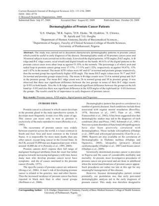
Fig 1: Determination of ATD angle, DAT angle and digital patterns
elucidate the possible diagnostic values of the ridge counts respectively. AT D triradii were also joined
derm atoglyphic features of Nigerian people with prostate as shown in Fig. 1 to determine the ATD an d DA T angles.
cancer. The various digits were designated as follow: Thumb-
i; Index finger-ii; Middle finger-iii; Ring finger-iv; Little
MATERIALS AND METHODS finger-v. L and R stand for left and right respectively.
Sixty (60) male subjects (50 years and above) Statistics: The students’ t-test, A NO VA and chi-square
comprising 30 males with prostate cancer and 30 normal were used for the statistical analysis in this study.
male subjects were selected at random from the
Department of Urology O f the Unive rsity of Port Harco urt RESULTS
Teaching Hospital (UPTH) between September and
December 2008. The clinical records of the patients were The percentages of the digital patterns in both
scrutinized properly to ensure that a set of the subjects did prostate cancer group and the norma l group are
have prostate cancer and the other set had not and w ere summarized in Table 1. Either ulnar loop or w horl had the
not likely to have the disease in future. All subjects w ere highest percentage in all digits of both ha nds in prostate
Nigerians by both parents and grand parents. cancer and n ormal grou ps. A lthoug h little differenc e in
Fingerprints were taken with white paper and purple ink values occurred but this was not significant. Next to either
pad. Hands were thoro ughly washed with water and soap Ulnar loop or W horl was Arch followed by radial loop
and dried before taking prints. This was done to remove which w as not observed in some digits in both groups.
dirt from the hands. There was significant difference in the mean ATD
Screening was done on the white duplicating paper angle between the two groups in both hands (Table 2)
containing the prints and viewed with the aid of a such that normal subjects had high er mean A TD angle
magnifying glass. No distinction was made between the than the prostate cancer p atients (p<0.05).The mean ATD
varieties of whorl(w) patterns, also tented arch was just angles were 44.55º, 40.98º, 43.65º and 40.95º for normal
recorded as an arch(A ). Loop was recorded as either and prostate cancer groups in right and left hand
ulnar loop(UL) or radial loop(R L). All the patterns are as respectively, although the difference between the right
defined by Penrose (1963). A straight line was drawn to and left hand was not significant.
join A and B triradii and B and C triradii and the number The mean dat angle (Table 3) w ere also significantly
of intersecting ridges counted. These give A-B and B -C different between the two groups with normal group
132
3. Curr. Res. J. Biol. Sci., 1(3): 131-134, 2009
Table 1: Percentage (%) frequen cies of digital patterns for each digit of both hands in prostate cance r (P) and norma l (N) subjects.
Right hand digits Prostate cancer=30; Normal =30
Ri Rii Riii Riv Rv
--------------------- ---------------------- ---------------------- --------------------- ----------------------------
Patterns P N P N P N P N P N
Arch 16 .7 20 .0 23 .3 23 .3 16 .7 20 .0 13 .3 3.3 10 .0 0.0
W horl 53 .3 43 .3 50 .0 26 .7 33 .3 26 .7 46 .7 46 .7 13 .3 13 .3
Ulnar loop 30 .0 36 .7 20 .0 43 .3 50 .0 53 .3 40 .0 50 .0 76 .7 86 .7
Radial loop 0.0 0.0 6.7 6.7 0.0 0.0 0.0 0.0 0.0 0.0
Left hand digits Prostate cancer=30; Normal=30
Li Lii Liii Liv Lv
-------------------- ---------------------- ----------------------- ----------------------- ----------------------------
Patterns P N P N P N P N P N
Arch 10 .0 20 .0 16 .7 23 .3 13 .3 10 .0 26 .7 6.7 13 .3 3.3
W horl 36 .7 33 .3 26 .7 36 .7 36 .7 26 .7 50 .0 36 .7 30 .0 16 .7
Ulnar loop 53 .3 46 .7 50 .0 36 .7 50 .0 63 .3 23 .3 56 .7 56 .7 80 .0
Radial loop 0.0 0.0 6.7 3.3 0.0 0.0 0.0 0.0 0.0 0.0
Tab le 2: Mean and s tanda rd erro r of pa lmar A TD angle s in prostate canc e r( P) DISCUSSION
and normal (N) subjects.
Righ t Palm Left P alm
---------------------------------- ------------------------------------------- Dermatoglyphic analysis of the digital patterns in
Norm al Prostate(cancer) Norm al Prostate(cancer) Dow n’s syndrome and normal individuals showed a
0
Mean( ) 44.5 40.98 43.65 40.95
Standard
statistically significant different of 96% loop pattern as
error 1.18 1.00 1.15 0.82 against 63.6% in normal (Boroffice, 1978).No such
P<0.05 difference was observe in the present study.
Table 3: Mean and standard error of palmar dat angles in prostate cancer(P) and
The average A -B ridg e cou nt in normal individu als
normal(N) subjects. was put at 34 while values higher than this were said to be
Righ t Palm Left P alm abnormal (Oladipo et al., 2007).
---------------------------------- -------------------------------------------
Norm al Prostate(cancer) Norm al Prostate(cancer)
The A-B ridge count observed in prostate cancer
Mean( 0) 59.08 40.98 58.28 60.07 group falls in the ra nge of the abnormal groups as it is
Standard higher in both hands than 34.
error 1.28 1.00 0.72 0.90
Normal ATD angles was equally put at 45º. An
P<0.05
average value that is far ab ove or below this value is
Tab le 4: Mean and standard err or of p alma r A-B ridge c oun ts in prostate cancer considered abnormal (Oladipo et al., 2007). Thus the
and normal groups.
values observed for prostate cancer were clearly abnormal
Groups Mean ±Standard error
------------------------------------------------------------- as these w ere far below the no rmal value 45º.Th is
Righ t palm Left p alm sugg ests that both A-B ridge count and AT D angles are
Prostate cancer 35.80±0.97 38.00±1.01 good parameters for the assessment of individuals who
Norm al 33.70±1.07 33.07±0.84
P<0.05
are likely going to show syndromes of prostate cancer
later in life.
Tab le 5: Mean and standard error of palm ar B-C ridge c oun ts in prostate cancer Apa rt from these parameters, the values of B-C ridge
and normal groups.
Groups Mean ±Standard error
count and dat angle could also be very good indication of
------------------------------------------------------------- prostate cancer trait as these values are significantly
Righ t palm Left p alm different betw een n ormal perso n and individuals with
Prostate cancer 29.47±1.08 30.77±0.82
Norm al 26.27±0.83 25.80±1.01
tendency to develop prostate cancer.
P<0.05 Thus, the presence of abnorm ally high A-B and B-C
ridge coun ts is a cha racteristic derm atoglyphic pattern of
showing higher value (59.08º) on the right palm than the prostate cancer which could be very u seful in its early
prostate cancer patients (40.98º).On the left palm, the diagnosis. These data is therefore recommended as a tool
normal group, however showed significantly lower which could be used for early diagnosis of pro state cancer
amo ngst N igerians.
value(58.28º) than the prostate cancer patients(60 .07º).
Analysis of the palmar A-B ridge count in Table 4
REFERENCES
showed that prostate cance r group had significantly higher
count than the normal group(p<0.05) in both hands. Boroffice, I.A., 1978. Down’s syndrome in Nigeria:
Similarly the B-C ridge count in Table 5 showed that derm atoglyphic analy sis of 50 cases. Nig. Med. J.,
prostate cancer group has significantly higher B-C ridge 8: 571-576.
count than the normal group in both hand (p<0.05) Foulds, L., 1975. Neoplastic Developmen t. New York
The mean A-B ridge coun ts on the right palm a nd left Academic Press. pp: 91.
palm of prostate cancer and normal groups were 35.80, Franceschini, P., A. Guala, D. Besana, G. Cara and D.
33.70, 38.00 and 33.70 respectively while those of BC Fanceschini, 2002. A men tally retarded fem ale w ith
ridge counts w ere 29.47, 26.27, 30.77 and 25.80 distinctive facial dy smo rphism , joint laxity,
respectively. clinodactly and abnormal dermatoglyphics.
133
4. Curr. Res. J. Biol. Sci., 1(3): 131-134, 2009
Genet.Couns., 13(1): 55-58. Potosky, A., B. Millar, P. Albetsen and B. Kramer, 1999.
Hoffman, R.M., D.L. Clanon, B. Henberg, J.J. Frank and The role of increasing detection in the rising
J.C. Peirce, 2000. U sing the free-to-total prostate incidence of prostate cancer. J. A m. M ed. A ssoc.,
specific antigen ratio to detect prostate cancer in men 273(7): 548-552.
with non-specific eleva tions of prostate-specific Potosky, A., B. Millar, P. Albetsen and B. Kramer, 2008.
antigen level. J. Gen. Intern. Med., 15(10): 739-748. Finasteride does not increase the risk of High-Grade
Hsing, A., W. Anand and P. Chokkalingain, 2006. prostate cancer. A Bias-Adjusted Modelling
Prosta te cancer epidemiology. Frontiers in A p p r o a c h . h t t p :/ / c an c e rp r e v e n t io n r e s e a rc h .
Biosciences, 11: 1388-1413. aacrjournals org/cgi/rapidpdf/1940-6207.
Oladipo, G.S. and B.M. Ogunno wo, 2004. Rex, A.P. and M. Preus, 1 982. A diagnostic Index for
Dermato glyph ic patern in Diabete Mellitus in South- down’s syndrome. J. Pediatr., 100(6): 903-906.
Eastern Nigeria population. Afr. J. Appl. Zool. Schmidt, S.K., D.P. Mukerjee and S.H. Ahmed, 1981.
Environ. Biol., 6: 6-8. Dermato glyph ic and cytoge netic studies in parents of
children with dow n’s syndrome. C lin. Genet.,
Oladipo, G.S., I.U. Gwunireama and J. Ighegbo, 2005.
20(3): 203-210.
Dermato glyph ic pattern of schizophrenics in South-
Steveson, R.E., B. Hane, J.F. Arena, M. M ay, L.
South Nige rian po pulation. J. Bio med . Afr., 8(2):
Lawrence H.A. Lubs and C.E. Schwartz, 1997. Arch
112-114.
finger prints, hypotonia and flexia associated with x-
Oladipo, G.S., O. Olabiyi, A.A. Oremosu, C.C.
linked mental retardation. J. M ed. Genet., 34(6):
Norohnna, A.O. Okanlawo and C.W . Paul, 2007. 465-469.
Sickle-cell anaemia in Nigera: D erma toglyp hic Than, M., K.A. Myat, S. Khadijah, N. Jamaludin and
analysis of 90 cases. Afr. J. Biochem., 1(4): 54-59. M.U. Isa, 1998. D ermatoglyphics of down’s
Oladipo, G.S., C.W. Paul, I.F. Bob-Manuel H.B. syndrome patients in Malaysia, a comparative study.
Fawehinmi and E .I. Ediba mod e, 200 9. Study of Anthropol. Anz. 56(4): 351-365.
digital and palmar dermatoglyphic patterns of Van-O s, J., P.W. Woodruff, L. Fananas, F. Ahmad, N.
Nigerian women with malignant mammary Shuriquie, R. Howard and R.M. Murray, 2002.
neoplasm. J. Appl. Biosci., 15: 829-834. Association between cerebral structural abnormalities
Penrose, L.S., 1963. Fing erprint, palm and chromosom es. and derm atoglyphic ridge co unt in schizophrenia.
Nature, pp: 933-938. Compr. Psychiat., 41(5): 380-384.
134