Fetal surgery for neural tube defects
•
3 likes•1,543 views

Fetal surgery for neural tube defects

Recommended
More Related Content
What's hot
What's hot (18)
Viewers also liked
Viewers also liked (6)
Similar to Fetal surgery for neural tube defects
Similar to Fetal surgery for neural tube defects (20)
More from South Dakota Pain Capable Unborn Child Protection Act
More from South Dakota Pain Capable Unborn Child Protection Act (20)
Recently uploaded
Recently uploaded (20)
Fetal surgery for neural tube defects
- 1. Best Practice & Research Clinical Obstetrics and Gynaecology Vol. 22, No. 1, pp. 175–188, 2008 doi:10.1016/j.bpobgyn.2007.07.004 available online at http://www.sciencedirect.com 11 Fetal surgery for neural tube defects Leslie N. Sutton * MD Chief Pediatric Neurosurgeona Professor of Neurosurgeryb a Department of Neurosurgery, Children’s Hospital of Philadelphia, 6th Floor Wood Bldg, 34th St. and Civic Center Blvd., Philadelphia, PA 19104, USA b University of Pennsylvania, School of Medicine, USA Open spina bifida remains a major source of disability despite an overall decrease in incidence. It is frequently diagnosed prenatally and can thus – potentially – be treated by fetal surgery. Animal studies and preliminary human studies strongly suggest that at least a portion of the neurological abnormalities seen in these patients are secondary, and occur in mid-gestation. It is estimated that approximately 400 fetal operations have now been performed for myelomeningocele world wide. Despite this large experience, the technique remains of unproven benefit. Preliminary re- sults suggest that fetal surgery results in reversal of hindbrain herniation (the Chiari II malforma- tion), a decrease in shunt-dependent hydrocephalus, and possibly improvement in leg function, but these findings might be explained by selection bias and changing management indications. A randomized prospective trial (the MOMS trial) is currently being conducted by three centers in the United States, and is estimated to be completed in 2009. Key words: fetal surgery; hydrocephalus; myelomeningocele; spina bifida. INTRODUCTION Despite advances in prevention, diagnosis, and intervention, neural tube defects (NTDs) remain a major source of morbidity and mortality in the United States and throughout the world. Daily consumption of 400 micrograms of folic acid before con- ception dramatically reduces the occurrence of neural tube defects, but prior to the institution of food fortification, only 29% of women of reproductive age in the United States were taking a supplement containing this amount.1 Although routine cereal grain fortification has resulted in a 19% decrease in prevalence, the prevalence values per 1000 births remains 4.18, 3.37, and 2.90 respectively for Hispanic, non-Hispanic * Department of Neurosurgery, Children’s Hospital of Philadelphia, 6th Floor Wood Bldg, 34th St. and Civic Center Blvd., Philadelphia, PA 19104, USA. Tel./Fax: 1 215 590 2780. E-mail address: sutton@email.chop.edu 1521-6934/$ - see front matter ª 2007 Elsevier Ltd. All rights reserved.
- 2. 176 L. N. Sutton white, and non-Hispanic black women.2 It is estimated that 23% of pregnancies in which the fetus is diagnosed with an NTD end in elective termination; the remainder are ultimately delivered. Furthermore, the prenatal management of spina bifida differs depending on the country: as a rule, there is more support for aggressive and intensive treatment in Asia and some regions of the United States than in Europe3, although immigration patterns might be changing this. Although folate supplementation and advances in care might be decreasing the mor- tality associated with spina bifida, the 5-year mortality remains 79 per 1000 spina bifida births.4 The mortality is as high as 35% among those with symptoms of brainstem dys- function secondary to the Chiari II malformation.5 In addition to sphincter dysfunction and lower extremity paralysis, 81% of affected children have hydrocephalus requiring treatment6, exposing them to the problems associated with shunts. Although 70% of af- fected individuals have an IQ above 80, only 37% are able to live independently as adults and one-third need daily care.7 No recent data are available, but in 1994 the cost of care exceeded $500 million per year (in 1992 dollars) in the United States alone.8 The increasing use of screening ultrasonography and amniocentesis has resulted in early detection of neural tube defects (NTDs), and the use of fetal MRI has improved accuracy of the diagnosis (Figure 1). Diagnosis is now common at 18 weeks of gesta- tion, allowing time for a thorough discussion of the likely outcome with parents. In ad- dition to the standard options of termination of the pregnancy and continuation of the pregnancy until term with cesarean or vaginal delivery, fetal closure of the defect as part of the Management of Myleomeningocle Study (MOMS) is an option for some families in the United States. Figure 1. T-2 weighed fetal MRI of a fetus with a myelomeningocele. The thin walled sac is intact, and the Chiari II malformation is present.
- 3. Fetal surgery for neural tube defects 177 Some have questioned the appropriateness of expending scarce medical recourses on a disease that is decreasing in incidence world-wide and for which termination (and even euthanasia in some countries9) is an option. Others have raised objections that the mother might feel pressured to consent to a procedure that is designed to benefit the fetus. The most cogent arguments, however, relate to the unproven benefits of fetal surgery for myelomeningocele. It is even unclear what benefits would need to be achieved to justify fetal surgery.10 There are biases inherent in any attempt to compare the outcome of currently treated fetal surgery patients with historical controls. To address this issue, fetal sur- gery groups at three institutions – The Children’s Hospital of Philadelphia (CHOP), Vanderbilt University, and the University of California at San Francisco (UCSF) – have agreed to conduct a randomized prospective study under the direction of the National Institutes of Health. The study opened in February of 2003 with the support of the American and Canadian pediatric neurosurgical communities, and about one- half of the proposed number of patients have been randomized. Until this study has been completed, fetal surgery remains of unproven benefit and is certainly not to be considered ‘standard of care’ in the legal sense.11 HISTORICAL PERSPECTIVE Spina bifida is considered a potential candidate for in-utero treatment because the condition is routinely detected before 20 weeks of gestation. Such an innovative sur- gical procedure would only have been considered in humans if there were substantial evidence that it might improve outcome relative to standard postnatal closure. ANIMAL STUDIES As outlined by George and Fuh12, the ideal animal model should develop as a sponta- neous mutant, should be surgically accessible, and the treated animals should survive long enough to assess outcome. Unfortunately, none of the current models of myelo- meningocele meet all of these criteria. The first potentially useful model of myelomeningocele was described in late-gesta- tion primates.13 The model most closely approximating the human form, however, was that described in fetal lambs.14 To mimic myelomeningocele, a laminectomy was per- formed exposing the spinal cord of the fetal lamb at 75 days of gestation, and the preg- nancy was allowed to continue. The lambs were delivered by cesarean section at 140 days of gestation. Clinically, the animals were paraplegic and incontinent, and the his- tology was strikingly similar to myelomeningocele in humans. When a latissimus dorsi flap was used to cover the exposed ‘placode’ in the fetal lamb at 100 days of gestation, however, the animals had near-normal motor function and the nerve tissue was relatively well preserved at birth. The concept of secondary neural tissue destruction and loss of function during pregnancy has also recently been supported by experiments using the curly tailed mouse15 and in fetal rabbits.16 The sheep model has been used to evaluate sphincter function, and fetal coverage of the exposed spinal cord appears to improve function.17 The mechanism of damage to the placode before birth remains unclear. The fact that aminotic fluid exchange might prevent neural tissue damage in a chick embryo model suggests that at least a portion of the damage might be from chemical neurotoxicity.18 It has been suggested that fetal meconium might play a role in this.19
- 4. 178 L. N. Sutton Laminectomy in early-gestation fetal sheep might result in the hindbrain hernia that is a component of the Chiari II malformation. Furthermore, early fetal closure of the defect might reverse the hernia.20 This has also been reproduced in a mouse model.21 These experiments did not reproduce all of the features of human myelodysplasia because there was no neural tube defect. They did, however, provide evidence of sec- ondary damage occurring within the uterine environment sufficient to justify human trials. HUMAN PATHOLOGY Pathologic studies of human embryos and fetuses with myelomeningocele in early ges- tation reveal an open but undamaged neural tube with almost normal cytoarchitecture, suggesting that neural degeneration occurs at some point during gestation (the ‘two- hit’ hypothesis). Osaka and co-workers22 found an everted neural plate in 18 embryos with classical caudal myelodysplasia; most of the membrane coverings were preserved. Interestingly, the Chiari II malformation was not seen in the embryos, whereas this malformation was present in the two fetuses with caudal myelodysplasia from the same series. Hydrocephalus was not present in the embryos, but was found in one fe- tus. Others23 have performed pathologic examination of the spinal cords of stillborn human fetuses with myelomeningocele. Varying degrees of neural tissue loss was seen at the site of the defect but normal dorsal and ventral horns were present at the prox- imal aspect of the lesion. More recently, George and Cummings24 found evidence of both abnormal patterning of neurons and secondary damage to the placode, as demonstrated by inflammation, gliosis, and fibrosis. Additional support for the two-hit hypothesis came from studies assessing leg func- tion in utero with serial sonograms. Korenromp et al25 noted normal movement of the hips and knees as early as 16 weeks of gestation in fetuses with myelomeningocele. Sival et al26 found that only one of 13 fetuses had abnormal leg movements prenatally but that abnormal leg motion was noted in 11 after birth. It is likely that some aspect of the intrauterine environment results in injury to the exposed spinal cord. Possible etiologies include chemical injury from the amniotic fluid, direct trauma, or trauma due to hydrodynamic pressure of the spinal fluid within the subarachnoid space or a hydro- myelic cavity. Studies using rat spinal cord tissue exposed to human amniotic fluid at various times during gestation indicated that late-gestation amniotic fluid could cause cell injury.27 Human pathologic specimens seem to support direct impact as the pri- mary cause of damage because the neural tissue is lost almost exclusively from the dorsal protruding portions of the cord.28 As pregnancy progresses, the volume of amniotic fluid decreases, which can result in more frequent contact of the spinal cord with the uterine wall. THE HUMAN EXPERIENCE WITH FETAL MYELOMENINGOCELE CLOSURE The first cases of in-utero spina bifida repair were performed in 1994 using an endo- scopic technique.29 This technique proved unsatisfactory and was abandoned. Percu- taneous fetoscopic patch coverage has been tried more recently in a small series of patients, and has also proved problematic.30 In 1997, in-utero closure of spina bifida defects was performed by hysterotomy at Vanderbilt University31 and at CHOP.32 The selection criteria were different at the two institutions. At Vanderbilt, patients
- 5. Fetal surgery for neural tube defects 179 were not excluded based on prenatal ventricular size, late gestational age, spinal level, or presence or absence of fetal leg motion by in-utero sonogram. At CHOP, a fetus was only considered for surgery if the gestational age at the time of the proposed sur- gery was 26 weeks or less, if the transatrial ventricular diameter was less than 16 mm (normal being less than 10 mm), if the estimated level of the lesion was S1 or above, and if there was convincing leg and foot motion on ultrasound and in the absence of foot or leg deformity. The early experience at these institutions suggested that compared with babies treated postnatally, those treated in utero had a decreased incidence of hindbrain her- niation33,34, and that ascent of the hindbrain structures could be demonstrated within 3 weeks of the fetal closure using serial MRI. It is clear that the radiographic appear- ance of hindbrain herniation (the Chiari II malformation) is improved by the procedure but the other manifestations of the Chiari complex, such as thinning of the corpus cal- losum and polymicrogyria, are not. It is not yet clear whether this translates into im- proved survival or functional outcome. Although the posterior fossa volume of the normal developing fetus has been measured using MRI35, the volume of the posterior fossa in fetal myelomeningocele patients has not. It is hypothesized that the volume is small and might be expanded by fetal surgery, but this remains unproven and is the subject of ongoing research. The overall fetal head size has been demonstrated to be small in myelomeningocele patients, and to increase to normal after fetal surgery; the significance of this is uncer- tain. It appears that the head enlargement is largely due to restoration of the cerebro- spinal volume, which is indicative of reversal of the hindbrain herniation.36 The fetal ventricles typically enlarge throughout gestation following fetal surgery. At present, however, no consideration is being given to placement of fetal shunts. With very short follow-up, it also appeared that this might have resulted in a decreased need for shunt- ing. With somewhat longer follow-up this effect has been maintained to some extent, although some infants who did not require shunts in the newborn period have re- quired shunts later on, usually within the first year. In the combined series of fetal sur- gery patients from CHOP and Vanderbilt, 104 patients followed for at least 1 year had an overall incidence of shunting of 54%, compared with 86% for a historical control group from CHOP.37 The effect was most evident for those with lumbar lesions, per- haps because of the larger number of these resulted in increased statistical power. The incidence of shunting in those patients who underwent fetal closure prior to 26 weeks of gestation was 42.7%, but was 75% in those who had fetal surgery after 25 weeks of gestation. It was hypothesized that early fetal closure of the spinal lesion eliminated the leakage of spinal fluid from the back, which put back-pressure on the hindbrain. This allowed reduction of the hindbrain hernia and relieved the obstruction of the outflow from the fourth ventricle. This apparent benefit might be due to selection bias or change in the indications for placing a shunt over time. Most infants and children who have undergone fetal myelomeningocele closure have persistent ventriculomegaly, but often do not have overt signs or symptoms of increased intracranial pressure. The prevailing opinion is that these patients do not re- quire shunts, but it is not yet known if the developmental and cognitive level of func- tion of these children would be improved by more aggressive treatment of the ventriculomegaly. Benefit in lower extremity function or sphincter continence has been difficult to demonstrate. Children with spina bifida treated with conventional postnatal closure have a level of neuologic function that correlates very well with the bony level of the defect as determined radiographically.6 The Vanderbilt series of early and late
- 6. 180 L. N. Sutton gestation fetal closures showed no improvement in leg function compared with histor- ical controls for comparable spinal level, but no attempt was made to ascertain the degree of leg function prenatally.38 The CHOP criteria demanded intact leg and foot motion to be present prior to fetal surgery, and only included early-gestation re- pairs. In our series, 57% had better-than-predicted leg function at birth in the thoracic and lumbar patients, but follow-up was short.39 There is concern that some of the early benefit in terms of leg function might be at risk. Virtually all of the postnatal lum- bosacral MRI studies of these patients suggest tethering, and recently some of the pa- tients have developed symptomatic epidermoid inclusion cysts, which have required repeat surgery.40 It is unclear at this point whether this is a problem unique to fetal closure, or simply that it is being found because of the careful surveillance that these patients are required to undergo. Clinically symptomatic tethering and epidermoid in- clusions are also seen in conventionally treated infants with myelomeningocele, partic- ularly in those who undergo intensive neurourological surveillance.41 It is presumed that if neurological functioning of the lower extremities is preserved by fetal closure, symptomatic cord tethering is likely to be even more of a problem, as there is more function to be lost. Interestingly, the six CHOP patients who have required re-explo- ration for tethering have all had intraoperative electrophysiological monitoring, and all have shown intact motor nerve conduction even to the lower sacral levels (unpub- lished data). The effect of fetal surgery on cognitive functioning has also been difficult to assess. It is known that the average IQ of children with myelomeningocele is significantly lower than that of control children, and that this appears to be due to the disease pro- cess itself, rather than associated complications such as shunt infection.42 Fetal surgery could theoretically improve outcome by reducing the incidence of hydrocephalus, or adversely impact outcome by increasing the incidence of prematurity. Preliminary data from CHOP showed a mean Mental Developmental Index of 90.8 in patients who un- derwent fetal surgery, which is probably not significantly different from postnatally treated individuals, and suggest no major effect of fetal surgery.43 An unexpected finding of fetal surgery is improved wound healing and decreased scar formation, resulting in a cosmetically more favorable back wound. This phenom- enon has been extensively studied and has been attributed to down regulation of a transforming growth factor-beta modulator44 and increased endothelial growth factor.45 Fetal surgery is not without risk. Perinatal mortality at CHOP has been 6% (3/50), due to extreme prematurity associated with intrauterine infection in one case.39 The mean gestational age at delivery was 34 weeks 4 days. There have been no maternal deaths in any fetal surgery series. No patient expe- rienced hysterotomy dehiscence or rupture. As fetal surgery requires a classic cesar- ean hysterotomy high in the uterus, all future pregnancies require cesarean delivery. No data have been presented to suggest diminished fertility in any of the women undergoing fetal surgery for this or any other condition. SURGICAL TECHNIQUE OF FETAL SURGERY The overriding concern in any fetal operation is maternal safety. Secondary goals are avoiding preterm labor and accomplishing the goals of surgery for the fetus. Technical difficulties associated with the small size of the fetus and fragility of the tissues gener- ally limit surgery before 18 weeks gestation, and after 30 weeks the risks of premature
- 7. Fetal surgery for neural tube defects 181 labor increase dramatically, so that at that point it usually is more reasonable to deliver the fetus first and then treat the abnormality ex utero. The trial currently underway requires that the fetus be less than 26 weeks gestation at the time of the surgery. Preoperative evaluation and counseling Fetal surgery requires the coordinated effort of many specialists, including pediatric surgeons, neurosurgeons, maternal–fetal medicine specialists, ultrasonographers, radi- ologists with MRI expertise, neonatologists, anesthesiologists, geneticists, nurses, social workers, and financial counselors. The issues associated with a serious birth defect are complex and emotionally charged, and ideally at least two preoperative ed- ucational sessions are held with the pregnant woman and her family. Initial screening is carried out with review of data already obtained locally by the treating obstetrician, supplemented by high-resolution ultrasound and MRI performed by the fetal team. Amniocentesis is performed to rule out associated genetic defects and congenital infection. It is imperative to exclude skin-covered dysraphic lesions such as lipomyelo- meningocele46 or myelocystocele.47 If the sac has a thick wall, no Chiari malformation is evident, and there is no elevation of amniotic fluid alpha-fetoprotein, one of these lesions should be suspected rather than an open myelomeningocele. As these forms of occult dysraphism are skin covered, they are unsuitable candidates for fetal intervention. The results of the preliminary studies are discussed in detail with the family. Mater- nal risk factors are assessed. If the maternal–fetal unit is deemed appropriate for fetal surgery, a second session is scheduled, in which members of the fetal team explain their roles and describe the potential risks associated with their portion of the pro- cedure. A formal meeting with a neonatal pediatrician is arranged, to discuss the im- plications of prematurity. Currently, fetal surgery for myelomeningocele is being offered in the United States only within the context of the MOMS trial. If the decision is made to proceed, a detailed consent form outlining the risks and potential benefits of the proposed procedure is signed, and the patient undergoes randomization. Control of labor Fetal surgeons have observed that the later in gestation the hysterotomy is performed, the more reactive the uterus becomes, increasing the risk of premature labor. The risk also appears to increase with larger uterine openings and with longer procedures. Pre- term labor is defined as labor before 37 weeks gestation. It is best considered a syndrome rather than a specific diagnosis because it can arise for a variety of reasons. As many as 30% of preterm labors are thought to result from intra-amniotic infections; such infections can occur after fetal surgery. Other risk factors include multiple ges- tation, a history of maternal smoking, and very young or older maternal age, which become important factors in the selection process for possible fetal surgery. In some cases, premature labor represents the need for the fetus to escape a hostile uterine environment, and aggressive measures to stop labor may be inappropriate. Contraindications to tocolysis include intrauterine infection, unexplained vaginal bleeding, and fetal distress. Otherwise, bed rest and hydration are commonly pre- scribed, but these are of unproved benefit. Drug therapy remains the mainstay in the prevention of premature labor, even though there are no reliable data to suggest that any of the available agents delay delivery for more than 48 hours.48 Magnesium
- 8. 182 L. N. Sutton sulfate is administered intravenously as a bolus and maintained intravenously. The ma- jor side effects are maternal nausea, weakness, headache, and pulmonary edema, and fetal hypotonia. Indometacin can be delivered orally or rectally. Side effects include ma- ternal nausea and an increase in bleeding time, and fetal ductus arteriosus constriction, tricuspid regurgitation, and right-sided heart failure; consequently, fetal echocardio- graphic monitoring is essential. Calcium channel blockers such as nifedipine can be given orally. Side effects include hypotension, tachycardia, and nausea. Deep haloge- nated anesthesia can provide intraoperative uterine relaxation, but it might produce fetal and maternal myocardial depression and decrease placental perfusion. Terbutaline sulfate is a b-adrenergic agonist that is usually administered by continuous subcutane- ous infusion by a pump. Side effects include maternal jitteriness, anxiety, vomiting, palpitations and pulmonary edema. Anesthesia Anesthetic considerations for fetal surgery include maternal, fetal, and uteroplacental factors.49 The mother receives an H2-antagonist the evening before and the morning of the operation. Before induction, an oral antacid is given to reduce the risk of acid aspiration, and a lumbar epidural catheter is placed for uterine relaxation and for post- operative analgesia. A rapid sequence induction and intubation are accomplished, and left uterine displacement is maintained to avoid caval compression. Anesthesia is main- tained with 0.5% expired isoflurane, 50% nitrous oxide, and a balance of oxygen. Be- fore the incision, the isoflurane is increased to 1% expired, and well before the uterine incision is increased to 2% and titrated to uterine relaxation. A few minutes before the uterus is opened, the nitrous oxide is discontinued. Ephedrine or phenylephrine is ad- ministered to maintain systolic arterial blood pressure >100 mmHg. Intravenous fluids are limited to 0.9% sodium chloride at a rate of 100 mL/h to avoid fetal hyperglycemia and maternal pulmonary edema. Neuromuscular blockade is provided with vecuro- nium, keeping in mind the increased sensitivity of the patient soon to receive magne- sium sulfate. The fetus might be given an intramuscular injection of a narcotic just prior to the incision, although the fetus receives satisfactory anesthesia via the placental cir- culation. During uterine closure, magnesium sulfate is given intravenously, followed by an infusion. The isoflurane is decreased to 0.5% expired, and nitrous oxide is restarted. Bupivacaine and long-acting morphine are given through the epidural catheter. After skin closure, the anesthetic agents are discontinued and the mother is extubated. The surgical procedure The uterus is exposed through a low transverse abdominal incision. The fetal and pla- cental positions are determined by ultrasound and the uterus is mobilized for optimal exposure. The hysterotomy is performed with monopolar cautery between large he- mostatic sutures, and enlarged with a uterine stapler, which simultaneously incises the uterine wall and lays down a layer of absorbable hemostatic clips to preserve the in- trauterine membranes. The hysterotomy is in the upper segment of the uterine corpus and fetal surgery necessitates cesarean delivery for all subsequent pregnancies because of the risk of uterine rupture. Every attempt is made to maintain intrauterine volume to prevent placental separation, contractions, and expulsion of the fetus. This is ac- complished by continuous high-volume perfusion of the amniotic cavity with warm Ringer’s lactate solution and by exposing only the portion of the fetus that is necessary.
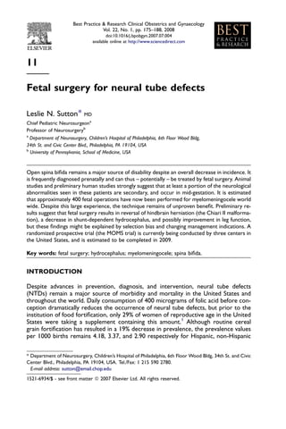
- 9. Fetal surgery for neural tube defects 183 The fetus is not removed from the uterus, and the surgery is performed through the hysterotomy opening (Figure 2). Care must also be taken throughout the procedure to avoid umbilical cord compression, which can occur at the margins of the hysterotomy. Fetal cardiac sonography is perfomed throughout the operation to warn of any threat to fetal circulation.50,51 After exposure of the fetus, a fetal narcotic injection is administered to supplement pain control. The concept of fetal pain is controversial52 but it is assumed that mater- nal anesthetic agents cross over the placental circulation and provide fetal anesthesia. The myelomeningocele closure is performed rapidly and as bloodlessly as possible, and is similar to the standard postnatal closure. Although the use of the operating mi- croscope is favored by some, loupes and a headlight provide a wider view and allow more mobility. The fringe of full-thickness skin is incised circumferentially with a num- ber 15 knife blade down to the fascia and the sac is mobilized medially to the facial defect, as in a standard closure. The sac is excised from the placode, with care taken to remove all epithelial tissue to prevent formation of an epidermoid inclusion cyst. No attempt is made to re-neurulate the placode, as the spinal cord tissue in the fetus is extremely friable. The closure is effected with dura, undermined fascia, or prefer- ably both. In the past, acellular human dermis graft material was used for the dural closure in some instances but recent reports of epidermoid inclusion cysts in some patients has prompted us to avoid this material for the deep layers. The skin is under- mined and every attempt is made to close it primarily with 4-0 PDS absorbable suture (Ethicon). When this is not possible, due to the size of the defect, acellular human dermis graft material can be used to complete the closure53, or lateral relaxing incisions might be performed. After completion of fetal surgery, the uterus is closed with a tight two-layer closure. A transparent dressing is used to allow postoperative sonographic monitoring. Postoperative management Initially, patients are observed in the high-risk obstetrical unit and subsequently kept on bed rest near the hospital. Tocolysis is maintained with magnesium sulfate Figure 2. Intraoperative view of a fetal myelomeningocele closure. The hysterotomy is lined with hemostatic clips. Note the exposed placode.
- 10. 184 L. N. Sutton intravenously and with indometacin rectal suppositories, followed by a calcium channel blocker; terbutaline is added if required. Infants are delivered by planned cesarean delivery at approximately 36 weeks gestation after fetal lung maturity is confirmed by amniocentesis, unless premature labor results in earlier delivery. Potential maternal complications include extrusion of the entire fetus or a fetal part through the hyster- otomy, bowel obstruction, pulmonary edema, placental abruption, and chorioamniitis. The major risk to the fetus is uncontrollable labor and premature delivery, thus expos- ing the child to the well-known risks of low birth weight. At CHOP, the babies have returned for a follow-up evaluation on a yearly basis. A yearly MRI of the brain and complete spine is obtained to assess status of the Chiari malformation, the size of the ventricles, and the presence of an epidermoid at the clo- sure site. Any deterioration in neurological function is cause for concern, as in any child with a myelomeningocele. Close communication is maintained with the neurosur- geon caring for the child in the community. THE MOMS TRIAL The biases inherent in assessing the outcomes of fetal surgery compared with histor- ical controls are obvious. After considerable discussion, a randomized three-center trial opened in February 2003 and is ongoing. The design of the trial required a number of compromises between the three centers involved: CHOP, Vanderbilt University, UCSF, and the sponsoring institution, the National Institute of Child Health and Human Development (NICHD). The study is an unblinded, randomized controlled clinical trial of 200 patients. Patients diagnosed with myelomeningocele between 16 and 25 weeks gestation are referred to the Data and Study Coordinating Center (DSCC) at George Washington University for initial screening and information (http://www.spinabifidamoms.com/english/ index.html). Those eligible and interested are assigned by the DSCC to one of the three fetal surgery units (CHOP, Vanderbilt, or UCSF), where final evaluation and screening are carried out. Patients who satisfy the eligibility criteria and consent to randomization are centrally randomized to one of the following two management protocols: 1. Intrauterine repair of the myelomeningocele at 18 to 25 weeks gestation, discharge to nearby accommodation on tocolytics when stable for preterm labor, weekly prenatal visits and biweekly ultrasounds conducted at the fetal surgery unit; cesarean delivery at 37 weeks gestation following demonstration of lung maturity. 2. Return to local perinatologist for prenatal care, with monthly ultrasounds reported to the fetal surgery unit; return to the fetal surgery unit at 37 weeks ges- tation for cesarean delivery following demonstration of lung maturity; neonatal re- pair of the myelomeningocele. The inclusion and exclusion criteria are listed in Box 1. Note that there are no exclu- sions based on ventricular size or status of fetal leg motion. As the primary end-point of the study is the need for a shunt at 1 year, it was felt that the presence or absence of fetal leg motion should not be an exclusion criterion. Furthermore, the experience to date suggests that few if any fetal candidates would have ventricles larger than 17 mm if only early gestation fetuses were eligible for the trial. The criteria for placing a shunt have been defined and, as many of these patients will be cared for primarily in their com- munities rather than the research center, it is important that neurosurgeons are aware of
- 11. Fetal surgery for neural tube defects 185 Box 1. Inclusion and exclusion criteria for participation in the Management of Myelomeningocele Study (MOMS) Inclusion criteria Myelomeningocele at level T1–S1 with hindbrain herniation. Lesion level will be confirmed by ultrasound, and hindbrain herniation will be confirmed by MRI scan at the fetal surgery unit Maternal age 18 years or older Gestational age at randomization of 18–25 weeks as determined by clinical in- formation and evaluation of first ultrasound. If the date of the patient’s last menstrual period (LMP) is deemed sure and her cycle is 26–32 days, and if the biometric measurements from the patient’s first ultrasound confirm this LMP within 10 days, the LMP will be used to determine gestational age. In all other cases (e.g. if the LMP is unsure, if she has an irregular cycle or her cycle is outside the 26- to 32-day window (or if the measurements from her first ul- trasound are more than 10 days discrepant from the subsequent ultrasound), the initial ultrasound determination will be used. Once the estimated date of conception has been determined for the purposes of the trial, no further revision is made Normal karyotype with written confirmation of culture results. Results by fluo- rescence in situ hybridization will be acceptable if the fetus is at 24 weeks ges- tation or more Exclusion criteria Nonresident of the United States Multifetal pregnancy Abnormal fetal echocardiogram Fetal anomaly other than myelomeningocele or an anomaly related to myelomeningocele Documented history of incompetent cervix Short cervix (20 mm measured by ultrasound) Preterm labor in the current pregnancy Past history of recurrent preterm labor Maternal-fetal Ah isoimmunization, Kell sensitization, or a history of neonatal alloimmune thrombocytopenia Maternal HIV or hepatitis-B status positive or unknown-because of the increased risk of transmission to the fetus during fetal surgery Uterine anomaly such as large or multiple fibroids or Mullerian duct abnormality Other maternal medical condition that is a contraindication to surgery or general anesthesia, including obesity No support person (e.g. husband, partner, mother) available for patient. Inabil- ity to comply with the travel and follow-up requirements of the trial. Inability to meet other psychosocial criteria (as determined by the case social worker) to handle the implications of surgery Maternal obesity
- 12. 186 L. N. Sutton these criteria. Secondary endpoints are neurologic function, cognitive outcome, and maternal morbidity. The follow-up studies will be conducted by centrally trained ob- servers who will be blinded to treatment arm and the overall management of the study will be conducted by the Biostatistics Center at George Washington under the auspices of the NICHD. As of March 2007, approximately 112 patients had been randomized. The clinical investigators are blinded to all results and no preliminary data are available. There is provision in the trial for interim analysis and thus far the study centers have been granted permission to continue. Accrual has been slower than expected but is con- tinuing. It is hoped that the trial will be completed before other institutions begin per- forming in-utero repair of spina bifida, which at this time remains of unproven benefit. Practice points Prenatal evaluation of a suspected fetus with myelomeningocle who is being considered for fetal surgery should include high-resolution ultrasound, MRI, and amniocentesis. A thick-walled sac, absence of hindbrain hernia, and lack of elevation of mater- nal or amniotic fluid alphafetoprotein should raise suspicion of an occult dysraphism such as lipomyelomeningocele or myelocystocele. Research agenda A randomized prospective trial of fetal surgery (the MOMS trial) is currently being conducted in the United States. The mechanism of reversal of hindbrain herniation remains undefined. Research is being conducted to determine the changes in fetal posterior fossa volume in myelomeningocele fetuses who undergo fetal surgery and those who do not. The indications for shunting myelomeningocle patients who have ventriculome- galy but no evidence of overt increased intracranial pressure remain undefined. The incidence of inclusion epidermoid cysts in fetal surgery patients and in postnatally closed patients is undefined. REFERENCES 1. Honein MA, Paulozzi LJ, Mathews TJ et al. Impact of folic acid fortification of the US food supply on the occurrence of neural tube defects. JAMA 2001; 285: 2981–2986. 2. Williams LJ, Mai CT, Edmonds LD et al. Prevalence of spina bifida and anencephaly during the transition to mandatory folic acid fortification in the United States. Teratology 2002; 66: 33–39.
- 13. Fetal surgery for neural tube defects 187 3. Oi S. Current status of prenatal management of fetal spina bifida in the world: worldwide cooperative survey on the medico-ethical issue. Childs Nerv Syst 2003; 19: 596–599. 4. Bol KA, Collins JS Kirby RS. Survival of infants with neural tube defects in the presence of folic acid fortification. Pediatrics 2006; 117: 803–813. 5. Wong LY Paulozzi LJ. Survival of infants with spina bifida: a population study, 1979-94. Paediatr Perinat Epidemiol 2001; 15: 374–378. *6. Rintoul N, Sutton L, Hubbard A et al. A new look at myelomenigoceles: functional level, vertebral level, shunting, and the implications for fetal intervention. Pediatrics 2002; 109: 409–413. 7. Oakeshott P Hunt GM. Long-term outcome in open spina bifida. Br J Gen Pract 2003; 53: 632–636. 8. Waitzman NJ, Romano PS Scheffler RM. Estimates of the economic costs of birth defects. Inquiry 1994; 31: 188–205. 9. Jochemsen H. Dutch court decisions on nonvoluntary euthanasia critically reviewed. Issues Law Med 1998; 13: 447–458. 10. Cochrane DD, Irwin B Chambers K. Clinical outcomes that fetal surgery for myelomeningocele needs to achieve. Eur J Pediatr Surg 2001; 11: S18–S20. 11. Lyerly AD, Cefalo RC, Socol M et al. Attitudes of maternal-fetal specialists concerning maternal-fetal surgery. Am J Obstet Gynecol 2001; 185: 1052–1058. 12. George TM Fuh E. Review of animal models of surgically induced spinal neural tube defects: implica- tions for fetal surgery. Pediatr Neurosurg 2003; 39: 81–90. 13. Michejda M. Intrauterine treatment of spina bifida. Primate model. Z Kinderchir 1984; 39: 259–261. *14. Meuli M, Meuli-Simmen C, Yingling C et al. In utero surgery rescues neurological function at birth in sheep with spina bifida. Nat Med 1995; 1: 342–347. 15. Stiefel D, Copp A Meuli M. Fetal spina bifida in a mouse model:loss of neural function in utero. J Neurosurg 2007; 3(106): 213–221. 16. Julia V, Sancho MA, Albert A et al. Prenatal covering of the spinal cord decreases neurologic sequelae in a myelomeningocele model. J Pediatr Surg 2006; 41: 1125–1129. 17. Yoshizawa J, Sbragia L, Paek BW et al. Fetal surgery for repair of myelomeningocele allows normal development of anal sphincter muscles in sheep. Pediatr Surg Int 2004; 20: 14–18. 18. Olguner M, Akgur FM, Ozdemir T et al. Amniotic fluid exchange for the prevention of neural tissue damage in myelomeningocele: an alternative minimally invasive method to open in utero surgery. Pediatr Neurosurg 2000; 33: 252–256. 19. Correia-Pinto J, Reis JL, Hutchins GM et al. In utero meconium exposure increases spinal cord necrosis in a rat model of myelomeningocele. J Pediatr Surg 2002; 37: 488–492. 20. Bouchard S, Davey MG, Rintoul NE et al. Correction of hindbrain herniation and anatomy of the vermis after in utero repair of myelomeningocele in sheep. J Pediatr Surg 2003; 38: 451–458. discussion 451–458. 21. Weber Guimaraes Barreto M, Ferro MM, Guimaraes Bittencourt D et al. Arnold-Chiari in a fetal rat model of dysraphism. Fetal Diagn Ther 2005; 20: 437–441. 22. Osaka K, Tanimura T, Hirayama A et al. Myelomeningocele before birth. J Neurosurgery 1978; 49: 711–724. 23. Hutchins G, Meuli M, Meuli-Simmen C et al. Acquired spinal cord injury in human fetuses with myelo- meningocele. Pediatr Pathol Lab Med 1996; 16: 701–712. 24. George TM Cummings TJ. The immunohistochemical profile of the myelomeningocele placode: is the placode normal? Pediatr Neurosurg 2003; 39: 234–239. *25. Korenromp MJ, van Gool JD, Bruinese HW et al. Early fetal leg movements in myelomeningocele. Lancet 1986; 1: 917–918. 26. Sival D, Begeer J, Staal-Schreinmachers A et al. Perinatal motor behaviour and neurological outcome in spina bifida aperta. Early Human Devel 1997; 50: 27–37. 27. Drewek M, Brunner J, Whetsell W et al. Quantitative analysis of the toxicity of human amniotic fluid to cultured rat spinal cord. Ped Neurosurg 1996; 27: 190–193. 28. Meuli M, Meuli-, Simmen C et al. The spinal cord lesion in human fetuses with myelomeningocele: implications for fetal surgery. J Ped Surg 1997; 32: 448–452. 29. Bruner J, Tulipan N Richards W. Endoscopic coverage of fetal open myelomeningocele in utero b(Letter). Am J Obstr Gyn 1997; 176: 256–257. 30. Kohl T, Hering R, Heep A et al. Percutaneous fetoscopic patch coverage of spina bifida aperta in the human–early clinical experience and potential. Fetal Diagn Ther 2006; 21: 185–193.
- 14. 188 L. N. Sutton *31. Tulipan N Bruner J. Myelomeningocele repair in utero: A report of three cases. Pediatric Neurosurgery 1998; 28: 177–180. *32. Adzick N, Sutton L, Crombleholme T et al. Successful fetal surgery for spina bifida. The Lancet 1998; 352: 1675–1676. *33. Bruner J, Tulipan N, Paschall R et al. Fetal surgery for myelomeningocele and the incidence of shunt- dependent hydrocephalus. JAMA 1999; 282: 1819–1825. *34. Sutton L, Adzick N, Bilaniuk L et al. Improvement in hindbrain herniation demonstrated by serial fetal magnetic resonance imaging following fetal surgery for myelomeningocele. JAMA 1999; 282: 1826–1831. 35. Chen SC, Simon EM, Haselgrove JC et al. Fetal posterior fossa volume: assessment with MR imaging. Radiology 2006; 238: 997–1003. 36. Danzer E, Johnson MP, Bebbington M et al. Fetal head biometry assessed by fetal magnetic resonance imaging following in utero myelomeningocele repair. Fetal Diagn Ther 2007; 22: 1–6. *37. Tulipan N, Sutton LN, Bruner JP et al. The effect of intrauterine myelomeningocele repair on the incidence of shunt-dependent hydrocephalus. Pediatr Neurosurg 2003; 38: 27–33. 38. Tubbs RS, Chambers MR, Smyth MD et al. Late gestational intrauterine myelomeningocele repair does not improve lower extremity function. Pediatr Neurosurg 2003; 38: 128–132. *39. Johnson MP, Sutton LN, Rintoul N et al. Fetal myelomeningocele repair: short-term clinical outcomes. Am J Obstet Gynecol 2003; 189: 482–487. *40. Mazzola CA, Albright AL, Sutton LN et al. Dermoid inclusion cysts and early spinal cord tethering after fetal surgery for myelomeningocele. N Engl J Med 2002; 347: 256–259. 41. Lais A, Kasabian NG, Dyro FM et al. The neurosurgical implications of continuous neurourological surveillance of children with myelodysplasia. J Urol 1993; 150: 1879–1883. 42. Nejat F, Kazmi SS, Habibi Z et al. Intelligence quotient in children with meningomyeloceles: a case- control study. J Neurosurg 2007; 106: 106–110. 43. Johnson MP, Gerdes M, Rintoul N et al. Maternal-fetal surgery for myelomeningocele: neurodevelop- mental outcomes at 2 years of age. Am J Obstet Gynecol 2006; 194: 1145–1150. discussion 1150–1152. 44. Beanes SR, Dang C, Soo C et al. Down-regulation of decorin, a transforming growth factor-beta mod- ulator, is associated with scarless fetal wound healing. J Pediatr Surg 2001; 36: 1666–1671. 45. Colwell AS, Beanes SR, Soo C et al. Increased angiogenesis and expression of vascular endothelial growth factor during scarless repair. Plast Reconstr Surg 2005; 115: 204–212. 46. Leung EC, Sgouros S, Williams S et al. Spinal lipoma misinterpreted as a meningomyelocele on antenatal MRI scan in a baby girl. Childs Nerv Syst 2002; 18: 361–363. 47. Midrio P, Silberstein H, Bilaniuk L et al. Prenatal diagnosis of terminal myelocystocele in the fetal surgery era: case report. Neurosurgery 2002; 50: 1152–1155. 48. Norwitz E, Robinson J Challis J. The control of labor. NEJM 1999; 341: 660–666. 49. Schwarz U Galinkin J. Anesthesia for fetal surgery. Semin Pediatr Surg 2003; 12: 196–201. 50. Rychik J, Tian Z, Cohen MS et al. Acute cardiovascular effects of fetal surgery in the human. Circulation 2004; 110: 1549–1556. 51. Keswani SG, Crombleholme TM, Rychik J et al. Impact of continuous intraoperative monitoring on outcomes in open fetal surgery. Fetal Diagn Ther 2005; 20: 316–320. 52. Lee SJ, Ralston HJ, Drey EA et al. Fetal pain: a systematic multidisciplinary review of the evidence. Jama 2005; 294: 947–954. 53. Danish SF, Samdani AF, Storm PB Sutton L. Use of allogeneic skin graft for the closure of large meningomyeloceles: technical case report. Neurosurgery 2006; 58. ONS-E376; discussion ONS-E376.