Powerful Google developer tools for immediate impact! (2023-24 C)
Spec operation manuaul
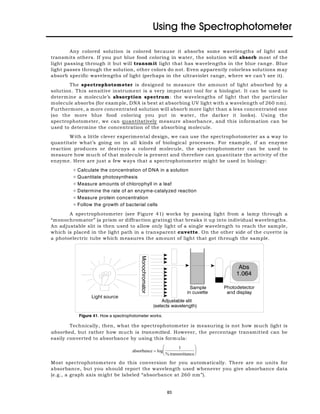
1. Figure 41. How a spectrophotometer works.
Using the Spectrophotometer
Any colored solution is colored because it absorbs some wavelengths of light and
transmits others. If you put blue food coloring in water, the solution will absorb most of the
light passing through it but will transmit light that has wavelengths in the blue range. Blue
light passes through the solution, other colors do not. Even apparently colorless solutions may
absorb specific wavelengths of light (perhaps in the ultraviolet range, where we can’t see it).
The spectrophotometer is designed to measure the amount of light absorbed by a
solution. This sensitive instrument is a very important tool for a biologist. It can be used to
determine a molecule’s absorption spectrum: the wavelengths of light that the particular
molecule absorbs (for example, DNA is best at absorbing UV light with a wavelength of 260 nm).
Furthermore, a more concentrated solution will absorb more light than a less concentrated one
(so the more blue food coloring you put in water, the darker it looks). Using the
spectrophotometer, we can quantitatively measure absorbance, and this information can be
used to determine the concentration of the absorbing molecule.
With a little clever experimental design, we can use the spectrophotometer as a way to
quantitate what’s going on in all kinds of biological processes. For example, if an enzyme
reaction produces or destroys a colored molecule, the spectrophotometer can be used to
measure how much of that molecule is present and therefore can quantitate the activity of the
enzyme. Here are just a few ways that a spectrophotometer might be used in biology:
N Calculate the concentration of DNA in a solution
N Quantitate photosynthesis
N Measure amounts of chlorophyll in a leaf
N Determine the rate of an enzyme-catalyzed reaction
N Measure protein concentration
N Follow the growth of bacterial cells
A spectrophotometer (see Figure 41) works by passing light from a lamp through a
“monochromator” (a prism or diffraction grating) that breaks it up into individual wavelengths.
An adjustable slit is then used to allow only light of a single wavelength to reach the sample,
which is placed in the light path in a transparent cuvette. On the other side of the cuvette is
a photoelectric tube which measures the amount of light that got through the sample.
Technically, then, what the spectrophotometer is measuring is not how much light is
absorbed, but rather how much is transmitted. However, the percentage transmitted can be
easily converted to absorbance by using this formula:
Most spectrophotometers do this conversion for you automatically. There are no units for
absorbance, but you should report the wavelength used whenever you give absorbance data
(e.g., a graph axis might be labeled “absorbance at 260 nm”).
83
2. 84
You will find two types of spectrophotometer in the Biology department. One is a fairly
simple “Spec-20” machine which measures only the transmittance or absorbance of visible light
(wavelengths above 340 nm) and can be used with square cuvettes, round cuvettes that look
like test tubes or even culture tubes used to grow bacteria. We also have computerized
SmartSpec spectrophotometers from BioRad. Both of these are UV-VIS spectrophotometers,
meaning that they can measure absorbance of either visible or ultraviolet light.
The BioRad spectrophotometers require 1 cm square cuvettes. Plastic or glass cuvettes
can be used for measuring absorbances in the visible range, but UV light is stopped by plastic
and glass, so UV absorbance (below 340 nm) must be measured in expensive, fragile quartz
cuvettes or in plastic cuvettes designed expressly for this purpose (which are never quite as
efficient as quartz cuvettes). Be sure you are using an appropriate cuvette for the volume of
solution you want to measure and for the wavelength you need to use!
Using the BioRad spectrophotometers for single-wavelength measurements
1. Turn on the unit using the rocker switch located on the back.
2. Press the l key for single-wavelength measurement.
3. Press 1, to read the absorbance at one wavelength, followed by ENTER.
4. Enter the wavelength that you wish to read.
5. Tell the instrument not to subtract a background measurement.
6. Prepare your blank cuvette and place it into the spectrophotometer.
3 Notice that the light path runs from front-to-back, so you want your cuvette
oriented correctly. The cuvette needs to be at least half full, so that your solution is
actually in the light path. Additionally, try not to touch the sides of the cuvettes where the
light will pass through—you don't want to measure the absorbance of your fingerprints!
7. Press the READ BLANK key and wait a moment while the zero is set. Then press the right arrow key
to move on.
3 The job of the blank is to tell the spectrophotometer what “zero” is. If you're
planning to measure a colored molecule dissolved in water, then the absorbance of plain
water would represent “zero,” and you should use water as your blank. However, you
might be dissolving your molecule in a solution which is colored to start with. In that case,
your blank should be the solvent with nothing added to it, so that the spectrophotometer
will subtract out the color of the solvent itself.
8. Remove your blank cuvette and insert the cuvette with your sample. Press the READ SAMPLE key.
Note the absorbance that is displayed.
9. Continue reading as many samples as you have.
Determining an absorption spectrum with the BioRad spectrophotometers
In some cases, you won’t know in advance the wavelength(s) absorbed by the solution
you’re interested in. In that case, the first step would be to measure its absorption spectrum
and find out experimentally what wavelengths are absorbed best. The spectrophotometer has
a program which will automatically try every wavelength and give you the absorbance at each
one. Here's the procedure:
1. After the spectrophotometer warms up, press the SCAN key.
2. Enter the wavelengths that you wish to scan.
3. Press ENTER to choose to not subtract a background setting.
4. Press ENTER to select the ‘slow’ scan.
3. 85
Figure 42. Diagram of a Spec-20
spectrophotometer.
5. Put your blank cuvette in the spectrophotometer and press READ BLANK.
6. Insert your sample cuvette and press the READ SAMPLE key. It will take a few moments to scan.
7. Press the PRINT key to get a small printout of your results. You can press PRINT again for your
partner’s notebook. Notice that the wavelengat which your sample had the maximal absorbance
(max) is listed near the top.
Following absorbance over time with the BioRad spectrophotometers
In some cases, you will want to measure how the absorbance of your solution changes
over time. This is particularlly useful for enzyme assays where your product (that absorbs light)
is being made. Rather than watching the clock and reading out the absorbance as time passes,
we can program these spectrophotometers to do that for us. Here's the procedure:
1. After the spectrophotometer warms up, press the KINETICS key.
2. Enter the wavelength that you wish to read.
3. Choose the total duration of the reaction. For example, if you want to collect the absorbance values
every five seconds for one minute, enter 60 seconds as the duration.
4. Choose the interval between successive readings. In the example above, this would be five seconds.
5. Press ENTER to choose to not subtract a background setting.
6. Put your blank cuvette in the spectrophotometer and press READ BLANK.
7. Insert your sample cuvette, start the reaction and press the READ SAMPLE key. Wait patiently while
the spectrophotometer records your data.
8. Press the PRINT key to get a small printout of your results. You can press PRINT again for your
partner’s notebook.
Using the Spec-20 spectrophotometer
1. Turn on the spectrophotometer by turning the left knob clockwise until it clicks. Use this knob to
adjust the pointer to zero on the transmittance scale (left end of the scale), or to set the digital display
to zero in transmittance mode.
2. Let the spectrophotometer warm up for 15 minutes.
3. Use the large knob on top of the spectrophotometer to set the
desired wavelength.
4. Again adjust the zero point with the left knob.
5. Place a blank in the sample holder and use the right knob to
adjust the reading to 100% transmittance (or zero on the
absorbance scale). Remove the blank.
6. If you are using the digital spectrophotometer, use the Mode
switch to choose absorbance or transmittance mode as needed.
7. Place your sample in the sample holder and read absorbance or
transmittance.
Determining concentration with a standard curve
There are two ways we could measure the concentration of some unknown solution
using the spectrophotometer. The first is to generate a standard curve: a graph of absorbance
vs. concentration for standard solutions whose concentrations are known. We then compare the
absorbance of an unknown solution (of the same molecule) to that curve. We don’t have to know
anything about the molecule in advance. Essentially, we are “calibrating” the spectrophotometer
to measure the concentration of the molecule we’re interested in.
4. 86
Figure 43. Standard curve used to determine the
concentration of an unknown solution
To make a standard curve, you’ll need at
least three or four different solutions with known
concentrations (called standards). You can come
up with these easily by taking a single solution of
known concentration and making several different
dilutions of it (see the section on measurements
and solutions on page 57). Then, measure the
absorbance of each known solution and draw a
graph, plotting absorbance vs. concentration, as
shown in Figure 43.
In this graph, concentration is placed on
the X-axis, because it’s the independent variable—
the one we’re manipulating. The absorbance is on
the Y-axis, because it’s the dependent variable: the
one whose value depends on the independent
variable.
The line on this graph represents the relationship between absorbance and
concentration. This should not be a “connect-the-dots” line: it needs to be a best-fit straight
line. Computer programs like Excel can calculate the equation of the best-fit line for you and
so can most scientific calculators. See the graphing section of this manual (page 43)for some
help with this. Generally, you want to include (0,0) as one of your points.
After generating a standard curve, you can measure the absorbance of the unknown
solution and compare it to the absorbances of the known solutions. You could do this by
eyeballing where the unknown would fall on the graph, but it’s more accurate to use the
equation of the best-fit line (in the form y = mx + b) to calculate the concentration. Here, y is the
absorbance, which you measure, x is the concentration that you want to know, and m and b
are the slope and intercept of the best-fit line, which Excel will tell you. By solving for x, you can
accurately calculate the concentration of the unknown.
In order for this calculation to be accurate, the absorbance of the unknown has to fall
within the range of absorbances of the known solutions—if it doesn’t, you may have to dilute
the unknown, or make different standards and try again. It’s important to note that the
relationship between absorbance and concentration is only linear within a certain range, and
maxonly when you use the right wavelength of light. The “right” wavelength is ë , or the
wavelength at which the molecule absorbs best. If you don’t know which wavelength to use, you
maxcan determine ë experimentally by measuring the absorption spectrum as described above.
maxThe peak wavelength is ë .
Determining concentration by the Beers-Lambert law
The second way to measure concentration using the spectrophotometer is really the
maxsame as the first, but with a shortcut. If you (or someone else) has already determined ë and
drawn a standard curve which shows a linear relationship between absorbance and
concentration, then you can calculate a number called the extinction coefficient, which you can
always use to find concentration for that molecule under those conditions.
The extinction coefficient simply relates concentration to absorbance, using a formula
called the Beers-Lambert law. In this equation, the absorbance is equal to the concentration of
the molecule of interest (c), times the length of the path the light takes through the cuvette (b;
always 1 cm for our spectrophotometers), times the extinction coefficient (g):
Abs = gbc
The extinction coefficient is the part that is specific for each compound and must be determined
experimentally. You can determine it from your standard curve by solving for g, because you
know the absorbance and concentration and can measure the width of the cuvette. Once you
know g, you can use it for other solutions of the same molecule.