1. ORTHOPHOS®
XG3D
•Visualizecanalanatomypriortotreatment
•MARSforbetterdiagnosisaroundmetal
•Easypatientpositioning
Learnmoreat
Sirona3D.com
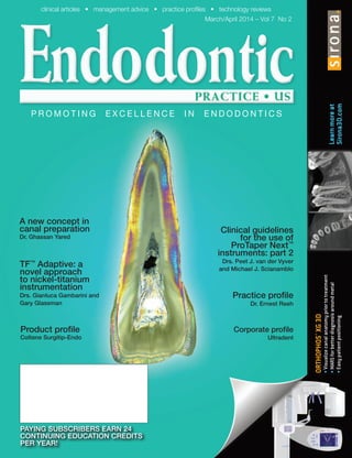
A new concept in
canal preparation
Dr. Ghassan Yared
PAYING SUBSCRIBERS EARN 24
CONTINUING EDUCATION CREDITS
PER YEAR!
TF™
Adaptive: a
novel approach
to nickel-titanium
instrumentation
Drs. Gianluca Gambarini and
Gary Glassman
clinical articles • management advice • practice profiles • technology reviews
March/April 2014 – Vol 7 No 2
P R O M O T I N G E X C E L L E N C E I N E N D O D O N T I C S
Corporate profile
Ultradent
Clinical guidelines
for the use of
ProTaper Next™
instruments: part 2
Drs. Peet J. van der Vyver
and Michael J. Scianamblo
Practice profile
Dr. Ernest Reeh
Product profile
Coltene Surgitip-Endo
2. simply better dentistry™
1145 Towbin Avenue Lakewood, New Jersey 08701
Visit us on the web at www.sswhiteburs.com
Simply Better Endodontics™
The V-Taper™
2H System is the most
efficient and effective endodontic file
system in the world.
Endodontist Recommended
The V-Taper™2H NITI rotary endodontic file system utilizes only 3 files to allow you to complete
most root canal cases. The V-Taper™2H performance enhanced system is the only system that
offers you these industry leading advantages, all in one file:
Conservation • Efficiency • Flexibility • Strength
20
( V06 )V06 )(
Conservative
V-Taper™2H files with their patented taper, are the most minimally invasive file
on the market today.
Efficient
Complete most root canals using only 2-3 V-Taper™2H files, fewer files means lower
cost per root canal treatment
Flexible
V-Taper™2H files are heat treated to increase flexibility to insure navigation of even
the most curved canals without ledging, transportation or zipping.
Strong
V-Taper™2H files are designed with a Parabolic Core, which was the strongest core
design in a recent test conducted at the University of Michigan.
The V-Taper™2H Rotary System advances the science of file design, manufacturing,
and materials.
“All dentin is not created equally,
EndoGuide® Burs and V-Taper™2H
Files foster conservation at the
heart of the tooth, peri-cervical
dentin. The preservation of
healthy dentin leads to longer
lasting restorations.”
– Dr. Eric Herbranson, Endodontist
25
( V06 )
17
Call for an in-office demo today.
800-535-2877
4. TABLE OF CONTENTS
Clinical
Clinical guidelines for the use
of ProTaper Next™
instruments:
part 2
Drs. Peet J. van der Vyver and
Michael J. Scianamblo illustrate the
use of ProTaper Next instruments
in difficult and challenging
endodontic cases........................12
MTA: the new material of choice
for pulp capping
Drs. Leendert (Len) Boksman and
Manfred (Manny) Friedman delve into
the benefits of MTA ....................20
The influence of mineral trioxide
aggregate (MTA) thickness on its
microhardness properties —
an in vitro study
Drs. Iris Slutzky-Goldberg, Lea
Sabag, and David Keinan test the
effect of the MTA thickness on its
microhardness properties............26
2 Endodontic practice Volume 7 Number 2
Corporate profile 10
Ultradent
Ultradent continues to keep a finger on the pulse of the endodontic specialty
ON THE COVER
0.2 mm tooth slice polarized photo
Maxillary central incisor Transversal slice.
Photo courtesy of Dr. Stanislav Heranin
Practice profile 6
Dr. Ernest Reeh
Focus on patients, family, academics, and endodontics
6. TABLE OF CONTENTS
Technology
3D Apical Cork — part 3
In the third article of this series,
Dr. Wyatt Simons reviews the
technological breakthroughs of
the Cork technique and system of
obturation with emphasis given to the
revolutionary 3D plugger ...............32
Continuing
education
A new concept in canal
preparation
Dr. Ghassan Yared discusses
canal preparation with only one
reciprocating instrument without prior
hand filing......................................36
TF™ Adaptive: a novel approach
to nickel-titanium instrumentation
Drs. Gianluca Gambarini and Gary
Glassman examine how to achieve
rotary motion when you want it — and
reciprocation when you need it......42
Product profile
Sonendo®
aims to transform the
future of endodontics ................50
Surgitip-endo aspirator tip for root
canals .........................................54
Endospective
Transcendent endodontics: the
seven key attributes
Dr. Rich Mounce reflects on the
qualities and equipment that can
improve future results....................52
Step-by-step
The Laschal FXP set incorporates
transferred oscillation technology
.....................................................56
Legal matters
Ethics, morals, and law in the
professional office
Dr. Bruce H. Seidberg discusses
how ethical and moral behavior are
governed by law ...........................57
Industry news ............60
Materials &
equipment ......................60
Anatomy matters
Influence on Fees, Reputation,
Longevity, and Because: part 10
Dr. John West discusses four more
reasons why anatomy should matter.
.....................................................61
4 Endodontic practice Volume 7 Number 2
3D Apical
Cork
32
7. ORTHOPHOS XG 3D
ORTHOPHOS XG 3D
The right solution for
your diagnostic needs.
Implantologists
will appreciate the
seamless clinical
workflow from initial
diagnostics, to treatment
planning, to ordering
surgical guides and final
implant placement.
Endodontists
will enjoy instantly
viewable 3D volumetric
images for revealing
and measuring canal
shapes, depths
and anatomies.
Orthodontists
will benefit from high-
quality pan and ceph
images for optimized
therapy planning.
General Practitioners
will achieve greater
diagnostic accuracy
for routine cases.
“With my Sirona 3D unit, I can see the anatomy of canals, calcification, extent of resorption, frac-
tures, and sizes of periapical radiolucencies, all of which influence treatment plans for my patients.
Combine that with the metal artifact reduction software that reduces distortions from metal objects,
my treatment process is a lot less stressful. My patients benefit from the technology and my
referrals appreciate the value.” ~ Dr. Kathryn Stuart, Endodontist - Fishers, Indiana
For more information, visit www.Sirona3D.com
or call Sirona at: 800.659.5977
The advantages of 2D & 3D in one comprehensive unit
ORTHOPHOS XG 3D is a hybrid system that provides clinical
workflow advantages, along with the lowest possible effective
dose for the patient. Its 3D function provides diagnostic accuracy
when you need it most: for implants, surgical procedures and
volumetric imaging of the jaws, sinuses and other dental anatomy.
www.facebook.com/Sirona3D
8. What can you tell us about your
background?
I have a bachelor’s degree in chemistry
with a minor in business. I was accepted
off of the alternate list for dental school and
then attained my DDS degree, graduating
third in my class and awarded the James
McCutcheon Gold Medal. (This award is
presented to the student who, over the 4
years of the DDS program, had shown to
possess, to an outstanding degree, those
qualities of scholarship, leadership, and
character, which may be expected to lead
to a distinguished position in the dental
profession.) I have taken specialty training
in endodontics and have a master’s degree
in Material Science, a PhD in Biophysics
with a related-field minor in engineering.
I am a Diplomate of the American Board
of Endodontics (Board Certified) and a
former faculty member at the University
of Minnesota Endodontics Department.
I have consulted for 3M and Carestream
Dental (formerly Kodak Imaging), and have
lectured both nationally and internationally
on a variety of science and dental topics,
as well as written peer-reviewed scientific
articles and abstracts.
Is your practice limited to
endodontics?
Yes.
Why did you decide to focus on
endodontics?
By my second year in dental school, I
developed a keen interest in endodontics.
I especially enjoyed the particular attention
to the fine detail and the high level of fine
motor control needed. I had done research
in chemistry and wanted to do research
in endodontics. As a result of frequent
discussions with the director of the
endodontics department, arrangements
were made for me to be able to conduct
research as a graduating second-year
dental student. I was able to present my
work at an International Association of
Dental Researchers (IADR) conference,
as well as have my name associated with
three other projects with which I was
involved. I continued my research interests,
and the following year also presented at
the IADR conference and had my name
associated with seven other projects that
I had also been involved in during the year
and summer break. After dental school,
I had wanted to go into endodontics as
a specialty, but felt I should appreciate
general dentistry before going to graduate
school. I worked in a private general dental
practice for 2 years and then attended
endodontic graduate school. I did research
during my endodontic residency and
published two papers as a result of my
graduate program research, as well as
winning a national research award for my
student research presentation award at
the American Association of Endodontists
(AAE) meeting. I have always had a passion
for endodontics and enjoy the challenges
my many colleagues send me.
How long have you been
practicing?
I graduated from dental school in 1984 and
practiced as a general dentist for 2 years.
I then spent 2 years in an endodontic
residency, graduating in 1988, and have
been practicing as an endodontist since
graduation. I went on to do a PhD after my
endodontic specialty training but made it a
requirement of my PhD program that I was
able to do private practice 1 day a week.
I worked for 7 years (during and after my
PhD program) for Boynton Health Services
as their endodontist. I then worked in
several group practices before setting up
our own endodontic practice with my wife,
who is also an endodontist. Currently, I
practice 2 to 3 days per week, and my wife
practices 2 days per week, so we both can
have some time to enjoy our four children
ages 11, 9, 9 (yes, twins), and 5 year olds.
They grow up so fast, and we both wanted
to have time with them.
Who has inspired you?
I have had several people who inspired me.
Initially, my dad, who told me when I was
deciding to either follow chemistry (when I
had standing job offers after graduation in
chemistry) or go into dentistry (which would
have been an unknown). My dad, who was
a man of few words, told me, “You have
always wanted to be a dentist,” (which I
had since I was about 8 years old), and he
said to me, “You never want to look back
Dr. Ernest Reeh
6 Endodontic practice Volume 7 Number 2
PRACTICE PROFILE
Focus on patients, family, academics, and endodontics
9. PRACTICEPROFILE
Volume 7 Number 2 Endodontic practice 7
at your life and say, ‘I wish I would have
…’” I have lived his “no regrets” philosophy
ever since.
My other mentors include the head
of the endodontics department in dental
school, who made doing research
possible; Dr. Don Collins, the Dean who
created a program when none existed to
make it possible for me to do research;
Dr. Gordon Thompson and my mentor
doing dental research, who was just
joining the endodontic faculty at the time;
and the abundantly enthusiastic Dr. Ken
Zakariasen.
In endodontic graduate school, I had
two people who had a major influence in
my life and career — Dr. Harold Messer,
a brilliant scientist and the head of the
endodontics division; and Dr. William “Bill”
Douglas, a renowned biomaterials expert
who opened his lab and mentored me
during my masters and PhD programs.
I learned many things, not only about
dentistry and research, but also about
teaching and mentoring.
What is the most satisfying aspect
of your practice?
I feel most satisfied when my doctors have
the confidence in me to refer tough cases
that challenge my skills and abilities. As I
like to say, “I enjoy challenges; I just hope
that not every case during the day will be a
tough one!” I feel satisfied when I can treat
cases that were thought to be untreatable
endodontically. I appreciate when I can be
a part of their team and be a part of the
treatment planning for cases.
Professionally, what are you most
proud of?
My master’s thesis work. I am sometimes
at an endodontic meeting, and a resident
will see my name tag and ask if I am the
guy who published the work on stiffness
of endodontically treated teeth. I reply that
I am. Then they tell me that I am “classic
literature in endo,” and I reply that I like to
think of myself as contemporary literature
in endo! I have been told that my work is
one of the 10 most referenced papers in
dentistry. That makes me very proud of the
work I did.
What do you think is unique about
your practice?
We offer a very personalized experience.
We work on one patient at a time, and
each gets our undivided attention for his
or her appointed time. We have created an
environment that is very calming from the
appearance of the office, to the music, to
the aromas, to the friendly interaction from
each staff member. Patients are exquisitely
numb, so care is done comfortably. Many
patients who tell us they are difficult to get
numb are surprised how easy care is for
them.
What has been your biggest
challenge?
Insurance. Need I say more?
What would you have become if
you had not become a dentist?
Probably a neurovascular surgeon. I enjoy
surgery, and I am very fine motor skill-
oriented, so it would be another good fit
for me.
What is the future of endodontics
and dentistry?
There is so much information that it is not
possible for one person to do everything
well. General dentists who take a lot of
programs in endodontics certainly advance
their skills and can do more and more
complex cases but at the expense of other
areas of dentistry. Most dentists want
some balance in their profession, so they
do not want to do more advanced cases.
Even those who have trained further still
need the help of their endodontist as there
is a lot to learn in a 2- to 3-year advanced
specialty degree. There continues to
be improvements, and it is part of the
endodontist’s job to explain the pros and
cons to our general dental colleagues.
Clearly, as knowledge continues to
Dr. Reeh’s team
10. 8 Endodontic practice Volume 7 Number 2
PRACTICE PROFILE
Top Favorites
There are two main aspects to my life: my
home and family, and my office and staff.
Hence, the list goes in two directions:
Family favorites
1. Date night with my lovely wife
2. Kid activities
3. Family game night (Something we
do one night per week usually on the
weekend.)
4. Our home (I like to just be home and
enjoy what we have done.)
5. Enjoying my hobbies
Office favorites
6./5. Quiet time (because it is so rare)
4. P5 Newtron®
(Satelec Acteon)
ultrasonics
3. Schick Elite Digital radiography
2. Carestream 9000D cone beam
computed tomography (three-
dimensional imaging)
1. Our staff (They are all great in their own
ways and are an integral part of my life.)
expand, the need for specialists becomes
increasingly important. Endodontics is not
about to be replaced by implants, and
we are now seeing that retaining natural
teeth is still first best compared to a good
second best of an implant as the pendulum
starts to swing away from replacing many
teeth to preserving natural teeth.
What are your top tips for main-
taining a successful practice?
There is no easy answer. First and foremost
is providing a high level of care, but that
is not enough. One has to create an
experience for the patient that proves the
value in the services provided. On top of it
all is maintaining a highly motivated, well-
trained staff that enjoy what they do. We
create an environment in which the staff
enjoys coming to work. Our staff currently
averages over 10 years with our office.
What advice would you give to
budding endodontists?
It is to not about all the devices. Cone
beam, torque-sensing motors, and so on
are all good, but it is about the patient.
One person once told me, “Patients don’t
care how much you know until they know
how much you care.” Keep the patient
experience in the forefront, doing what is
right, and the rest will follow naturally.
What are your hobbies, and what
do you do in your spare time?
My biggest hobby is my family. I love to
spend time with my wife and kids, from
little stuff like building a snowman to going
to bigger things like going skiing. Four kids
always take a lot of effort, but it’s worth it.
I hope to share in my kids’ hobbies to the
extent they want to include me. My kids
enjoy chess, and I am the chess master
for the chess club at their school. They
wanted to try downhill skiing, so we took
up skiing this year. I like cross-country
skiing also, but none of them shows much
interest, so I haven’t done any for a while. I
have a few hobbies of my own that I enjoy.
I enjoy automotives through reading car
magazines (I subscribe to three), going
to the auto show (usually with a friend as
the kids typically don’t want to go), going
to advanced driving skills courses; and I
have an old car that I tinker with. I enjoy
camping. I made a campsite down a path
in our backyard. The kids and I go camping
a couple of times each summer, as well as
Cub Scout camps. (Mom typically doesn’t
like to camp but, on occasion, is a good
sport and joins us.) I have a wood shop
in my basement and have a number of
projects that I like to do. I am currently
working on a chessboard and chess pieces
with my boys. I also enjoy cooking. I do not
like being a short-order cook preparing
multiple different meals for the kids and
grown-ups, but unfortunately, that is most
of what I do presently. It is just where we
are at in our lives. When I get a chance,
I like to cook and bake and am known
for the cheesecakes that I make and for
presentation of dishes.
Dr. Reeh’s family
EP
11. OPERATORY COMPUTER
MICROSCOPE
ONLINE PORTAL
IMAGING EQUIPMENT
We’re that something you’ve been searching for. Ask yourself:
WHAT IF I could surround myself with employees committed to clinical and practice excellence?
WHAT IF my support team was exceptional because of complete commitment to my vision?
WHAT IF I had immediate, meaningful, and quality contact with my referrals every minute of every day?
WHAT IF I had hundreds of world-class endodontists
mentoring me daily, helping me to become more successful?
WHAT IF my patients recognized immediately that my
practice was centered around quality?
C a l l 1 - 8 7 7 - 4 3 5 - 7 8 3 6 o r e m a i l u s a t s a l e s @ t d o 4 e n d o . c o m f o r a f r e e d e m o o f t h i s t i m e - s a v i n g p r o g r a m .
MAKE IT POSSIBLE WITH
TDO SOFTWARE
12. History of Ultradent
Ultradent Products, Inc., began when one
forward-thinking dentist, Dr. Dan Fischer,
set out to create dental products more
effective than those that were currently
available. Dissatisfied with many dental
options, Dr. Fischer hoped to develop
better products that were not only
advanced for their time but would also set
future industry standards.
It all began in 1974, when, upon graduating
from Loma Linda University and starting
his own dental practice in the Salt Lake
Valley, Dr. Fischer noticed a dire need
for more rapid, profound hemostasis.
At the time, no products existed on the
market that predictably controlled bleeding
and sulcular fluid, which made getting
accurate impressions and the overall
practice of high-quality operative dentistry
a challenge. Experimenting with various
chemistries after-hours and often drawing
his own blood to test their hemostatic
effects, Dr. Fischer discovered a solution
that, when combined with his innovative
scrubbing technique, achieved rapid,
profound hemostasis every time. This
product, known as Astringedent®
, is now
considered Ultradent’s flagship product.
In order to share it with the industry, Dr.
Fischer founded Ultradent in 1978.
What began as a family-only business
quickly grew as word of Ultradent’s
groundbreaking, high-quality products
spread. In 35 short years, the company
expanded from a small home operation
to the 220,000-square-foot facility in
South Jordan, Utah, that Ultradent calls
home today. Ultradent’s headquarters
houses 1,100 employees and continues
to expand, breaking ground last fall
Ultradent
10 Endodontic practice Volume 7 Number 2
CORPORATE PROFILE
By maintaining close
relationships with top endodontic
researchers at several domestic
and international universities, as
well as by keeping several highly
skilled dentists on staff, Ultradent
continues to keep a finger on
the pulse of this important and
rapidly growing area of dentistry.
products, application device materials,
and techniques. Ultradent’s product family
now includes world-class adhesives,
composites, tooth whitening systems,
and endodontic products that are used by
clinicians around the world, including dental
professionals in large group practices,
dental and veterinary labs, private practices,
government agencies, and universities.
Over the last decade, Ultradent expanded
its reach to orthodontics, serving as the
parent company for Opal Orthodontics.
Also headquartered in South Jordan, Utah,
Opal Orthodontics houses its own on-site
orthodontic clinic.
A minimally invasive philosophy
Dr. Fischer has said, “Respecting and
preserving our patients’ dentate throughout
their life: this should be among our principal
responsibilities. I believe to my bootstraps
in respecting human tissues to the ultimate
degree, in preserving mineral mother dentin,
and in respecting supporting tissues as
well. It comes down to first and foremost,
remembering the fabulous human behind
that oral cavity.” He goes on, “The more I
cut the tooth, the more I weaken the tooth,
and the more of the tooth I cut, and the
more times I cut, the sooner I will kill the
tooth. Trauma to the tooth is additive, even
over decades!”
Ultradent strives to offer the latest and
greatest in technology, and Dr. Fischer’s
passion for a minimally invasive approach to
dentistry has and will continue to guide the
development of every new product created
in the future. It was this very approach that
led to the creation of Ultradent’s extensive
line of endodontic products available to
clinicians today.
Ultradent endodontics
Endo-Eze®
AET classic stainless steel files,
Ultradent’s extensive line of endodontic
products and solutions, were born out
the necessity for a successful endodontic
protocol that met the minimally invasive
philosophy that Dr. Fischer so passion-
ately advocates. The result: the Endo-
Eze Anatomic Endodontic Technology
(AET) technique. Because of the 30°
reciprocating motion of the handpiece, the
system produces less-invasive root canal
therapies, as the combination of motion
Dr. Dan Fischer
on a 10,000-square-foot building to
create space for increased molding and
manufacturing.
One of the most vertically integrated
dental companies in the world, Ultradent
manufactures over 90% of its products
(which includes over 500 materials,
devices, and instruments) at its Utah
campus. Instead of saving on production
costs through outsourcing, which many
U.S. manufacturers do, Dr. Fischer firmly
believes in the opposite. He says, “The
more one outsources, the more one ships
production, or R&D, or other aspects to
other parts of the world, the more one loses
touch with what has made them who they
are.” Ultradent exports approximately 70%
of its products internationally to countries in
nearly every continent in the world.
Ultradent currently holds dozens
of patents and trademarks on unique
13. CORPORATEPROFILE
Volume 7 Number 2 Endodontic practice 11
and files proved able to better follow the
natural canal anatomy. This stands in
contrast to the popular rotary NiTi systems,
which are not designed to replicate the
naturally formed canal, but to prepare
the canal in a round, conical shape. By
following the natural canal shape, the files
minimize excess removal of healthy tooth
structure.
Ultradent offered a number of
endodontic products before introducing
AET and has created a number of market-
leading devices and chemistries since its
introduction. A few of these are outlined
here:
NaviTip®
In the year 2000, Ultradent introduced
NaviTip — the very first endodontic tip
capable of delivering irrigants to just
about any part of the root canal system.
Today, with the world’s smallest and most
technologically sophisticated cannula for
irrigation and delivery, NaviTip remains
unsurpassed in its performance.
NaviTip features a unique rigidity at the
handle of the tip, making it strong enough
to be introduced as deep as needed in the
canal. And the annealed and rounded tip
end gives it the ability to navigate down the
tiny intricate curvatures of any canal of any
tooth.
Available in four lengths (17 mm, 21
mm, 25 mm, and 27 mm), three gauges
(29 ga, 30 ga, and 31 ga), and even with a
flocked end to help clean debris or product
out of a canal, NaviTip is available in an
option to suit any need a clinician may
have. The NaviTip even has a version with
sideport openings that deliver irrigants
toward the canal walls rather than toward
the apex, which minimizes the risk of
expressing strong chemistries past the
apex.
Endo-Eze®
Arios™
and Endo-Eze AET
TiLOS™
Building on the success of the AET files,
Ultradent developed the Endo-Eze TiLOS
system with several well-respected
American and international specialists.
This very simple technique uses both
stainless steel and NiTi files in combination
with traditional hand files. If the clinician
prefers, the TiLOS system can be used
without traditional hand files as well. The
award-winning TiLOS system is available
in convenient, autoclavable, preconfigured
patient kits as well as refills. The simplest
is the RediPack, which contains the files
needed to treat about 90% of endodontic
cases. The TiLOS technique still uses a
reciprocating handpiece, which provides
a “milling” rather than “drilling” motion.
Experience has shown that a milling motion
reduces the amount of file separation
that occurs. And of course, the TiLOS
instruments and technique follow the
minimally invasive Ultradent philosophy
that the company has been built on.
More and more clinicians are
discovering Ultradent’s Engine Pack,
which contains three engine-driven files.
This preconfigured kit is perfect for the
preflaring of canals — something every
clinician does, but often requires gathering
the necessary files from different kits to do
so. The Engine Pack contains all the files
needed for this preflaring procedure, it’s
autoclavable, very economical, and it can
be integrated into any technique currently
being taught today.
The pulse of the endodontists
For many years, Ultradent has developed
and provided endodontic equipment such
as files, delivery tips, irrigants, handpieces,
sealer, and gutta percha with the goal of
simplifying and elevating the quality of
endodontic outcomes. By maintaining
close relationships with top endodontic
researchers at several domestic and
international universities, as well as by
keeping several highly skilled dentists
on staff, Ultradent continues to keep a
finger on the pulse of this important and
rapidly growing area of dentistry. Ultradent
proudly offers the latest and most cutting-
edge metals, file types, and technologies,
while continually working to refine and
work toward less-invasive endodontic
solutions and protocol. To learn more
about the endodontic products mentioned
or the wide array of additional endodontic
solutions provided by Ultradent, please
visit ultradent.com, or call 800-552-5512.
This information was provided by Ultradent.
EP
14. In part 1 of this series, published in the
January/February issue of Endodontic
Practice US, the authors outlined the clinical
guidelines for the use of the ProTaper
Next™
(Dentsply/Maileffer) instruments.
(ProTaper Next is only available in North
America through DENTSPLY Tulsa Dental
Specialties.)
There are five instruments in the
system, but most canals can be prepared
by using only the first two instruments. The
first instrument in the system is the ProTaper
Next X1, with a tip size of 0.17 mm and a 4%
taper. This instrument is used after creation
of a reproducible glide path by means of
hand instruments or PathFile™
rotary files
(DENTSPLY Tulsa Dental Specialties). The
ProTaper Next X1 is always followed by the
second instrument: the ProTaper Next X2
(0.25 mm tip and 6% taper). The ProTaper
Next X2 can be regarded as the first
finishing file in the system, as it leaves the
prepared root canal with adequate shape
and taper for optimal irrigation and root
canal obturation. The ProTaper Next X1
and X2 have an increasing and decreasing
percentage tapered design over the active
portion of the instruments. The last three
finishing instruments are the ProTaper Next
X3 (0.30 mm tip with 7% taper), ProTaper
Next X4 (0.40 mm tip with 6% taper),
and the ProTaper Next X5 (0.50 mm tip
with 6% taper). These instruments have a
decreasing percentage taper from the tip
to the shank. The ProTaper Next X3, X4,
and X5 can be used to either create more
taper in a root canal or to prepare larger
root canal systems.
The advantages of the ProTaper Next
system include the following:
• The instruments are manufactured
from M-Wire that contributes toward more
flexible instruments, increased safety, and
protection against instrument fracture
(Gutmann and Gao, 2012), allowing the
clinician to treat more complex root canal
systems with a high level of success.
• The instruments have a bilateral
symmetrical rectangular cross section with
an offset from the central axis of rotation
(except in the last 3 mm of the instrument,
D0-D3), creating an asymmetric rotary
motion. The exception is the ProTaper
X1, which has a square cross section in
the last 3 mm to give the instruments a bit
more core strength in the narrow apical
part. The asymmetric rotary motion allows
the instrument to experience a rotational
phenomenon known as precession or
swagger (Scianamblo, 2011). According to
Van der Vyver and Scianamblo (2013), this
design characteristic includes six benefits:
1. It further reduces (in addition to
the progressive tapered design) the
engagement between the instrument and
the dentin walls because only two cutting
Clinical guidelines for the use of ProTaper Next™
instruments: part 2
12 Endodontic practice Volume 7 Number 2
CLINICAL
Drs. Peet J. van der Vyver and Michael J. Scianamblo illustrate the use of ProTaper Next instruments
in difficult and challenging endodontic cases
Figure 1A: Preoperative radiograph of a maxillary right
second premolar
Dr. Peet J. van der Vyver is extraordinary professor at
the Department of Odontology, School of Dentistry,
University of Pretoria and Private Practice, Sandton,
South Africa (see www.studio4endo.com for more).
Michael J Scianamblo, DDS, is an endodontist and
the developer of Critical Path Technology. He is a
postgraduate and fellow of the Harvard School of Dental
Medicine and has served as a faculty member of the
University of the Pacific and the University of California,
Schools of Dentistry in San Francisco.
Figure 1B: Length determination radiograph. Note the
“S”-shaped canal configuration
Figure 1C: Postoperative radiograph after canal obturation
with GuttaCore obturators
15. CLINICAL
Volume 7 Number 2 Endodontic practice 13
points make contact with the canal wall at
any time. This will contribute to a reduction
in taper lock, screw-in effect, and stress on
the file.
2. It ensures debris removal in a coronal
direction because the off-center cross
section allows for more space around the
flutes of the instrument. This will lead to
improved cutting efficiency, as the blades
will stay in contact with the surrounding
dentin walls. Root canal preparation is
done in a very fast and effortless manner.
3. The swaggering (asymmetric) rotary
motion of the instrument initiates activation
of the irrigation solution during canal
preparation, improving debris removal.
4. It reduces the risk of instrument fracture
because there is less stress on the file and
more efficient debris removal.
5. Every instrument is capable of cutting
a larger envelope of motion (larger canal
preparation size) compared to a similarly
sized instrument with a symmetrical mass
and axis of rotation. This allows the clinician
to use fewer instruments to prepare a root
canal to the adequate shape and taper to
allow for optimal irrigation and obturation.
6. There is a smooth transition between
the different sizes of instruments because
the design ensures that the instrument
sequence itself expands exponentially.
The aim of this article is to illustrate
the use of ProTaper Next instruments
in complex and challenging endodontic
cases. The preparation technique for
minimally invasive root canal preparation
with ProTaper Next instruments will also be
discussed.
“S”-shaped root canals
A major challenge in endodontics is the
treatment of “S”-shaped or bayonet-
shaped root canals. This type of root canal
configuration can be present in root canal
systems of maxillary laterals, canines, and
premolars, as well as mandibular molars
(Rueben, et al., 2008). The authors would
recommend using PathFile No. 3 (ISO tip
0.19 mm) (after PathFile Nos. 1 and 2) in
these challenging root canal systems as
the final glide path preparation file. This
will increase the glide path size before
introducing the ProTaper Next X1, resulting
in less engagement as the file travels down
the canal curvatures.
Figure 2A: Preoperative radiograph of
a maxillary right first molar
Figure 2B: Length determination
radiograph. Note the “S”-shaped canal
configuration in the distobuccal root
canal
Figure 2C: Postoperative radiograph
after glide path preparation with
PathFiles and canal preparation with
ProTaper Next X1 and X2. Obturation
was done with GuttaCore obturators.
Note maintenance of “S”-shaped
curvature in the obturated distobuccal
root canal system
16. Case report 1
The patient, a 41-year-old female,
presented with irreversible pulpitis on her
maxillary right second premolar (Figure
1A). The length determination radiograph
revealed an “S”-shaped canal configuration
(Figure 1B). The canal was negotiated and
glide path enlarged using PathFile Nos. 1,
2, and 3. Canal preparation was done with
ProTaper Next X1 and X2.
In this case, emphasis was placed
on using a backstroke, outward brushing
motion with the ProTaper Next instruments
to remove restrictive dentin in the canal,
allowing the instruments to progress
apically. The canal was obturated (Figure
1C) with a size 20 GuttaCore™
obturator
to working length followed by another X2
GuttaCore obturator (DENTSPLY Tulsa
Dental Specialties) to ensure adequate
obturation of the oval coronal part of the
root canal system.
14 Endodontic practice Volume 7 Number 2
CLINICAL
Case report 2
A 45-year-old male patient presented
with severe pain on his maxillary right first
molar. A preoperative periapical radiograph
revealed placement of a deep amalgam
restoration (Figure 2A).
The length determination radiograph
revealed an “S”-shaped canal configuration
in the distobuccal root canal (Figure 2B).
The root canals were negotiated to working
length, and the glide paths enlarged using
PathFile Nos. 1 and 2. PathFile No. 3 was
used in the distobuccal root canal. Canal
preparation was done with ProTaper Next
X1 and X2 in all three root canals.
After gauging with a size 25 nickel-
titanium hand instrument, it was decided to
enlarge the palatal root canal to a ProTaper
Next X3. All three root canals were
obturated with matching ProTaper Next
gutta-percha cones using the Calamus®
Dual Obturation Unit (DENTSPLY Tulsa
Dental Specialties) (Figure 2C). Note the
maintenance of the “S”-shaped curvature
in obturated distobuccal root canal system.
Challenging curvatures in the
apical third of root canals
Apical root canal curvatures must always
be respected and never straightened. Ac-
cording to Catellucci (2005), straightening
these curves would mean displacing the
apical foramen from its original position,
which can lead to treatment failure. Other
problems that can be encountered when
treating curved canals include ledge
formation, perforation, zip formation, and
file separation (Ingle, 2005).
It is very important to identify canal
curvatures during initial canal negotiation
in order to avoid the above-mentioned
preparation errors. The greater the angle
of curvature and the smaller the radius
of curvature, the more complex the
Figure 3A: Non-vital mandibular left first molar and
inadequately root canal treated mandibular right second
molar
Figure 3B: Initial length determination radiograph. Note
that the files were short in all the root canals in the
mandibular second molar
Figure 3C: Periapical radiograph demonstrating the fit of
the plastic inserts of ProTaper obturators to the corrected
working length (mandibular second molar) after canal
negotiation with C+ and K-files and preparation with
ProTaper Next
Figure 3D: Final result after the canals were obturated with ProTaper
obturators
Figure 3E: Periapical radiograph (30°
mesial angulated) demonstrating respect
of the original canal anatomy after
canal preparation with ProTaper Next
instruments
Figure 3F: Six-month follow-up periapical radiograph
illustrating periapical healing
17. CLINICAL
Volume 7 Number 2 Endodontic practice 15
management and treatment will be (Pruett,
Clement, Carnes, 1997).
Again, the authors would recommend
using all three PathFiles in these challenging
root canal systems to enlarge the glide
path prior to canal preparation. It is also
important to note that the reduced apical
tapers of the ProTaper Next instruments
(compared to ProTaper Universal) are ideal
for maintaining apical curvatures or “S”-
shaped root canals.
Case report 3
The patient, a 27-year-old male, presented
with a non-vital mandibular left first molar
and an inadequately root canal treated
mandibular right second molar (Figure 3A).
Access cavities were prepared, and the
previous gutta percha was removed from
the canals of the second molar.
A length determination radiograph
revealed sharp apical curvatures in the last
few millimeters of the mesial and distal roots
of the mandibular first molar. It was also
noted that the working length was short in
the canals of the second molar (Figure 3B).
A combination of C+ and K-files were used
to negotiate the canals in the mandibular
second molar to full working length. A
reproducible glide path was established
in all the root canals, and the glide paths
enlarged to ISO 0.19 mm using PathFiles.
The coronal two-thirds of the canals
were prepared with ProTaper Next X1 and
X2 using a backstroke, outward brushing
motion to remove restrictive dentin in
the canals, allowing the instruments to
progress towards the apical third. The
apical third of the root canals were prepared
with a controlled push-pull motion, allowing
the instruments to progress up to working
length.
The prepared root canals were gauged
with a size 25 nickel-titanium hand file. The
file was snug at working length except in
the distal canal of the lower first molar. This
canal was enlarged with a ProTaper Next X3
instrument. Figure 3C shows radiographic
confirmation of the working length and the
fit of the plastic carriers of size 25 ProTaper
obturators (without gutta percha). All the
canals were obturated (Figure 3D) with
size 25 ProTaper obturators, except the
distal root canal in the lower first molar
that received a size 30 ProTaper obturator.
Figure 4A: Preoperative radiograph of non-vital maxillary
left first and second molars
Figure 4B: Length determination radiograph for the
maxillary first molar
orders@engineeredendo.com www.engineeredendo.com
The Finishing File is the most cost effective
and simplest way to clean a canal!
THE FUTURE HAS RETURNED.
THE ORIGINAL PLASTIC
ENDODONTIC ROTARY
FINISHING FILE IS BACK.
MANUFACTURER DIRECT.
MADE IN THE U.S.A.
18. 16 Endodontic practice Volume 7 Number 2
CLINICAL
Figure 3E demonstrates the final result
after obturation, and Figure 3F illustrates
healing of the periapical pathology around
the roots on a 6-month postoperative
radiograph.
Minimally invasive canal
preparation
According to Gutmann (2013), minimally
invasive endodontic (MIE) procedures can
range from diagnosis to making a decision
to treat (or not to treat) the case. They also
include the following:
1. Minimal removal of dentin during
access cavity preparation (Clark, Khademi,
2010), enlarging and shaping of the root
canal system to retain as much as sound
dentin as possible
2. Retention of tooth structure during
disassembly and retreatment procedures
We have to accept that if access
openings are too restricted, it can impact
on the final result of treatment. Gutmann
(2013) further suggests that efforts should
be made to minimize the excess removal
of cervical tooth structure in the canal
orifice through the use of Peeso reamers,
Figure 5A: ProTaper Next
X1 is introduced into the
canal and used in a push-
pull motion. Restrictive
dentin is removed on the
outstroke, pulling motion.
The push-pull motion was
repeated a few times until
the instrument progressed
approximately 4 mm
(arrow). The instrument
was removed from the root
canal; the flutes cleaned;
and the canal irrigated,
recapitulated, and re-
irrigated
Figure 5B: The file was
reintroduced into the
root canal, and the same
protocol repeated. The
instrument now progressed
up to the apical third of the
root canal (arrow)
Figure 5C: The last cutting
cycle carried the file up to
working length (arrow)
Gates Glidden burs, and orifice-opening
instruments.
These instruments tend to straighten
the canal and weaken the root canal walls,
predisposing them to cracks and, in some
cases, can even lead to root canal wall-
stripping defects. For some clinicians, it
might be an option not to brush excessively
with ProTaper Next instruments but
rather to use the “push-pull” preparation
technique.
Case report 4
The patient, a 39-year-old male, presented
with non-vital maxillary first and second
molars (Figure 4A). He also reported that
his previous dentist, for pain relief, did
emergency root canal treatments on both
teeth.
The temporary filling on the upper first
molar was removed, and four root canal
orifices located and explored (mesiobuccal,
mesiobuccal 2, distobuccal, and palatal).
Figure 4B shows a periapical radiograph
confirming the working lengths that were
electronically measured with the Propex
Pixi™
apex locator (Dentsply/Maillefer).
Reproducible glide paths were
established by using a size 10 K-file by
hand, followed by mechanically enlarging
the glide paths in all four root canals using
PathFile Nos. 1, 2, and 3. All four root
canals were prepared with ProTaper Next
using the following technique, resulting
in minimally invasive canal preparations.
In order to explain the technique, we will
outline the preparation steps for one of the
mesiobuccal root canals.
ProTaper Next X1 was introduced into
the canal and used in a push-pull motion.
Restrictive dentin was removed on the out-
stroke, pulling motion. The push-pull mo-
tion was repeated a few times until the in-
strument progressed approximately 4 mm
(Figure 5A). The instrument was removed
from the root canal; the flutes cleaned; and
the canal irrigated, recapitulated, and re-ir-
rigated. The file was re-introduced into the
root canal, and the same protocol repeat-
ed (Figure 5B). After three cutting cycles
of 4 mm each, the full working length was
reached (Figure 5C).
ProTaper Next X2 was introduced
and used following the same protocol.
20. 18 Endodontic practice Volume 7 Number 2
CLINICAL
References
Castellucci A, ed. Endodontics Volume II.
Florence, Italy: IL Tridente; 2005.
Clark D, Khademi J. Modern molar endodontic
access and directed dentin conservation. Dent
Clin North Am. 2010;54(2):249-273.
Gutmann JL. Minimally invasive dentistry
(Endodontics). J Conserv Dent. 2013;16(4):282-
283.
Gutmann JL, Gao Y. Alteration in the inherent
metallic and surface properties of nickel-titanium
root canal instruments to enhance performance,
durability and safety: a focused review. Int Endod
J. 2012;45(2):113-128.
Ingle JI. Root canal preparation. In: PDQ
Endodontics. BC Decker, ed. Hamilton, Ontario:
PMPH-USA; 2005: 129.
Pruett JP, Clement DJ, Carnes DL Jr. Cyclic
fatigue testing of nickel-titanium endodontic
instruments. J Endod. 1997;23(2):77-85.
Reuben J, Velmurugan N, Vasanthi S,
Vijayalakshm P. Endodontic management of a
maxillary second premolar with an S-shaped root
canal. J Conserv Dent. 2008;11(4):168-170.
Scianamblo MJ, inventor. US patents 6942484,
7094056,7955078, 20060228669. 2011.
Van der Vyver PJ, Scianamblo. Clinical guidelines
for the use of ProTaper Next instruments: part
one. Endod Practice. 2013;16(4):33-40.
Figure 6A: ProTaper Next X3 gutta-percha cone and three size 20
GuttaCore verifiers fitted to working lengths prior to obturation
Figure 6B: Postoperative result after obturation
After two cutting cycles of 4 mm each,
full working length was reached. A size
25/02 nickel-titanium hand file was used
to gauge the apical foramen. The file fitted
snug at working length, and shaping was
complete.
The mesiobuccal, mesiobuccal 2, and
distobuccal canals were prepared up to
ProTaper Next X2, and the palatal canal
was prepared up to ProTaper Next X3.
Because the instruments were used in a
push-pull motion instead of a deliberate
brushing motion, the canal shapes
were generally smaller in size and more
conservative. The concept of larger apical
sizes has been advocated to improve
bacterial reduction. However, maintaining
smaller sizes (> 20 < 40) would seem
desirable for the preservation of radicular
dentin in the majority of cases and to rather
focus on improved methods for cleaning
and disinfecting root canal systems
(Gutmann, 2013).
The palatal canal was obturated
with a ProTaper Next X3 gutta-percha
cone using the Calamus Dual Obturation
Unit (Dentsply/Maillefer). It was decided
to obturate the two mesiobuccal and
distobuccal canals with GuttaCore cross-
linked gutta-percha carriers.
It must be noted that because of the
more conservative canal preparations
obtained with the push-pull preparation
protocol, it was not possible to passively fit
a size X2 GuttaCore verifier (size 025) up to
working length in the prepared root canals.
Only size 20 GuttaCore verifiers fitted
passively, without resistance to working
length (Figure 6A). The selected root canals
were then obturated using three size 20
GuttaCore obturators. Figure 6B shows
the final result after obturation. Carrier-
based obturation also forms part of the
MIE concept due to the minimal amount
of application forces involved during the
obturation process onto the remaining root
structure. EP
22. The use of MTA (Angelus, Londrina,
Brazil/Clinician’s Choice Dental Prod-
ucts, New Milford, Connecticut) (Figure 1)
has revolutionized endodontics, since its
introduction to dentistry in 1993.1
(It has
been on the dental market since about
1998.) In the years since, it has proven
to be an exceptional material with a wide
range of clinical uses, all scientifically and
clinically proven.2-4
Initially recommended as a material for
filling root end surgical preparations and
for perforation repair, this material is also
advocated for immediate apical sealing
in teeth with open apices,5
pulpotomies,
apexification, or apexogenesis in vital
teeth with open apices,6-9
and other
endodontic and reparative procedures.
The extraordinary success in perforation
repair since its introduction has motivated
its use in these many other areas. This
article will look at the success, practicality,
and scientific basis for use in pulp capping
procedures, particularly in permanent
teeth, as MTA has been described very
recently as “the material of choice”10
for this
treatment.
Properties of MTA
MTA stands for mineral trioxide aggregate,
denoting the three dominant oxides in the
material’s composition — namely, calcium,
aluminum, and selenium. Its particle sizes
are strictly controlled during manufacturing,
as they all need to be less than 10 microns,
so that the material may be completely
hydrated. MTA has a similar mechanism
of action to calcium hydroxide11
in that the
main component of the material, calcium
oxide, when in contact with a humid
environment, is converted into calcium
hydroxide.12
This results in a high pH of
12.5, making its surroundings inhospitable
for bacterial growth, and producing an
antibacterial effect for a long period
of time. But unlike calcium hydroxide
products, such as DYCAL®
(Dentsply,
York, Pennsylvania), MTA Angelus
(Angelus Dental Solution, Londrina,
Brazil/Clinician’s Choice Dental Products,
New Milford, Connecticut) has very low
solubility, so it maintains a hard, excellent
marginal seal. Finally, unlike most dental
materials, MTA actually needs moisture to
set, so it thrives in a moist environment. Of
MTA: the new material of choice for pulp capping
20 Endodontic practice Volume 7 Number 2
CLINICAL
Drs. Leendert (Len) Boksman and Manfred (Manny) Friedman delve into the benefits of MTA
Figure 1: MTA Angelus (Clinician’s Choice Dental
Products)
Dr. Leendert (Len) Boksman, DDS, BSc, FADI, FICD,
is a paid part-time consultant to Clinician’s Choice
and is a former Associate Professor with tenure in the
Department of Operative Dentistry, Faculty of Dentistry
University of Western Ontario. He has recently retired
from private practice, but consults on a part-time basis,
lectures nationally and internationally, and publishes
extensively in the field of Restorative Dentistry. He
volunteers as an Adjunct Clinical Professor at UTech
School of Oral Health Sciences Dental Faculty Kingston,
Jamaica. He can be reached at lenpat28@gmail.com.
Manfred (Manny) Friedman, BDS, BChD, maintains
a private practice limited to endodontics in London,
Ontario, and is an adjunct clinical professor in the
Division of Restorative Dentistry at the Schulich School
of Medicine and Dentistry at the University of Western
Ontario. He can be reached at ndofriedman@rogers.
com
Figure 2: Preoperative radiograph of carious pulp
exposure on tooth No. 30
Figure 3: Radiograph of periapical radiolucency
Figure 4: One year follow-up with healthy pulp and
resolution of the periapical lesion
Figure 5: Preoperative
radiograph of pulp exposure
Figure 6: MTA placed after
caries removal and pulp
exposure
Figure 7: Dentin bridge
formation after 40 days
23. CLINICAL
Volume 7 Number 2 Endodontic practice 21
Figure 8: Preoperative radiograph of clinical case Figure 9: Clinical presentation of the lesion after rubber
dam isolation
Figure 10: Initial rough cavity outline
Figure 11: Use of short shank bur interferes with vision Figure 12: Relative lengths of short vs. long burs Figure 13: Increased visibility of lesion with long shank bur
TYPHOON
ACCESSORY FILES
CONTROLLED MEMORY
NiTi™
TECHNOLOGY
Navigate the canal and find working length quickly with
X-PLORER Canal Navigation Files. Available in 15/.01,
20/.01, 20/.02, and 25/.02.
The INSTIGATOR 25/.08 (21mm) Orifice Opener File
beautifully and efficiently shapes and enlarges the
coronal area of the root canal.
Up to 600% more resistant to fatigue
failure with 2-3 times the torsional
strength of regular NiTi files.*
Unlike traditional NiTi files that try to straighten
within the canal, TYPHOON™
files adapt
perfectly to the canal path. These balanced
lateral cutting forces along the length of
the canal dramatically reduce ledging and
transportation – effortlessly navigating
even the most tortuous of canals.
* Shen Y, Qian W, Abtin H, Gao Y, Haapasalo M. Effect of Environment of Fatigue Failure of Controlled
Memory Wire Nickel-Titanium Rotary Instruments. J Endod 2012;38:376-380
1-800-265-3444 • www.clinicianschoice.com
AAE
Booth #229
24. Figure 17: Bleeding of the pulp exposure controlled
22 Endodontic practice Volume 7 Number 2
CLINICAL
the commercially available MTA products,
MTA Angelus is well suited for pulp capping
procedures due to its setting time of 10
minutes, compared with the 4-hour setting
time of the other commercially available
MTA. It is also packaged in airtight bottles,
allowing the practitioner to use only what is
exactly needed without introducing undue
moisture into the remainder.
Use of MTA for direct pulp capping
This combination of desirable qualities
makes MTA “the material of choice” for
cases of pulp exposure in both primary
teeth and permanent teeth13,14
(Figures
2-4). Pulpal exposure is inevitable when
excavating many large carious lesions.
While many dentists are hesitant to
perform direct pulp capping procedures
due to previously unpredictable results
Figure 18: MTA is placed by ultrasonic vibration of plastic
instrument
Figure 19: First increment of MTA placed
Figure 20: Second increment of MTA fully covers pulp
exposure
Figure 21: LC Glass Ionomer is placed with a Skini syringe Figure 22: Initial placement of the light cured glass
ionomer
with conventional materials, MTA is a
more predictable and reliable material for
direct pulp capping teeth, with reversible
pulpitis, as borne out by numerous clinical
and histological studies.15-19
Mente, et al.,
recently concluded, “MTA appears to be
more effective than calcium hydroxide for
maintaining long-term pulp vitality after
direct pulp capping.”20
Numerous other
studies show much promise in the long-
term health of pulps that have been capped
using MTA, and years of clinical use have
demonstrated the superlative ability of this
material in dentin bridge formation (Figures
5-7).21,22
MTA clinical case presentation
A young female patient presented to the
dental office with a large carious exposure
on the distal of tooth No. 30, as evidenced
by the radiograph in Figure 8. Since there
was no evidence of periapical rarefaction
and no spontaneous pain, it was decided to
place a direct pulp cap, if after excavating
the caries, the bleeding could be controlled
without the use of hemostatic agents. After
delivering a mandibular block, and isolation
with the rubber dam (Paro Dam – Clinician’s
Choice Dental Products, New Milford,
Connecticut), the clinical photograph of
the distal caries is shown in Figure 9.
The initial outline form was created using
a pear-shaped 332 carbide bur followed
by removal of the soft caries with a round
carbide bur (Figure 10). When excavating
deep caries and using a regular length
bur (Figure 11), the head of the handpiece
interferes with adequate vision of the caries
removal process. As evidenced by Figure
12, the use of a long shank bur (Figure 13)
Figure 14: Photo of pulp exposure Figure 15: NaOCl placed over pulp exposure Figure 16: Cotton used to dry area
26. 24 Endodontic practice Volume 7 Number 2
CLINICAL
Figure 23: Valo LED curing light used to set the glass ionomer
Figure 24: Triodent V3 ring used to create separation for
tight contact
Figure 25: UltraEtch is placed on enamel first
may complicate access for distal molars,
but the distancing of the head of the
handpiece from the occlusal cavo-surface
margins allows better visualization of the
caries removal process. The final removal
of the caries is accomplished with the use
of a new sterile diamond round bur, which
causes less tissue damage to the pulp than
the round carbide bur (which also will be
contaminated by the caries excavation).
The initial carious pulp exposure is shown
in Figure 14. A cotton pledget soaked
in 5½% sodium hypochlorite (NaOCl) is
placed over the pulp tissue and removed
when the bleeding has stopped (Figure
15). The area is delicately dried with the
use of tissue in cotton pliers (Figure 16).
At this point in the procedure, the area is
not washed, nor air dried. With the area
decontaminated with the bleach and the
bleeding stopped (Figure 17), the MTA
(Angelus Dental Solution, Londrina, Brazil/
Clinician’s Choice Dental Products, New
Milford, Connecticut) is prepared by mixing
the powder and liquid according to the
manufacturer’s instructions. The MTA is
picked up by a plastic instrument, carried
to the exposure site, and is deposited by
vibrating the plastic instrument with an
ultrasonic tip (Figure 18). Figure 19 shows
the first increment placed. Similarly, a
second increment is carried to the exposure
site and is deposited by the vibration of
the ultrasonic (Figure 20). The vibration
simplifies the placement of the MTA with
the material smoothly flowing from the
plastic instrument and adapting well to the
tooth structure facilitating a good seal. To
protect the MTA during its setting, a light-
cured glass ionomer (Fuji 2 LC GC America,
Alsip, Illinois) is injected precisely over the
MTA site with a Skini Syringe and Endo-
Eze®
canula (Ultradent Products, Salt Lake
City, Utah) (Figures 21, 22) and fully light
cured with a Valo®
broad spectrum curing
light (Figure 23). After careful cutback of the
glass ionomer cement and a cleaning of all
the margins, a Triodent contoured matrix
band was placed, followed by the insertion
of a Wave-Wedge. The Wave-Wedge does
not cause separation but only serves to
adapt the matrix gingivally. A Triodent V3
green molar ring (Triodent) was placed to
create tooth separation, and the band was
burnished with a ball burnisher to confirm
contact with tooth No. 31 (Figure 24).
Ultra-Etch®
was placed for 15 seconds
over the glass ionomer, remaining dentin,
and enamel margins (Figures 25, 26),
gently washed, and lightly dried. A single
Figure 26: Then the entire cavity is flooded by phosphoric
acid
Figure 27: MPa bonding agent is placed as a single coat
and light cured
Figure 28: Initial fill with Cosmedent Nano
27. coat of the fifth-generation bonding agent
MPa (Clinician’s Choice Dental Products,
New Milford, Connecticut) was applied
with a micro-brush (Figure 27), air thinned,
and the ethanol solvent evaporated.
After light curing with the Valo, the A2
Cosmedent Nano composite (Cosmedent)
was incrementally placed, first laterally to
decrease the C factor vectors, light cured,
and then the center valley filled in, adapted,
and light cured (Figure 28). After initial
recapitulation of the occlusal anatomy with
a 7802 bur (Figure 29), the rubber dam
was removed, and a diamond impregnated
Groovy Occlusal polishing point (Clinician’s
Choice Dental Products, New Milford,
Connecticut) (Figure 30) was used to
create the final polish of the Nano-filled
composite. The final restoration is shown
in Figure 31 with the final postoperative
radiograph (Figure 32) showing the close
adaptation of the MTA, glass ionomer and
the Cosmedent Nano.
Summary statement
The clinical and research evidence clearly
support the use of MTA as the “new” pulp
capping material of choice.
References
1. Lee SJ, Monsef M, Torabinejad M. Sealing ability
of a mineral trioxide aggregate for repair of lateral root
perforations. J Endod. 1993;19(11):541-544.
2. Parirokh M, Torabinejad M. Mineral trioxide
aggregate: a comprehensive literature review--Part
I: chemical, physical, and antibacterial properties. J
Endod. 2010;36(1):16-27.
3. Torabinejad M, Parirokh M. Mineral trioxide
aggregate: a comprehensive literature review-part II:
leak-age and biocompatibility investigations. J Endod.
2010;36(2):190-202.
4. Parirokh M, Torabinejad M. Mineral trioxide
aggregate: a comprehensive literature review-Part III:
Clinical applications, drawbacks, and mechanism of
action. J Endod. 2010;36(3):400-413.
5. Kratchman SI. Perforation repair and one-step
apexification procedures. Dent Clin North Am.
2004;48(1):291-307.
6. Shayegan A, Petein M, Abbeele AV. Beta-tricalcium
phosphate, white mineral trioxide aggregate, white
Portland cement, ferric sulfate, and formocresol
used as pulpotomy agents in primary pig teeth.
Oral Surg Oral Med Oral Pathol Oral Radiol Endod.
2008;105(4):536-542.
7. Holland R, de Souza V, Murata SS, Nery MJ,
Bernabé PF, Otoboni Filho JA, Dezan Júnior E. Healing
process of dog dental pulp after pulpotomy and pulp
covering with mineral trioxide aggregate or Portland
cement. Braz Dent J. 2001;12(2):109-113.
8. Ng FK, Messer LB. Mineral trioxide aggregate
as a pulpotomy medicament: an evidence-based
assessment. Eur Arch Paediatr Dent. 2008;9(2):58-73.
9. Chacko V, Kurikose S. Human pulpal response to
mineral trioxide aggregate (MTA): A histological study. J
Clin Pediatr Dent. 2006;30(3):203-210.
10. Parirokh M, Torabinejad M. Mineral trioxide
aggregate: a comprehensive literature review--Part III:
Clinical applications, drawbacks, and mechanism of
action. J Endod. 2010;36(3):400-413.
11. Castellucci A. The use of mineral trioxide aggregate
in clinical and surgical endodontics. Dent Today.
2003;22(3)74-81.
12. Duarte MA, Demarchi AC, Yamashita JC, Kuga
MC, Fraga Sde C. pH and calcium ion release of 2 root-
end filling materials. Oral Surg Oral Med Oral Pathol
Oral Radiol Endod. 2003;95(3):345-347.
13. Farsi N, Alamoudi N, Balto K, Al Mushayt A.
Clinical assessment of mineral trioxide aggregate (MTA)
as direct pulp capping in young permanent teeth. J Clin
Pediatr Dent. 2006;31(2):72-76.
14. Tuna D, Olmez A. Clinical long-term evaluation of
MTA as a direct pulp capping material in primary teeth.
Int Endod J. 2008;41(4):273-278.
15. Pitt Ford TR, Torabinejad M, Abedi HR, Bakland
LK, Kariyawasam SP. Using mineral trioxide aggregate
as a pulp-capping material. J Am Dent Assoc.
1996;127:1491-1494.
16. Faraco IM Jr, Holland R. Response of pulp of dogs
to capping with mineral trioxide aggregate or a calcium
hydroxide cement. Dent Traumatol. 2001;17(4):163-166.
17. Bogen G, Kim JS, Bakland LK. Direct pulp
capping with mineral trioxide aggregate. J Am Dent
Assoc. 2008;139:305-315.
18. Bodem O, Blumenshine S, Zeh D, Koch MJ.
Direct pulp capping with mineral trioxide aggregate
in a primary molar: a case report. Int J Paediatr Dent.
2004;14(5):376-379.
19. Mussolino de Queiroz A, Assed S, LeonardoI MR,
Nelson-Filho P, Bezerra da Silva LA. MTA and calcium
hydroxide for pulp capping. J Appl Oral Sci. 2005;13(2).
20. Mente J1, Geletneky B, Ohle M, Koch MJ,
Friedrich Ding PG, Wolff D, Dreyhaupt J, Martin N,
Staehle HJ, Pfefferle T. Mineral trioxide aggregate or
calcium hydroxide direct pulp capping: an analysis of
the clinical treatment outcome. J Endod. 2010;36(5).
21. Min KS, Park HJ, Lee SK, Park SH, Hong CU,
Kim HW, Lee HH, Kim EC. Effect of mineral trioxide
aggregate on dentin bridge formation and expression
of dentin sialoprotein and heme oxygenase-1 in human
dental pulp. J Endod. 2008;34(6):666-670.
22. Asgary S, Parirokh M, Eghbal MJ, Ghoddusi J,
Eskandarizadeh A. SEM evaluation of neodentinal
bridging after direct pulp protection with mineral
trioxide aggregate. Aust Endod J. 2006;32(1):26-30.
EP
Figure 29: Initial trimming with 7802 multi-fluted finishing bur
Figure 30: After occlusal adjustment, a Groovy polishing
point is used
Figure 31: Final clinical result
Figure 32: Postoperative radiograph of MTA pulp cap
CLINICAL
Volume 7 Number 2 Endodontic practice 25
28. Abstract
Aim: The purpose of this study was to
test the effect of the MTA thickness on the
microhardness properties.
Materials and method: A total of 30
roots from extracted single canal human
teeth were divided into 3 groups of 4-mm,
6-mm, and 10-mm long root sections. After
canal preparation, white MTA (ProRoot®
,
DENTSPLY Tulsa Dental Specialties) was
delivered into the root canal space using
an MTA carrier. The microhardness was
measured after 4 weeks using a Vickers
Diamond Microhardness Test for each
sample. Statistical analysis included one-
way analysis of variance and the t-test at a
5% level of significance.
Results: The 10-mm thick ProRoot MTA
was significantly harder than the 6-mm
or 4-mm material (p < 0.0001); there was
no statistical difference in microhardness
between the 4-mm thick and the 6-mm
thick material (p > 0.05).
Conclusions: MTA was found suitable
for filling the entire root canal space in
compromised cases on the basis of its
microhardness.
Introduction
Mineral trioxide aggregate (MTA) was
first described in the dental literature by
Lee, et al.1
MTA is composed of three
powdered ingredients, which are 75%
Portland cement, 20% bismuth oxide, 5%
gypsum, and trace amounts of SiO2
, CaO,
MgO, K2
SO4
, and Na2
SO4
.1
There are four
major components in Portland cement:
tricalcium silicate, dicalcium silicate,
tricalcium aluminate, and tetracalcium
aluminoferrite. The self-setting properties
of calcium silicate cements are attributed
to the progressive hydration reaction of
the orthosilicate ions.3
Calcium silicate
hydrate gel polymerizes and hardens over
time, forming a solid network, which is
associated with an increased mechanical
strength.4
MTA proved to be superior to
materials, such as amalgam, IRM, and
Super-EBA, in both biocompatibility
and sealing ability.5-10
Many applications
were suggested for the clinical use of
this material, among them as a root end-
filling material,11
root perforation repair,12,13
and a direct pulp capping following
pulpotomy.14-15
MTA is also commonly
used for one-step apexification.16
In an
in vivo study, which compared MTA and
calcium hydroxide ability to stimulate
root-end closure in necrotic permanent
teeth with immature apices, none of the
MTA-treated teeth showed any clinical or
radiographic pathosis.17
It was also shown
that root canal-treated teeth obturated with
MTA exhibit higher fracture resistance than
untreated teeth.18
The compressive strength and
surface microhardness of calcium silicate
cements tend to increase with time.19
The
effect of condensation pressure on the
surface hardness of ProRoot demonstrate
a negative correlation, in which higher
condensation pressures produce lower
surface hardness values.20
This may be
related to forcing the liquid out of the mix
prior to setting resulting in alteration to
the powder liquid ratio. However, higher
condensation pressures resulted in fewer
voids and micro-channels when analyzed
by SEM.20
The microhardness of MTA can be
influenced by the pH values of the mixing
medium, and even the mixing techniques
used.21
More porosity and unhydrated
structure were observed in White MTA
(WMTA) exposed to low pH values.22,23
The placement of MTA is technique-
sensitive, and according to several studies,
the application of ultrasonic energy may
improve its sealing properties.24,25
It
was also demonstrated that acid etch
applied 4 hours after mixing MTA with
water significantly reduced its resultant
compressive strength compared with
the controls, but these differences were
not significant after 24 and 96 hours.19
However, newer formulas of trioxide
aggregate may set even after 15 minutes
that may make it more resistant to acid
etching.
The manufacturer recommends to
place 3- to 5-mm thick MTA. This is in
accordance with the results of previous
studies, which suggested a minimum
thickness of 3-4 mm when the material
was used as a root-end filling material26
to
prevent apical leakage.27
In another in vitro
study, which tested white and gray MTA for
microhardness, a 5-mm thick barrier was
significantly harder than a 2-mm barrier,28
regardless of the type of MTA used. This
may be attributed to a sufficient bulk of
material that can also get hydration through
all its thickness.
The purpose of this study was to
measure the microhardness of MTA in vitro
in relation to its thickness and to compare
the microhardness when the material was
used only as root-end filling (4- or 6-mm
thick) or for obturation of the entire root
canal (10 mm).
Materials and methods
An in vitro examination of MTA
microhardness in extracted teeth was
carried out using the technique previously
described by Valois and Costa.27
The influence of mineral trioxide aggregate (MTA)
thickness on its microhardness properties —
an in vitro study
26 Endodontic practice Volume 7 Number 2
CLINICAL
Drs. Iris Slutzky-Goldberg, Lea Sabag, and David Keinan test the effect of the MTA thickness on its
microhardness properties
Dr. Iris Slutzky-Goldberg is from the Department of
Endodontics, The Hebrew University-Hadassah School
of Dental Medicine, Jerusalem, Israel.
Dr. Lea Sabag is a graduate student, The Hebrew
University-Hadassah School of Dental Medicine,
Jerusalem, Israel.
When this study was carried out, Dr. David Keinan was
a postgraduate student, Department of Endodontics,
The Hebrew University-Hadassah School of Dental
Medicine, Jerusalem, Israel. He now works in the
Department of Endodontics, Medical Corps, Tel
Hashomer Hospital, Ramat-Gan, Israel.
29.
30. 28 Endodontic practice Volume 7 Number 2
CLINICAL
Thirty extracted, single-canal human
teeth, stored in tap water at 4° C, were used
for this study. The crowns were separated
from the roots at the cemento-enamel
junction and were divided according to
length into three groups of 10 teeth each:
standardized to 10 mm, 6 mm, and 4 mm.
The canals were instrumented with Gates
Glidden burs No. 1-No. 4 (Dentsply
Maillefer Switzerland) in a crown-down
manner until the No. 1 size bur could pass
through the apical foramen. The specimens
were then prepared with K-files until an ISO
size 90 file could be visualized 1 mm past
the apex. Irrigation with 10 ml 3% sodium
hypochlorite was used throughout the
instrumentation, followed by a final flush
of 5 ml. To provide a simulated periapical
environment, the root segments were
placed in saline, as previously described by
Lee, et al.1
Following previously described
procedures white MTA (Dentsply Maillefer
Switzerland) was delivered into to the canal
space by using ultrasonically vibrated
pluggers,29
and the teeth were sealed
with Coltosol®
F (Coltène Whaledent), a
premixed non-eugenol provisional filling
material. The MTA was allowed to set at
37° C and 100% humidity for 24 hours.
All samples were stored in tap water for 4
weeks at 37° C and 100% humidity. The
samples were then removed, sectioned
longitudinally with a diamond bur, and
embedded in resin. The samples were
then polished with a variable speed grinder
polisher (IsoMet®
-6 Buehler Düsseldorf,
Germany).
Microhardness measurements were
carried out using a Vickers Diamond
Microhardness Tester MHT-1 (Matsuzawa,
Tokyo,Japan)oneachsample.Theindenter
exerted 500g pressure for 15 seconds on
the set material, producing one impression
with two orthogonal diagonals. The
samples were evaluated under an optical
microscope (Olympus Optical Microscope,
Hamburg, Germany) at 10X magnification;
digital images were captured and imported
into a Photo Shop Pro version 5.01 (Jasc
Software, Inc., Minneapolis, Minnesota).
Indentation size was measured in microns.
Microhardness was calculated
according to the following equation:
F - Pressure in kg applied to the material
d - average of the two diagonals in
millimeters
The results were statistically analyzed
by one-way analysis of variance and the
t-test. Significance was set at 5%.
Results
More measurements were carried out in
the 10-mm samples (N = 37) than in the
4-mm (N = 15) or 6-mm (N = 15) samples,
as the 10-mm length roots allowed more
indentation sites (Table 1).
As can be seen in the 10-mm thickness
group (N = 37), the indentation size was
between 76-146 microns (average, 92 ±16
microns). In the 6-mm thickness group (N
= 15), indentation size was 90-123 microns
(average 107 ±12 microns). In the 4-mm
thickness group (N = 15), the indentation
size was 96.5-133.5 microns (average 110
±11 microns).
The microhardness in the 10-mm
group was an average 1131 ±254 MPa; for
the 6-mm group,823 ± 182 MPa; and for
the 4-mm group, 760 ± 146 MPa.
Statistical analysis showed that the 10-
mm thick MTA was significantly harder than
the 6-mm or 4-mm thick MTA (p < 0.0001);
no statistical difference in microhardness
was found between the two other groups
(p > 0.05).
Discussion
Initially, mineral trioxide aggregate
was introduced for the repair of root
perforations.1
As hard tissue induction is
one of its exceptional properties, it has
been recommended for use as an apical
barrier in the treatment of immature teeth
with necrotic pulps and open apices.30
The setting and hardening of calcium
silicate cements are hydration reactions
and require water.31
We used ProRoot
white MTA, since gray MTA may cause
discoloration when placed in the coronal
area or near the CEJ in anterior teeth.32
There are several composition differences
between gray MTA and white MTA. White
MTA contents of Al2
O3
, MgO, and Fe2
O3
are much less than in gray MTA.33
The
particle size distribution of white MTA is
approximately 8 times smaller than that
of gray MTA, and this could provide more
surface area for hydration reactions and
greater early strength.33
The minimal thickness recommended
in the literature for ProRoot MTA when
used as root-end filling material is 3 mm26
and; for apexification, 4 mm.34
Five-mm
thick ProRoot MTA was recommended as
an apical barrier, based on findings that
showed that 5-mm MTA was significantly
harder than 2-mm thick MTA.28
The results
of our study did not show any statistical
difference between the 6-mm and 4-mm
thick samples, suggesting that with
regard to microhardness, a minimum MTA
thickness of 4 mm may be sufficient for
apical closure.
The higher microhardness demon-
strated in the 10-mm group as compared
with the 4-mm group or the 6-mm group
was a surprising finding of the study. MTA
requires moisture for setting.9
An in vitro
study by Budig and Eleazer35
had shown
that even dry MTA packed into the root
canal space can set by outside moisture
penetrating through the root when soaked
in saline for 72 hours. Therefore, it should
have been expected that there will not be
any statistical difference in microhardness
between the 4- and 6-mm long samples.
One possible explanation for the better
results of the 10-mm long samples can
Group 1
10 mm
Group 2
6 mm
Group 3
4 mm
Number of items 37 15 15
Mean value (MPa) 1131 823 760
Highest value (MPa) 1568.69 1099.46 975.55
Lowest value (MPa) 423.28 602.92 509.73
Standard deviation 254 182 146
Confidence interval 95% [1059-1203] [709,6-936,1] [646,3-872,8]
Median 1177 795 756
Average absolute
deviation from median
160 154 120
Table 1: Microhardness of MTA samples
31. Available in:
Economical 15 Pack
Operatory 5 Pack
A BIOACTIVE AND
BIOCOMPATIBLE
DENTIN SUBSTITUTE
a restorative dentin substitute
• Root perforations
• Resorption
• Direct & indirect
pulp caps
• Pulpotomies
• Deep Caries
• Apexification
Incomplete prior endodontic therapy,
periapical demineralization and an
iatrogenic furcal perforation
View of the furcal perforation Biodentine is passively placed over
furcal perforation and smoothed
3 mos. later the tooth is asymptomatic,
functional and firm.
Photos courtesy of Dr. Francois Bronnec, DDS, MSc
FREE CE CREDIT
on Biodentine, visit
SeptodontLearning.com
Contact your dealer
for a SPECIAL OFFER
on Biodentine
For more clinical cases and other information, visit septodontusa.com/products/biodentine
32. 30 Endodontic practice Volume 7 Number 2
CLINICAL
be related to a higher pH remaining in the
longer sample following irrigation with the
basic sodium hypochlorite. Nekoofar, et
al.,21
had already demonstrated the effect
of the pH on the physical properties of
MTA.
The results of this study imply that MTA
can be used for obturation of the entire
root canal, as previously suggested by
Whiterspoon, et al.29
This holds true for the
coronal fragment of a horizontally fractured
tooth,36
for short-length canals as well as
in compromised cases, such as treatment
of a necrotic immature tooth37
or young
permanent teeth after traumatic injury.38
The superior healing properties of MTA,
which are attributed to its osteoconductive
and cementogenic properties, appear to
render the use of MTA for filling of the entire
root canal system with improved healing
rate,39
and in compromised cases, such
as internal root resorption.40
Furthermore, it
was also found that MTA resisted bacterial
leakage to a higher degree than did gutta
percha and sealer when used as an
obturation material.38
The use of MTA for
filling the entire root canal system may also
serve to reinforce the root.41
Furthermore,
sealing the entire root canal with MTA will
enable completion of the root filling in one
visit, a reduction in treatment time, thereby
facilitating the timely restoration of the
tooth.29
The microhardness test is non-
destructive, and any further consequences
of any changes of strength in the superficial
layers will affect the possibility of the
material to fail over time.42
One should also
bear in mind that microhardness is only one
of the physical properties that should be
examined when considering the ability of
MTA to serve as a total root filling material
in compromised cases. The prudent
clinician has also to recognize the fact that
removal of the set material, especially in
curved canals may be impossible.43
Further study, including long-term
success, is required to determine the
suitability of MTA as a root canal obturation
material.
Conclusions
Based on the results obtained from this
in vitro study, the 10-mm thick ProRoot
MTA exhibits greater microhardness than
the 4-mm thick or 6-mm thick material.
No statistical difference in microhardness
was observed between the 4-mm and the
6-mm thick groups. On the basis of its
microhardness, it appears that ProRoot
MTA is suitable for root canal obturation in
selected compromised cases.
References
1. Lee SJ, Monsef M, Torabinejad M. Sealing ability of a mineral
trioxide aggregate for repair of lateral root perforations. J Endod.
1993;19(11):541–544.
2. Sarkar NK, Caicedo R, Ritwik P, Moiseyeva R, Kawashima
I. Physicochemical basis of the biologic properties of mineral
trioxide aggregate. J Endod. 2005;31(2):97−100.
3. Gandolfi MG, Van Landuyt K, Taddei P, Modena E, Van
Meerbeek B, Prati C. Environmental scanning electron
microscopy connected with energy dispersive x-ray analysis and
Raman techniques to study ProRoot mineral trioxide aggregate
and calcium silicate cements in wet conditions and in real time. J
Endod. 2010;36(5):851–857.
4. Zhao W, Wang J, Zhai W, Wang Z, Chang J. The self-
setting properties and in vitro bioactivity of tricalcium silicate.
Biomaterials. 2005;26(31):6113–6121.
5. Torabinejad M, Watson TF, Pitt Ford TR. Sealing ability of
a mineral trioxide aggregate when used as a root end filling
material. J Endod. 1993;19(12):591–595.
6. Torabinejad M, Hong CU, McDonald F, Pitt Ford TR. Physical
and chemical properties of a new root-end filling material. J
Endod. 1995;21(7):349-353.
7. Fischer EJ, Arens DE, Miller CH. Bacterial leakage of mineral
trioxide aggregate as compared with zinc-free amalgam,
intermediate restorative material, and Super-EBA as a root-end
filling material. J Endod. 1998;24:176–179.
8. Adamo HL, Buruiana R, Schertzer L, Boylan RJ. A comparison
of MTA, Super-EBA, composite and amalgam as root-end filling
materials using a bacterial microleakage model. Int Endod J.
1999;32(3):197–203.
9. Torabinejad M, Hong CU, Pitt Ford TR, Kettering JD.
Cytotoxicity of four root end filling materials. J Endod.
1995;21(10):489–92.
10. Camargo SE, Camargo CH, Hiller KA, Rode SM, Schweikl
H, Schmalz G. Cytotoxicity and genotoxicity of pulp capping
materials in two cell lines. Int Endod J. 2009;42(3):227–237.
11. Torabinejad M, Smith PW, Kettering JD, Pitt Ford TR.
Comparative investigation of marginal adaptation of mineral
trioxide aggregate and other commonly used root-end filling
materials. J Endod. 1995;21(6):295–299.
12. Ford TR, Torabinejad M, Abedi HR, Bakland LK, Kariyawasam
SP. Using mineral trioxide aggregate as a pulp capping material. J
Am Dent Assoc. 1996;127(10):1491–1494.
13. Main C, Mirzayan N, Shabahang S, Torabinejad M. Repair
of root perforations using mineral trioxide aggregate: a long-term
study. J Endod. 2004;30(2):80–83.
14. Pitt Ford TR, Torabinejad M, McKendry DJ, Hong CU,
Kariyawasam SP. Use of mineral trioxide aggregate for repair of
furcal perforations. Oral Surg Oral Med Oral Pathol Oral Radiol
Endod. 1995;79:756–763.
15. Holland R, de Souza V, Murata SS, Nery MJ, Bernabé PF,
Otoboni Filho JA, Dezan Júnior E. Healing process of dog dental
pulp after pulpotomy and pulp covering with mineral trioxide
aggregate or Portland cement. Braz Dent J. 2001;12(2):109–113.
16 . Kratchman SI. Perforation repair and one-step apexification
procedures. Dent Clin North Am. 2004;48(1):291-307.
17. El-Meligy OA, Avery DR. Comparison of apexification with
mineral trioxide aggregate and calcium hydroxide. Pediatr Dent.
2006;28(3):248-253.
18. Bortoluzzi EA, Souza EM, Reis JM, Esberard RM, Tanomaru-
Filho M. Fracture strength of bovine incisors after intra-radicular
treatment with MTA in an experimental immature tooth model. Int
Endod J. 2007;40(9):684–691.
19. Kayahan MB, Nekoofar MH, Kazandağ M, Canpolat C,
Malkondu O, Kaptan F, Dummer PM. Effect of acid-etching
procedure on selected physical properties of mineral trioxide
aggregate. Int Endod J. 2009;42(11):1004–1014.
20. Nekoofar MH, Adusei G, Sheykhrezae MS, Hayes SJ, Bryant
ST, Dummer PM. The effect of condensation pressure on selected
physical properties of mineral trioxide aggregate. Int Endod J.
2007;40(6):453-461.
21. Nekoofar MH, Aseeley Z, Dummer PM. The effect of various
mixing techniques on the surface microhardness of mineral
trioxide aggregate. Int Endod J. 2010;43(4):312–320.
22. Saghiri MA, Lotfi M, Saghiri AM, Vosoughhosseini S,
Aeinehchi M, Ranjkesh B. Scanning electron micrograph and
surface hardness of mineral trioxide aggregate in the presence of
alkaline pH. J Endod. 2009;35(5):706-710.
23. Shie MY, Huang TH, Kao CT, Huang CH, Ding SJ. The effect
of a physiologic solution pH on properties of white mineral
trioxide aggregate. J Endod. 2009;35(1):98-101.
24. Hachmeister DR, Schindler WG, Walker WA 3rd, Thomas DD.
The sealing ability and retention characteristics of mineral trioxide
aggregate in a model of apexification. J Endod. 2002;28(5):386–
390.
25. Lawley GR, Schindler WG, Walker WA III, Kolodrubetz D.
Evaluation of ultrasonically placed MTA and fracture resistance
with intracanal composite resin in a model of apexification. J
Endod. 2004;30(3):167-172.
26. Lamb EL, Loushine RJ, Weller RN, Kimbrough WF, Pashley
DH. Effect of root resection on the apical sealing ability of mineral
trioxide aggregate. Oral Surg Oral Med Oral Pathol Oral Radiol
Endod. 2003; 95(6):732-735.
27. Valois C, Costa ED Jr. Influence of the thickness of mineral
trioxide aggregate on sealing ability of root-end fillings in
vitro. Oral Surg Oral Med Oral Pathol Oral Radiol Endod.
2004;97(1):108-111.
28. Matt GD, Thorpe JR, Strother JM, McClanahan SB.
Comparative study of white and gray mineral trioxide aggregate
(MTA) simulating a one- or two-step apical barrier technique. J
Endod. 2004;30(12):876-879.
29. Witherspoon DE, Small JC, Regan JD, Nunn M. Retrospective
analysis of open apex teeth obturated with mineral trioxide
aggregate. J Endod. 2008;34(10):1171–1176.
30. Bakland LK. Management of traumatically injured pulps in
immature teeth using MTA. J Calif Dent Assoc. 2000;28(11):855–
858.
31. Maltese C, Pistolesi C, Bravo A, Cella F, Cerulli T, Salvioni D.
Effects of setting regulators on the efficiency of an inorganic acid
based alkali-free accelerator reacting with a Portland cement.
Cements and Concrete Research. 2007;37:528–3536.
32. Asgary S, Parirokh M, Eghbal MJ, Brink F. Chemical
differences between white and gray mineral trioxide aggregate. J
Endod. 2005;31(2):101–103.
33. Asgary S, Parirokh M, Eghbal M, Stowe S, Brink F. A
qualitative X-ray analysis of white and grey mineral trioxide
aggregate using compositional imaging. J Mater Sci Mater Med.
2006;17(2):187−191.
34. Giuliani V, Baccetti T, Pace R, Pagavino G. The use of MTA
in teeth with necrotic pulps and open apices. Dent Traumatol.
2002;18(4):217-221.
35. Budig CG, Eleazer PD. In Vitro Comparison of the Setting
of dry ProRoot MTA by moisture absorbed through the Root. J
Endod. 2008;34(6):712-714.
36. Erdem AP, Ozdas DO, Dincol E, Sepet E, Aren G. Case Series:
root healing with MTA after horizontal fracture. Eur Arch Paediatr
Dent. 2009;10(2):110–113.
37. Mohammadi Z, Yazdizadeh M. Obturation of immature
non-vital tooth using MTA. Case report. N Y State Dent J.
2011;77(1):33-35.
38. Al-Kahtani A, Shostad S, Schifferle R, Bhambhani S. In-vitro
evaluation of microleakage of an orthograde apical plug of
mineral trioxide aggregate in permanent teeth with simulated
immature apices. J Endod. 2005;31(2):117–119.
39. Bogen G, Kuttler S. Mineral trioxide aggregate obturation: a
review and case series. J Endod. 2009;35(6):777–790.
40. Jacobovitz M, de Lima RK. Treatment of inflammatory internal
root resorption with mineral trioxide aggregate: a case report. Int
Endod J. 2008;41(10):905-912.
41. Cauwels RG, Pieters IY, Martens LC, Verbeeck RM. Fracture
resistance and reinforcement of immature roots with gutta
percha, mineral trioxide aggregate and calcium phosphate
bone cement: a standardized in vitro model. Dent Traumatol.
2010;26(2):137-142.
42. Kang JS, Rhim EM, Huh SY, Ahn SJ, Kim DS, Kim SY,
Park SH. The effects of humidity and serum on the surface
microhardness and morphology of five retrograde filling materials.
Scanning. 2012;34(4):207-214.
43. Boutsioukis C, Noula G, Lambrianidis T. Ex vivo study of
the efficiency of two techniques for the removal of mineral
trioxide aggregate used as a root canal filling material. J Endod.
2008,34(10):1239-1242.
EP
33. Have you seen the light?
Introducing the all New Evolution XR6
LED: With over 80,000LUX, 5800K for
a pure daylight look and over 50,000
hours of bulb life, you will see clearer,
sharper images every time.
In addition to superior clarity, the XR6
also offers all APOchromatic Lenses, a
new ergonomic design for an almost
effortless movement, and comes at a
highly competitive price.
Don’t be left in the dark,
try a Seiler Microscope today!
FREE 30 DAY DEMO!
“As an Endodontist, I have often stated that if you take away
my dental microscope I shouldn’t even show-up to work. My
philosophy is to employ logical, well thought out, products
that improve our final results. Seiler microscopes now provide
a great new LED high quality light source and even better
movement – while still maintaining a competitive price in the
market.”
~ Barry Musikant, DDS — New York, NY
Visit us at the AAE,
Booth #723
Check out our new extended
reach pantographic arm with
8” of additional length providing a
smooth, flawless movement
800-489-2282 • www.seilermicro.com
Focus on Precision