
Empfohlen
Weitere ähnliche Inhalte
Was ist angesagt?
Was ist angesagt? (20)
Andere mochten auch
Andere mochten auch (10)
Ähnlich wie semen analysis-in fertility management
Ähnlich wie semen analysis-in fertility management (20)
Kürzlich hochgeladen
Kürzlich hochgeladen (20)
semen analysis-in fertility management
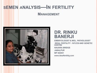
- 1. SEMEN ANALYSIS—IN FERTILITY MANAGEMENT DR. RINKU BANERJI EMBRYOLOGIST & MOL. PATHOLOGIST IDEAL FERTILITY : IVF,ICSI AND GENETIC CENTRE NAUDRA BRIDGE JABALPUR MP 482001 www.idealfertility.com
- 2. WHY IS THIS TEST REQUESTED? ― TO KNOW THAT A MAN CAN BECOME GENETIC FATHER OR NOT‖
- 4. LIVE
- 5. WHO IS THE LUCKY ONE??
- 6. ―Opportunities are equal for all. But the difference is ---- successful person gives result unsuccessful person gives only reasons
- 7. ―THESE ARE THE RESULTS‖
- 9. HERE WE ARE!
- 10. TO ANSWER THE QUERIES----- What is the problem?? Where is the problem???
- 11. MALE REPRODUCTIVE ORGAN TESTIS IS FACTORY, PRODUCT IS SPERM 11 DPANKAR MANAGEMENT PITUITARY SUPPLY ROUTE VASMESSANGERS FSH FACTORY TESTIS PENIS MARKET SEMEN
- 12. M ALE INFERTILITY IF PRODUCT (SPERM) IS NOT COMING TO THE MARKET (SEMEN) OR OF POOR QUALITY: REASONS MIGHT BE: 1.MALFUNCTIONING OF MANAGER (HYPOTHALAMO PITUITARY) :PRETESTICULAR 2.MALFUNCTIONING OF FACTORY (TESTIS) : TESTICULAR 3. MALFUNCTIONING IN SUPPLY LINE (Obstruction in epi/Vas) : POST TESTICULAR 12 DPANKAR
- 13. SO PROBLEM MAY BE Pre-testicular- Testicular- Post-testicular-
- 14. BESIDES SEMEN ANALYSIS : We can advice to Go for andrological evaluation Which includes- FSH testicular volume by usg semen fructose
- 15. MALE INFERTILITY FSH IS LOW ( < 4 mIU/ml ) : Means adequate messangers is not released from hypothalamus( Hypogonadotrophic Hypogonadism ) :PRETESTICULAR TESTICULAR VOLUME WILL BE LOW TREATMENT IS POSSIBLE BY INJECTING GONADOTROPINS 15 DPANKAR
- 16. MALE INFERTILITY FSH IS RAISED (normal :4-14 mIU/ml): twice than normal : Means testis is not functioning well hence (Hypothal -Pituit) sends large amount of messangers Concomitantly the volume of the Testis will be Low (normal >12 cc) DIAGNOSIS : PRIMARY TESTICULAR FAILURE TREATMENT NOT POSSIBLE ( ? ) 16 DPANKAR
- 17. FSH IS IN NORMAL RANGE (4-14 ) TESTICULAR VOLUME IS NORMAL (> 12 CC ) FRUCTOSE IS ABSENT IN EJACULATE DIAGNOSIS : OBSTRUCTION IN OUTFLOW most common at vaso-epididymal junction TREATMENT : SURGICAL BY-PASS OF OBSTRUCTION OR RETRIEVAL OF SPERMS FROM TESTIS AND DOING icsi 17 DPANKAR
- 18. PARTS OF SEMINAL FLUID Source of secretions Volume Characteristics TESTES,EPIDIDYMES,VASA- DEFERENTIA 0.1-0.2 CC SPERM PRESENT ACCESSORY GLANDS: Urethral and bulbourethral glands 0.1-0.2cc Viscous, clear Prostate 0.5-1.0cc Acidic, watery Seminal vesicles 1.0-3.0cc Gelatinous, fructose positive Complete ejaculate 2.0-5.0cc Liquefies in 20m-25m
- 20. BUT THERE ARE TOO MANY VARIATIONS LIKE:
- 21. VARIATION IN SPERM COUNT
- 22. So a standard range has been created which as a pathologist we can examine and predict IF ANY PARAMETER IS ABNORMAL— WE can select them for medical intervention.
- 23. COLLECTION & ABSTINENCE 1ST fraction---sperm rich next fraction----vesicular fluid Abstinence 2-7 days
- 24. EXAMINATION LIQUEFACTION appearance VOLUME VISCOSITY pH Motility Concentration Morphology Vitality Cellular element(nonspecific) Macroscopic examination Microscopic examination
- 25. Semen analysis should begin with a simple inspection soon after liquefaction. 30 minutes, to 1 hour after ejaculation, to prevent dehydration or changes in temperature from affecting semen quality.
- 26. Delayed liquefaction making semen evaluation difficult. In these cases, additional treatment, mechanical mixing or enzymatic digestion may be necessary. 1. Some samples can be induced to liquefy by the addition of an equal `volume of physiological medium (e.g. Dulbecco’s phosphate-buffered saline; followed by repeated pipetting. 2. Inhomogeneity can be reduced by repeated (6–10 times) gentle passage through a blunt gauge 18 (internal diameter 0.84 mm) or gauge 19 (internal diameter 0.69 mm) needle attached to a syringe. 3. Digestion by bromelain, proteolytic enzyme may help to promote liquefaction
- 27. Comment: These treatments may affect seminal plasma biochemistry, sperm motility and sperm morphology, and their use must be recorded.
- 28. INCOMPLETE LIQUEFACTION Dysfunction of seminal vesicle Or prostate (fibrinogen from prostatic fluid degrades fibrin in coagulum)
- 29. APPEARANCE Normal—grey,opalescent Yellow –in jaundice Reddish –in haemospermia-indicative of infection/inflammation/ductal obstruction
- 30. VOLUME The volume is best measured by weighing the sample in the vessel in which it is collected. Collect the sample in a pre- weighed, clean, disposable container. Weigh the vessel with semen in it. Subtract the weight of the container. Calculate the volume from the sample weight, assuming the density of semen to be 1 g/ml
- 31. VOLUME the volume can be measured directly. Collect the sample directly into a modified graduated glass measuring cylinder with a wide mouth. These can be obtained commercially. Read the volume directly from the graduations (0.1 ml accuracy).
- 32. •Comment 1: Low semen volume is characteristic of obstruction of the ejaculatory duct or congenital bilateral absence of the vas deferens (CBAVD) condition in which the seminal vesicles are also poorly developed. •Comment 2: Low semen volume can also be the result of collection problems (loss of a fraction of the ejaculate), partial retrograde ejaculation or androgen deficiency. •Comment 3: High semen volume may reflect active exudation in cases of active inflammation of the accessory organs. WHY IS IT IMPORTANT
- 33. Note: Measuring volume by aspirating the sample from the specimen container into a pipette or syringe, or decanting it into a measuring cylinder, is not recommended, because not all the sample will be retrieved and the volume will therefore be underestimated. The volume lost can be between 0.3 and 0.9 ml
- 35. viscosity 1. After liquefaction, the viscosity of the sample can be estimated by gently aspirating it into a wide-bore (approximately 1.5 mm diameter) plastic disposable pipette,allowing the semen to drop by gravity and observing the length of any thread. 2. A normal sample leaves the pipette in small discrete drops. 3. If viscosity is abnormal,the drop will form a thread more than 2 cm long. 4. The viscosity should be recorded as abnormal when the thread exceeds 2 cm. 5. In contrast to a partially unliquefied sample, a viscous semen specimen exhibits homogeneous stickiness and its consistency will not change with time. High viscosity can be recognized by the elastic properties of the sample, which adheres strongly to itself when attempts are made to pipette it. 6. The methods to reduce viscosity are the same as those for delayed liquefaction
- 36. Comment: High viscosity can interfere with determination of sperm motility, sperm concentration, detection of antibody-coated spermatozoa and measurement of biochemical markers
- 37. PH The pH of semen reflects the balance between the pH values of the different accessory gland secretions, mainly the alkaline seminal vesicular secretion and the acidic prostatic secretion. The pH should be measured after liquefaction at a uniform time, preferably after 30 minutes, but in any case within 1 hour of ejaculation since it is influenced by the loss of CO2 that occurs after production.
- 38. In azoospermic male: If the pH is <7.0 in a semen sample with low volume , there may be ejaculatory duct obstruction or congenital bilateral absence of the vas deferens pH >7.8--indicates dysfunctional seminal vesical : Semen pH increases with time, as natural buffering decreases, so high pH values may provide little clinically useful information
- 39. Depth of wet preparations=VOL. OF SAMPLE X AREA OF SPREAD USUALLY WE NEED A DEPTH OF 20 um FOR ASSESSMENT chamber depth of less than 20 um constrains the rotational movement of spermatozoa (Le Lannou et al., 1992; Kraemer et al., 1998). If the chamber is too deep, it will be difficult to assess spermatozoa as they move in and out of focus AREA OF COVER SLIP VOL. DEPTH 22mm X 22mm 10 ul 20.7 um 21mm X 26mm 11ul 20.1 um 18mm X 18mm 6.5ul 20.1 um . WET PREPARATION ON SLIDE
- 40. The volume of semen and the dimensions of the cover slip must be standardized,
- 41. WET PREPARATION At a glance we get idea about: sperm density sperm morphology any other cells besides sperm agglutination debris
- 42. NUMBER OF SPERMATOZOA PER HPF IN WET PREPARATION estimate the number of spermatozoa per HPF (×200 or ×400). One HPF is equivalent to approximately 16 nl (at ×200) or 4nl (at ×400) If spermatozoa are observed, count them, determine the necessary dilution
- 43. Note: If there are 100 spermatozoa per high-power field (HPF) of 4 nl in the initial wet preparation, there are theoretically 25 per nl (25 000 per ul or 25 millions/ ml).
- 44. NON-SPECIFIC AGGREGATION OF SPERMATOZOA IN SEMEN Views of spermatozoa aggregated with an epithelial cell (a), debris (b) or spermatozoa (c, d).
- 45. DEGREE OF AGGLUTINATION 1.Isolated (<10sperm/ agglutinate, manyfree sperm) 2.Moderate (10–50sperm/ agglutinate, freesperm) 3.Large(agglutinates >50sperm, somespermstill free) 4.Gross(all spermagglutinated, and agglutinates interconnected A. Head-to-head B. Tail-to-tail (heads are seen to be free and move clear of agglutinates) C. Tail-tip-to-tail-tip D. Mixed (clear headto- head and tail-to-tail agglutinations) E. Tangle (heads and tails enmeshed. Heads are not clear of agglutinates as they are in tailto- tail agglutination)
- 46. MOTILITY
- 47. Progressive motility (PR): spermatozoa moving actively, either linearly or in a large circle, regardless of speed. Non-progressive motility (NP): all other patterns of motility with an absence of progression, e.g. swimming in small circles, the flagellar force hardly displacing the head, or when only a flagellar beat can be observed. Immotile (IM): no movement MOTILITY REPORTED AS (WHO)
- 48. Initially scan the SLIDE for Pro. Motile cells count Nonpro. motile spermatozoa IMmotile spermatozoa With experience, it may be possible to score all three categories of sperm movement at one time, and to score larger areas of the grid. MOTILITY
- 49. Lower reference limit The lower reference limit for TOTAL MOTILITY (PR + NP) is 40% (5th centile, The lower reference limit for PROGRESSIVE MOTILITY (PR) is 32% (5th centile, Comment: The total number of progressively motile spermatozoa in the ejaculate is of biological significance. This is obtained by multiplying the total number of spermatozoa in the ejaculate by the percentage of progressively motile cells.
- 50. CASE 1: 100% IMMOTILE SPERM
- 51. REPORT In this case –checked for any infection,agglutination In this case Gave antibiotic t/t prophylactically Did HOS test –then ICSI was done in 8 oocytes With HOS positive Sperms
- 52. COUNT BY NEUBAUER CHAMBER(RECOM. BY WHO) OR MAKLER’S CHAMBER,CASA-TOO EXPENSIVE AND HAVE LITTLE ADVANTAGE OVER STANDARD SEMEN ANALYSIS
- 53. THE IMPROVED NEUBAUER HAEMOCYTOMETER The improved Neubauer haemocytometer has two separate counting chambers, each of which has a microscopic 3 mm × 3 mm pattern of gridlines etched on the glass surface. It is used with a special thick coverslip (thickness number 4, 0.44 mm), which lies over the grids and is supported by glass pillars 0.1 mm above the chamber floor. Each counting area is divided into nine 1 mm × 1 mm grids. These grids are referred to by the numbers
- 54. 1 2 3 4 5 6 7 8 9 Sperm counted in no 5 grid 5 squares to 25 squares
- 55. With a depth of 100 um, each grid holds 100 nl. Four of these grids (nos 1, 3, 7 and 9) contain four rows of four squares, each holding 6.25 nl; two grids (nos 2 and 8) contain four rows of five squares, each of 5 nl; two grids (nos 4 and 6) contain five rows of four squares, each of 5 nl; and squares the central grid (number 5) contains five rows of five squares, each of 4nl. (correspond to wet preparation) Each of the 25 squares of the central grid (number 5) is subdivided into 16 smaller squares . 1 2 3 4 5 6 7 8 9
- 56. Thus, grids 1, 2, 3, 7, 8 and 9 each have four rows holding 25 nl per row, while grids 4, 5 and 6 each have five rows holding 20 nl per row. Depending on the dilution and the number of spermatozoa counted, different areas of the chamber are used for determining sperm concentration. For 1 + 19 (1:20) and 1 + 4 (1:5) dilutions, rows from grid number 5 are assessed and, when necessary, from grids numbers 4 and 6 For 1 + 1 (1:2) dilutions,all nine grids can be assessed if necessary to achieve a count of 200 spermatozoa 1 2 3 4 5 6 7 8 9
- 57. As the central grid (number 5) of the improved Neubauer chamber holds 100 nl, there would be 2500 spermatozoa within it. Diluting the sample 1 + 4 (1:5) would reduce the background and the sperm number to about 500 per grid, which is sufficient for an acceptably low sampling error. If there are 10 spermatozoa per HPF of the wet preparation, there would be 2.5 per nl and 250 per central grid. Diluting the sample 1 + 1 (1:2) as suggested would reduce the background and the sperm number to about 125 per grid; this would give 375 in the three grids numbered 4, 5 and 6—again, this is suffi cient for an acceptably low sampling error.
- 58. COUNT only intact spermatozoa (having head andtail) . Do not count motile pinheads Do not count immature germ (round) cells. Do not assess overlapping spermatozoa and those lying with the head on edge; these cannot be analysed adequately. They should not be present in a good smear , but may occur when debris and a large amount of particulate material are present (such as in viscous sample. These samples should be washed and the slides prepared before staining.
- 59. COUNT :WHAT I DO Assess the sperms concentration from slide And make a dilution of 1:20 sperm according to the chart given below. Charge the Neubauer chamber Count the squares in the central grid usually 5 X –multiply by 106 per ml.
- 60. MULTIPLICATION FACTOR Make a dilution and count as follows: e.g. if 6 spematozoa are counted in 25 squares with semen diluted 9 X(1+9) Then count =6/10 x 106 per ml or 600000 sperms per ml Ref: ICMR AND WHO COLLABORATING CENTRE FOR RESEARCH IN HUMAN REPRODUCTION Dilution factor Correction factor for no. of squares counted 1+9 1+19 1+49 25 10 05 02 10 04 02 0.8 05 02 01 0.4 No. of squares
- 61. COUNTING
- 62. the sperm concentration X volume of the whole ejaculate== sperm number The lower reference limit for sperm concentration is 15 × 106/ ml The lower reference limit for total sperm number is 39 × 106 spermatozoa /ejaculate (5th centile, 95% CI 33–46 × 106).
- 63. Normozoospermia Normal ejaculate Oligozoospermia Sperm concentration fewer than 15x106/ml Asthenozoospermia <40% spermatozoa with total motility(pro. And non- prog) or <32% spermatozoa with pro. movement Teratozoospermia Fewer than 30% spermatozoa with normal morphology Oligoasthenoteratoz oospermia Signifies disturbance of all three variables (combination of only two prefixes can be used) Azoospermia No spermatozoa in the ejaculate Aspermia No ejaculate Certain terms used in reporting
- 64. Acrosome should be 40-70 % of head size
- 65. GLOBOZOOSPERMIA
- 66. WHY REPORTING 100% GLOBOZOOSPERMIA IMPORTANT? Even if we do icsi with globo. Sperm There is fertilization failure –why?
- 67. Sperm vitality, is estimated by assessing the membrane integrity of the cells, If <10% progressively motile spermatozoa----do vitality assessment . The percentage of live spermatozoa is assessed by identifying those with an intact cell membrane, from dye exclusion or by hypotonic swelling. The dye exclusion method damaged plasma membranes, such as those found in non-vital (dead) cells, allow entry of stains The hypo-osmotic swelling test presumes that only cells with intact membranes (live cells) will swell in hypotonic solutions. SPERM VITALITY
- 68. HOS TEST This is useful when staining of spermatozoa must be avoided, e.g. when choosing spermatozoa for ICSI. Spermatozoa with intact membranes swell within 5 minutes in hypo-osmotic medium as indicated by curling of the tail While dead sperms shows no change all flagellar shapes are stabilized by 30 minutes
- 69. 30 minutes incubation for routine diagnostics; but 5 minutes incubation when spermatozoa are to be processed for therapeutic use. Preparing the reagents 1. Swelling solution for diagnostic purposes: dissolve 0.735 g of sodium citrate dihydrate and 1.351 g of D-fructose in 100 ml of purified water. Freeze 1-ml aliquots of this solution at –20 °C. 2. For therapeutic use: dilute the medium to be used 1 + 1 (1:2) with sterile, purified water. 8. Tally the number of unswollen (dead) and swollen (vital) cells with the aid of a laboratory counter. 9. Evaluate 200 spermatozoa in each replicate, in order to achieve an acceptably low sampling error
- 70. VOLUME 1.5 ml (1.4-1.7) TOTAL COUNT /EJACULATION 39 million (33-46) Count/ml 15 million/ml(12-16) Total MOTILITY 40 %(38-42) % NORMAL FORMS 4 % (3.0-4.0) SOME LOWER REF. VALUE (WHO)PARAMETERS Pro. Motility % 32% (31-34) Vitality 58% (55-63) WBC( peroxidase positive) <1.0 million/ml .
- 71. KRUGER’S CRITERIA FOR SPERM MORPHOLOGY For this test, freshly ejaculated sperm are smeared on a slide and stained using a morphology staining product for human sperm. Sperm are judged as normal based on the following criteria: Head must be oval in shape with smooth contours, 5-6 µm in length and 2.5 to 3.5 µm wide with the acrosome taking up 40-70% of the head. Neck and mid-piece must have no abnormalities and a cytoplasmic droplet (a remnant from sperm production) if present must not be larger than half the size of the head. Tail must not be coiled or bent and should not have a droplet at the end.
- 72. KRUGER’S CRITERIA After 200 individual sperm are counted at a magnification of 1,000 times, the percent normal forms is calculated. The prognosis is based on the following scale: >=15% normal:Normal range - Good prognosis 5-14% normal:Sub optimal range - Prognosis is fair to good, however, the lower the percent normal, the lower the chance of successful fertilization 0-4% normal:Poor prognosis - Will usually need IVF with intracytoplasmic sperm injection (ICSI)
- 75. DAY 1 EMBRYO – 2 PN STAGE
- 76. DAY 2 AND DAY 3 EMBRYO 4-cell 8 cell
- 77. COMPACTION STAGE
- 78. DAY 5 BLASTOCYST (HATCHING)
- 81. THANK YOU FROM ALL OF US
Hinweis der Redaktion
- LIFE BEGINS-----
- How I count?