
Empfohlen
Weitere ähnliche Inhalte
Was ist angesagt?
Was ist angesagt? (19)
Ähnlich wie Regional Protein Changes in Dyssynchronous Heart Failure
Ähnlich wie Regional Protein Changes in Dyssynchronous Heart Failure (20)
Regional Protein Changes in Dyssynchronous Heart Failure
- 1. Deborah DiSilvestre, Elliot R. McVeigh, Gordon F. Tomaselli and David A. Kass David D. Spragg, Christophe Leclercq, Morteza Loghmani, Owen P. Faris, Richard S. Tunin, Regional Alterations in Protein Expression in the Dyssynchronous Failing Heart Print ISSN: 0009-7322. Online ISSN: 1524-4539 Copyright © 2003 American Heart Association, Inc. All rights reserved. is published by the American Heart Association, 7272 Greenville Avenue, Dallas, TX 75231Circulation doi: 10.1161/01.CIR.0000088782.99568.CA 2003;108:929-932; originally published online August 18, 2003;Circulation. http://circ.ahajournals.org/content/108/8/929 World Wide Web at: The online version of this article, along with updated information and services, is located on the http://circ.ahajournals.org/content/suppl/2003/08/25/01.CIR.0000088782.99568.CA.DC1.html Data Supplement (unedited) at: http://circ.ahajournals.org//subscriptions/ is online at:CirculationInformation about subscribing toSubscriptions: http://www.lww.com/reprints Information about reprints can be found online at:Reprints: document.Permissions and Rights Question and Answerthis process is available in the click Request Permissions in the middle column of the Web page under Services. Further information about Office. Once the online version of the published article for which permission is being requested is located, can be obtained via RightsLink, a service of the Copyright Clearance Center, not the EditorialCirculationin Requests for permissions to reproduce figures, tables, or portions of articles originally publishedPermissions: by guest on February 25, 2013http://circ.ahajournals.org/Downloaded from
- 2. Regional Alterations in Protein Expression in the Dyssynchronous Failing Heart David D. Spragg, MD*; Christophe Leclercq, MD*; Morteza Loghmani, BS; Owen P. Faris, BS; Richard S. Tunin, MS; Deborah DiSilvestre, MS; Elliot R. McVeigh, PhD; Gordon F. Tomaselli, MD; David A. Kass, MD Background—Left ventricular (LV) mechanical dyssynchrony induces regional heterogeneity of mechanical load and is an independent predictor of mortality and sudden death in heart failure (HF) patients. We tested whether dyssynchrony also induces localized disparities in the expression of proteins involved with mechanical stress, function, and arrhythmia susceptibility. Methods and Results—Eleven dogs underwent tachycardia-induced HF pacing, either from the right atrium or high right ventricular free wall. Whereas global LV dysfunction was similar between groups, LV contractile coordination assessed by tagged MRI was markedly dyssynchronous with right ventricular pacing but synchronous with right atrial pacing. In dyssynchronous failing hearts, the lateral LV endocardium displayed a 2-fold increase in phosphorylated erk mitogen-activated protein kinase expression (with no change in phospho-p38 or phospho-jnk), a 30% decline in sarcoplasmic reticulum Ca2ϩ -ATPase, an 80% reduction in phospholamban, and a 60% reduction in the gap junction protein connexin43, relative to neighboring myocardial segments. In contrast, hearts from both right atrial–paced HF dogs and an additional 4 noninstrumented control animals showed minimal regional variability in protein expression. Conclusions—LV dyssynchrony in failing hearts generates myocardial protein dysregulation concentrated in the late-activated, high-stress lateral endocardium. Such molecular polarization within the LV creates transmural and transchamber expression gradients of calcium handling and gap junction proteins that may worsen chamber function and arrhythmia susceptibility. (Circulation. 2003;108:929-932.) Key Words: heart failure Ⅲ pacing Ⅲ molecular biology Ⅲ sarcoplasmic reticulum The pathophysiology of heart failure (HF) involves abnor- malities of stress response kinase signaling, neurohu- moral stimulation, excitation-contraction coupling, gap junc- tion and ion channel function, and chamber remodeling.1,2 In addition, recent studies have shown that left ventricular (LV) mechanical coordination is frequently disturbed in HF, par- ticularly in the setting of interventricular conduction delay. HF patients with interventricular conduction delay and con- comitant ventricular dyssynchrony have mortality and sudden cardiac death rates above those predicted from the apparent severity of systolic dysfunction, HF etiology, functional class, or concurrent medical therapy.3,4 Acute mechanical discoordination induced by single-site right ventricular (RV) or LV pacing depresses systolic function, worsens myocardial efficiency, and leads to marked increases in wall stress heterogeneity.5,6 Stress is highest in late-activated myocardial regions because of both exaggerated stretch in early systole (secondary to septal contraction) and late systolic con- traction against increased afterload. Chronic discoordination leads to chamber remodeling of both early- and late-activated segments.7 However, its effect on myocardial protein expression remains unknown. In the present study, we tested the hypothesis that stress polarization due to dyssynchrony induces regional expression disparities of key proteins involved with stress response, muscle mechanics, and electrophysiology. Using HF models with or without superimposed LV discoordination, we studied tissue levels of the mitogen-activated protein kinases, sarcoplasmic reticulum Ca2ϩ -ATPase (SERCA2a) and phospho- lamban (PLB), and the gap junction protein connexin43 (Cx43). We reveal marked dyssynchrony-dependent transmural and tran- schamber protein expression gradients, suggesting a novel mecha- nism for the enhanced morbidity seen in discoordinate LV failure. Methods Model Generation Adult mongrel dogs (Bruce Rotz Kennels, Shippensburg, Pa) were paced (210 to 250 bpm) from the right atrium (RA; nϭ5) or RV free Received January 27, 2003; de novo received May 27, 2003; revision received July 14, 2003; accepted July 18, 2003. From the Division of Cardiology, Department of Medicine (D.S., C.L., M.L., R.T., D.D., G.T., D.A.K.), Johns Hopkins University, Baltimore, Md; and the National Heart, Lung and Blood Institute/Laboratory of Cardiac Energetics (O.F., E.M.), National Institutes of Health, Bethesda, Md. Dr Kass serves as a consultant to Guidant, Inc, which provided funding for this study. *These authors contributed equally to the work. Cine images are available in the online-only Data Supplement at http://www.circulationaha.org. Correspondence to David A. Kass, Halsted 500, Johns Hopkins Hospital, 600 N Wolfe St, Baltimore, MD 21287. E-mail dkass@jhmi.edu © 2003 American Heart Association, Inc. Circulation is available at http://www.circulationaha.org DOI: 10.1161/01.CIR.0000088782.99568.CA 929 by guest on February 25, 2013http://circ.ahajournals.org/Downloaded from
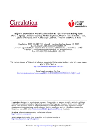
- 3. wall (nϭ6), as described previously,8 or were left uninstrumented (nϭ4). At terminal study, dogs were anesthetized (10 to 15 mg/kg thiopental, 1% to 2% isoflurane); LV pressures measured by micro- manometer (Millar); and the hearts retroperfused with cold cardio- plegia, excised, dissected into endocardial and mid/epicardial seg- ments from the interventricular septum and LV lateral wall, and frozen in liquid nitrogen. All procedures followed USDA guidelines and the protocol was approved by the Animal Care and Use Committee of the Johns Hopkins Medical Institutions. Assessment of Mechanical Dyssynchrony A subgroup of 4 dogs was studied by magnetic resonance (MR) imaging to document the effect of activation site on LV coordination. MR 3D tagged images were obtained (GE Signa 1.5T) during either RA or RV free-wall stimulation, and mechanical dyssynchrony indexed by a circumferential uniformity ratio estimate (CURE), as described previously.9 Perfectly synchronous or dyssynchronous contraction patterns yield CURE indices of 1 or 0, respectively. Western Blot Analysis Frozen myocardium was homogenized in lysis buffer (Cell Signaling Technology), and equivalent samples (confirmed by coprobing for glyceraldhyde-3-phosphate dehydrogenase or calsequestrin) were loaded for gel electrophoresis. After transfer to nitrocellulose, membranes were blocked and probed overnight at 4°C with primary antibodies for p-p38, p-erk, p-jnk (Cell Signaling Technology; 1:1000 dilution), PLB, SERCA2a (Affinity BioReagents; 1:5000 dilution), or Cx43 (Chemicon Intl; 1:1000 dilution). Membranes were incubated with HRP-conjugated secondary antibodies (Cell Signaling Technology and Upstate Biotechnology; 1:10 000 dilu- tions) for 1 hour at room temperature. Protein levels were detected by chemiluminescence and autoradiography, quantified using NIH ImageJ software, and blot intensity normalized such that lateral endocardial signal equaled 100 arbitrary units. Statistical Analysis Results are expressed as meanϮSEM. Statistical significance was assessed by ANOVA with post hoc Tukey tests for multiple comparisons. Results Mechanical Dysfunction and Dyssynchrony: RV Versus RA Pacing Models RA and RV pacing models generated similar degrees of global LV dysfunction, as reflected by depression of dP/dtmax (normal values in anesthetized dog are Ϸ2000 mm Hg/s) and elevation of LV end-diastolic pressure (Table). However, the two models generated markedly different degrees of LV mechanical coordination. Whereas rapid RA pacing pre- served nearly uniform contraction at all LV regional seg- ments, high RV free wall pacing induced substantial LV dyssynchrony, with early interventricular septal contraction and coincident lateral LV stretch followed by delayed lateral LV contraction and septal stretch (CURE index; Table). Cine images based on tagged MRI circumferential strain analysis depicting this dyssynchrony can be viewed in the online-only Data Supplement at http://www.circulationaha.org. Dyssynchronous HF and Regional Protein Expression The Figure displays representative Western blots and sum- mary data for regional protein expression of activated MAPKs, SERCA2a, PLB, and Cx43 in failing hearts with preserved (RA pacing) or disturbed (RV pacing) LV coordi- nation. With RV pacing, p-erk increased nearly 100% in the posterolateral LV endocardium relative to the other segments assessed (PϽ0.009). In contrast, p-erk levels did not vary significantly by region in RA pacing HF models. p-jnk and p-p38 levels did not vary regionally in either RV- or RA- paced models. Total levels for all three MAP kinase proteins were also not significantly different among the four regions (data not shown). The late-activated LV endocardium also demonstrated significant reductions in SERCA2a and PLB protein expres- sion in dyssynchronous HF. SERCA2a declined Ϸ30% rela- tive to the other regions (PϽ0.001), and PLB fell even more (Ϸ80%; PϽ0.001). In contrast, RA pacing did not induce regional disparities in the expression of either protein. Fi- nally, the same posterolateral endocardial region displayed marked downregulation of Cx43 expression (Ϸ60% relative to other segments; PϽ0.001) in RV-paced dogs. Once again, this localized abnormality was not observed in HF with preserved LV coordination, as shown by the RA-paced data. In noninstrumented control hearts, MAPK and Cx43 levels did not vary significantly by region. However, both SERCA2a and PLB levels were moderately and globally reduced in LV endocardial versus epicardial tissue (by 20% and 30%, respectively; Pϭ0.03), consistent with prior data.12 This transmural gradient was significantly lower than that observed in the lateral endocardium of dyssynchronous HF hearts. Discussion The present study provides novel evidence that mechanical dyssynchrony spatially polarizes ventricular protein expres- sion, particularly within the late-activated lateral wall, gen- erating transmural and transventricular gradients in p-erk, SERCA2a, PLB, and Cx43 levels. Similar expression gradi- ents were not seen in HF with equivalent global dysfunction but preserved LV coordination, indicating the importance of dyssynchrony to this process. We propose that such region- ally specific molecular polarization may contribute to hetero- geneous electromechanics and underlie enhanced arrhythmia susceptibility in HF patients with LV dyssynchrony. Mechanical load heterogeneity in the setting of LV disco- ordination was examined by Prinzen et al5 using MR tagged imaging and stress modeling. The authors predicted that late-activated ventricular segments were subjected to greatest stress because of locally enhanced preload (secondary to early systolic stretch) and afterload (due to late systolic contraction against high LV cavity pressures), and correlated LV Dysfunction and Coordination Parameters in RA and RV Pacing–Induced HF RV-Paced Dogs (Dyssynchronous) (nϭ6) RA-Paced Dogs (Synchronous) (nϭ5) P dP/dtmax, mm Hg/s 1295Ϯ250 1141Ϯ183 NS LV end-diastolic pressure, mm Hg 26Ϯ6 32Ϯ5 NS CURE index (dyssynchrony), dimensionless 0.52Ϯ0.14 0.90Ϯ0.04 Ͻ0.05 930 Circulation August 26, 2003 by guest on February 25, 2013http://circ.ahajournals.org/Downloaded from
- 4. increased wall stress to increased regional blood flow, nutri- ent consumption, and hypertrophy.5,6 The endocardium is particularly subject to stress redistribution because of myo- cardial fiber orientation, direct cavity pressure load, and a greater compromise in blood flow.10 Biomechanical stress on the posterolateral endocardium in dyssynchronous HF is uniquely elevated, therefore, and may explain the distribution of molecular abnormalities in our study. Several investigations have examined regional changes in protein expression in HF. Using a canine model of LV hypertrophy, Dosch et al11 reported higher atrial natriuretic peptide levels in LV basal and midwall segments, with peak expression in basal endocardium. Prestle et al12 described SERCA2a and PLB transmural expression gradients in failing human hearts, with Ϸ25% lower levels in the LV free wall endocardium. The present study supports the latter findings, but by contrasting models of LV failure differing only in degree of mechanical LV coordination, firmly identifies local biomechanical input (rather than systemic hemodynamics or neurohumoral signaling) as a key regulator of protein expres- sion. We believe this is the first study providing a possible molecular mechanism for the enhanced arrhythmia suscepti- bility seen in dyssynchronous HF. We did not test the functional impact of the molecular abnormalities observed, as this will require extensive isolated myocyte analysis from different layers and regions of the heart. However, prior data support such changes as poten- tially important. For example, increased SERCA2a/PLB lev- els have been associated with improved SERCA2a-mediated calcium sequestration and enhanced systolic function.13 In this regard, the increase in the lateral endocardium SERCA2a/PLB ratio (Ͼ3-fold) may reflect a regionally targeted adaptive mechanism to counter higher stress. Ele- ments of the PLB promoter (eg, GATA box) are responsive to biomechanical stretch and might explain the disparate changes seen in septal (stretched) versus lateral (higher- stress) SERCA2a/PLB expression.14,15 To our knowledge, however, specific regulators of SERCA2a and PLB that might drive reduced expression in response to mechanical stress have not been identified. Cx43 downregulation has been linked with arrhythmia susceptibility in a variety of models, likely because of unidirectional conduction delay and Histograms and representative Western blots detailing regional expression of ac- tivated MAPKs (A), calcium-handling pro- teins (B), and Cx43 (C), in RA-paced and RV-paced models. Expression is pre- sented in arbitrary units (A.U.). Septum-RV and Septum-LV indicate the RV- and LV-facing aspects of the inter- ventricular septum, respectively. Note the increased scale used to display p-erk expression, and that sample blot for RV p-p38 shows duplicate lanes for each myocardial segment. p-erk was signifi- cantly increased in RV-paced lateral endocardium, relative to other myocar- dial segments (*PՅ0.05 vs Septum-RV and LV-Lateral Wall Mid/Epicardium; Pϭ0.01 vs Septum-LV), whereas SERCA2a (†PϽ0.02 vs Septum-LV and Septum-RV; PϽ0.001 vs LV-Lateral Wall Mid/Epicardium), PLB (‡PϽ0.001 vs Septum-RV and LV-Lateral Wall Mid/Epi- cardium; PϽ0.02 vs Septum-LV), and Cx43 (¶PϽ0.01 vs Septum-LV and Septum-RV; PϽ0.001 vs LV-Lateral Wall Mid/Epicardium) were relatively decreased. Spragg et al Dyssynchrony and Myocardial Protein Changes 931 by guest on February 25, 2013http://circ.ahajournals.org/Downloaded from
- 5. facilitated reentry.16,17 Furthermore, MAPK activation and Cx43 downregulation may be connected, as constitutively activated p-jnk potently reduces Cx43 levels in mouse myo- cardium, and p-erk has been implicated in Cx43 downregu- lation in liver epithelial cells.18,19 Ongoing studies are ad- dressing the physiological importance of the expression changes revealed in the present study, as well as expanding the analysis by means of subproteomic and transcriptome analysis. In conclusion, LV mechanical dyssynchrony superimposed with tachycardia-induced HF induces marked regional het- erogeneity of protein expression at the site of greatest hemodynamic load. These observations raise the possibility that cardiac resynchronization therapy may not only improve ventricular mechanics, but also modulate regional myocardial protein expression. Further studies with more chronic HF models are needed to determine the extent to which cardiac resynchronization can reverse molecular polarization and impact net cardiac function and arrhythmia susceptibility. Acknowledgments This work was supported by grants from the National Institutes of Health (P50-HL52307) and from Guidant, Inc. References 1. Hoshijima M, Chien KR. Mixed signals in heart failure: cancer rules. J Clin Invest. 2002;109:849–855. 2. Marban E. Cardiac channelopathies. Nature. 2002;415:213–218. 3. Baldasseroni S, Opasich C, Gorini M, et al. Left bundle-branch block is associated with increased 1-year sudden and total mortality rate in 5517 outpatients with congestive heart failure: a report from the Italian network on congestive heart failure. Am Heart J. 2002;143:398–405. 4. Iuliano S, Fisher SG, Karasik PE, et al. QRS duration and mortality in patients with congestive heart failure. Am Heart J. 2002;143:1085–1091. 5. Prinzen FW, Hunter WC, Wyman BT, et al. Mapping of regional myo- cardial strain and work during ventricular pacing: experimental study using magnetic resonance imaging tagging. J Am Coll Cardiol. 1999;33: 1735–1742. 6. van Oosterhout MF, Arts T, Bassingthwaighte JB, et al. Relation between local myocardial growth and blood flow during chronic ventricular pacing. Cardiovasc Res. 2002;53:831–840. 7. van Oosterhout MF, Prinzen FW, Arts T, et al. Asynchronous electrical activation induces asymmetrical hypertrophy of the left ventricular wall. Circulation. 1998;98:588–595. 8. Kaab S, Nuss HB, Chiamvimonvat N, et al. Ionic mechanism of action potential prolongation in ventricular myocytes from dogs with pacing- induced heart failure. Circ Res. 1996;78:262–273. 9. Leclercq C, Faris O, Tunin R, et al. Systolic improvement and mechanical resynchronization does not require electrical synchrony in the dilated failing heart with left bundle-branch block. Circulation. 2002;106: 1760–1763. 10. Yin FC. Ventricular wall stress. Circ Res. 1981;49:829–842. 11. Dosch JC, Szwerc MF, Lin JC, et al. Pressure overload induces heter- ologous expression of the atrial natriuretic factor (ANF) gene. IUBMB Life. 2001;52:315–319. 12. Prestle J, Dieterich S, Preuss M, et al. Heterogeneous transmural gene expression of calcium-handling proteins and natriuretic peptides in the failing human heart. Cardiovasc Res. 1999;43:323–331. 13. Minamisawa S, Hoshijima M, Chu G, et al. Chronic phospholamban- sarcoplasmic reticulum calcium ATPase interaction is the critical calcium cycling defect in dilated cardiomyopathy. Cell. 1999;99:313–322. 14. Pikkarainen S, Tokola H, Majalahti-Palviainen T, et al. GATA-4 is a nuclear mediator of mechanical stretch-activated hypertrophic program. J Biol Chem. 2003;278:23807–23816. 15. McTiernan CF, Frye CS, Lemster BH, et al. The human phospholamban gene: structure and expression. J Mol Cell Cardiol. 1999;31:679–692. 16. Gutstein DE, Morley GE, Tamaddon H, et al. Conduction slowing and sudden arrhythmic death in mice with cardiac-restricted inactivation of connexin43. Circ Res. 2001;88:333–339. 17. Kitamura H, Ohnishi Y, Yoshida A, et al. Heterogeneous loss of con- nexin43 protein in nonischemic dilated cardiomyopathy with ventricular tachycardia. J Cardiovasc Electrophysiol. 2002;13:865–870. 18. Petrich BG, Gong X, Lerner DL, et al. c-Jun N-terminal kinase activation mediates downregulation of connexin43 in cardiomyocytes. Circ Res. 2002;91:640–647. 19. Ruch RJ, Trosko JE, Madhukar BV. Inhibition of connexin43 gap junc- tional intercellular communication by TPA requires ERK activation. J Cell Biochem. 2001;83:163–169. 932 Circulation August 26, 2003 by guest on February 25, 2013http://circ.ahajournals.org/Downloaded from