

Empfohlen
Weitere ähnliche Inhalte
Was ist angesagt?
Andere mochten auch
Mehr von muhammed Yasar
ppt
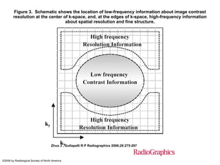
- 1. Figure 3. Schematic shows the location of low-frequency information about image contrast resolution at the center of k-space, and, at the edges of k-space, high-frequency information about spatial resolution and fine structure. Zhuo J , Gullapalli R P Radiographics 2006;26:275-297 ©2006 by Radiological Society of North America
Hinweis der Redaktion
- Figure 3. Schematic shows the location of low-frequency information about image contrast resolution at the center of k-space, and, at the edges of k-space, high-frequency information about spatial resolution and fine structure.