1. Analysis of Human Tyrosyl-DNA Phosphodiesterase I
Catalytic Residues
Amy C. Raymond1,2
, Marc C. Rideout2
, Bart Staker2
, Kathryn Hjerrild2
and Alex B. Burgin Jr1,2
*
1
Biology Department, San
Diego State University, 5500
Campanile Drive, San Diego
CA 98182-4614, USA
2
deCODE genetics
BioStructures Group, 7869 NE
Day Road West, Bainbridge
Island, WA 98110, USA
Tyrosyl-DNA phosphodiesterase I (Tdp1) is involved in the repair of
DNA lesions created by topoisomerase I in vivo. Tdp1 is a member of the
phospholipase D (PLD) superfamily of enzymes and hydrolyzes 30
-phos-
photyrosyl bonds to generate 30
-phosphate DNA and free tyrosine in
vitro. Here, we use synthetic 30
-(4-nitro)phenyl, 30
-(4-methyl)phenyl, and
30
-tyrosine phosphate oligonucleotides to study human Tdp1. Kinetic
analysis of human Tdp1 (hTdp1) shows that the enzyme has nanomolar
affinity for all three substrates and the overall in vitro reaction is diffusion-
limited. Analysis of active-site mutants using these modified substrates
demonstrates that hTdp1 uses an acid/base catalytic mechanism. The
results show that histidine 493 serves as the general acid during the initial
transesterification, in agreement with hypotheses based on previous
crystal structure models. The results also argue that lysine 495 and
asparagine 516 participate in the general acid reaction, and the analysis
of crystal structures suggests that these residues may function in a proton
relay. Together with previous crystal structure data, the new functional
data provide a mechanistic understanding of the conserved histidine,
lysine and asparagine residues found among all PLD family members.
q 2004 Elsevier Ltd. All rights reserved.
Keywords: tyrosyl-DNA phosphodiesterase I; phospholipase D
superfamily; topoisomerase I; DNA repair; proton relay*Corresponding author
Introduction
DNA topoisomerases are ubiquitous enzymes
that catalyze changes in DNA topology by altering
the linkage of DNA strands.1
Eukaryotic topo-
isomerase I (TopoI) uses an active-site tyrosine
residue to cleave one strand of DNA forming a 30
-
phosphotyrosine intermediate. This opening of the
DNA backbone is necessary to allow the removal
of superhelical tension that is generated during
replication and transcription. The phosphodiester
DNA backbone is restored when the 50
-hydroxyl,
generated during cleavage, attacks the 30
-phospho-
tyrosyl phosphodiester.2
Because the rate of
re-ligation is normally much faster than the rate of
cleavage, the steady-state concentration of topo-
isomerase–DNA adducts is extremely low. This is
important to maintain the integrity of the genome;
however, TopoI–DNA adducts can accumulate in
the presence of naturally occurring DNA damage
such as nicks,3
abasic sites,4
modified bases,5
modi-
fied sugar molecules6
or as a result of exposure to a
variety of chemotherapeutic drugs.7,8
Tyrosyl-DNA
phosphodiesterase I (Tdp1) has been shown to act
as a specific repair enzyme for TopoI lesions
in vivo,9
and catalyzes the hydrolysis of the phos-
phodiester bond between the 30
end of DNA and a
single tyrosine residue in vitro.10
Tdp1 is therefore
an important DNA repair enzyme and a potential
molecular target for new anti-cancer drugs.11
Tdp1 is a member of the phospholipase D (PLD)
superfamily and is conserved from yeast to man.12
The PLD superfamily represents an extremely
diverse family of enzymes, including phospholipid
hydrolases, cardiolipin synthases, phosphatidyl
serine synthases, poxvirus envelope proteins,
endonucleases, and a Yersinia murine toxin.13,14
0022-2836/$ - see front matter q 2004 Elsevier Ltd. All rights reserved.
E-mail address of the corresponding author:
aburgin@decode.com
Abbreviations used: Tdp1, tyrosyl-DNA
phosphodiesterase I; hTdp1, human tyrosyl-DNA
phosphodiesterase I; TopoI, topoisomerase I; PLD,
phospholipase D; yTdp1, yeast Tdp1; 4-nitro, 30
-(4-
nitro)phenyl phosphate DNA; 4-methyl, 30
-(4-
methyl)phenyl phosphate DNA.
doi:10.1016/j.jmb.2004.03.013 J. Mol. Biol. (2004) 338, 895–906
2. Despite this diverse array of substrates and bio-
logical functions, it had been shown that all
members of this family shared a signature
HxKx4D motif.15
Tdp1 has two such motifs,
marked by histidine 263/lysine 265 (H263/K265)
and histidine 493/lysine 495 (H493/K495). Sur-
prisingly, discovery of the Tdp1 subfamily showed
that the aspartic acid (D) is not conserved and that
Tdp1 represents a unique subclass within the PLD
superfamily.12
Characterization of the Tdp1
orthologs also showed that two asparagine resi-
dues are more highly conserved (N283 and N516).
Previous mechanistic and structural studies of
other phospholipase D superfamily members
argue that Tdp1 uses a two-step general acid/base
reaction mechanism to cleave 30
-phosphotyrosyl
bonds.12,16,17
On the basis of crystal structure
models, it has been proposed that in the first step
of the reaction H263 acts as a nucleophile forming
a 30
-phosphohistidine Tdp1 covalent intermediate,
and that H493 protonates the phenoxy anion of
the tyrosine leaving group.18,19
Free Tdp1 and 30
-
phosphate DNA are presumably generated by
hydrolysis of the 30
-phosphohistidine intermediate
in a second step. This two-step general acid/base
reaction is summarized in Figure 1. It has also
been proposed that K265, N283, K495 and N516
are all involved in substrate binding and transition
state stabilization.18
Here, we use a new series of Tdp1 substrate
analogs to show that Tdp1, and presumably other
members of the PLD superfamily, use a general
acid/base catalytic mechanism. This functional
data validates the proposed roles of H263 and
H493. Surprisingly, the results show that K495 and
N516 participate directly or indirectly in protonat-
ing the tyrosine leaving group during the first
transesterification reaction. This result suggests
that the conserved lysine and asparagine residues
may have multiple functions during the course of
the reaction. A comparison of hTdp1 crystal struc-
tures before and after substrate binding, suggests
a mechanism for how these residues participate in
the general acid reaction.
Results
Although Tdp1 has been shown to be involved
in the repair of topoisomerase I–DNA covalent
adducts, the natural in vivo substrate is not
known. In vitro, Tdp1 hydrolyzes 30
-phospho-
tyrosyl DNA, but does not cleave 50
-phospho-
tyrosyl DNA or 30
-phosphoseryl DNA.10
Single or
double-stranded oligonucleotides containing a 30
-
tyrosine residue or a small topoIB peptide frag-
ment (four to eight amino acid residues) are also
efficient substrates in vitro.12,20
Oligonucleotides
containing larger peptide fragments (.11 amino
acid residues) are hydrolyzed very slowly and it is
presumed that in vivo the topoisomerase is proteo-
lyzed to one (i.e. tyrosine residue) or a few amino
acid residues before it is ultimately acted upon by
Tdp1.10
Tdp1 can cleave 30
-tyrosine or 30
-(4-nitro)-
phenyl mononucleotides,10,21
but these substrates
are relatively poor. The preference for oligonucleo-
tide substrates versus mononucleotide substrates is
expected, since three conserved Tdp1 residues
make specific hydrogen bond contacts to two phos-
phodiester bonds upstream of the cleavage site.
Structural data also suggest that there might be a
conserved stacking interaction (F259) with an
upstream base.18
Unlike contacts between Tdp1
and the DNA portion of the substrate, binding
interactions with the leaving group are less well
defined. For example, one crystal structure shows
that a topoI fragment of eight amino acid residues
makes three hydrogen bond contacts to Tdp1;18
however, these residues are not conserved and the
conformation of the peptide is significantly
different from the conformation of the correspond-
ing region found in the crystal structures of
topoisomerase I.22
In addition, yeast Tdp1 (yTdp1)
yielded identical kinetics (KM and kcat) with
oligonucleotide substrates containing four amino
acid residues or a single tyrosine residue
(kcat=KM ¼ 1 £ 106
M21
s21
).20
Taken together, these
results show that the only feature of a leaving
group that is shared by all efficiently cleaved sub-
strates is a single phenol moiety.
This observation prompted us to synthesize and
compare two oligonucleotide substrates that
would result in minimal leaving groups when
cleaved by Tdp1: 4-nitro-phenol and 4-methyl-
phenol. These two leaving groups, diagrammed in
Figure 2a, were chosen because they would
occupy very similar space within the enzyme
active site, but the quality of the two leaving
groups is significantly different. The presence of a
strong electron withdrawing group (–NO2) lowers
the pKa of the phenoxy anion, creating a better
leaving group, whereas a methyl group at the 4
position increases the pKa of the phenoxy anion
similar to the b-methylene carbon atom of tyrosine.
Figure 1. Two-step reaction mechanism for Tdp1. In
the first transesterification step, the Tdp1 nucleophile
H263 attacks the substrate 30
-phosphotyrosyl bond. The
tyrosine phenoxide is protonated by the Tdp1 general
acid H493. In the second step, the 30
-phosphohistidine
intermediate is hydrolyzed to produce 30
-phosphate
DNA and free Tdp1.
896 Functional Analysis of Tdp1 Catalytic Residues
3. To test the efficiency of these new substrates we
compared them to identical oligonucleotides
containing a 30
-phospho-tyrosine residue. Sub-
strates were 50
-end labeled and incubated with
hTdp1 D1-148. All crystal structures of hTdp1
have been obtained using this N-terminal deletion
and we have independently confirmed previous
studies12,23
demonstrating that full-length hTdp1
and hTdp1 D1-148 have indistinguishable specific
activities (data not shown). The N terminus of
Tdp1 is extremely variable in size and the amino
acid identity among Tdp family members, which
argues that this region is not important for
activity.12
hTdp1 D1-148 was used for all of the
kinetic studies described below so that the com-
parison to the structural data would be more
direct. After incubation with the enzyme, the reac-
tion products were resolved on a denaturing
sequencing gel. As expected, all the three sub-
strates are converted to 30
-phosphate DNA
products that co-migrate in the gel (Figure 2b).
The 30
-tyrosyl phosphate DNA substrate migrates
more slowly in the gel than the 30
-(4-nitro)phenyl
phosphate or 30
-(4-methyl)phenyl phosphate DNA,
abbreviated 4-nitro and 4-methyl, due to the slight
increase in molecular mass. The identities of all
three starting materials and the Tdp1-mediated
cleavage products (30
-phosphate DNA) were con-
firmed by mass spectrophotometric analysis of
unlabelled reactions (data not shown).
To determine how well hTdp1 recognizes and
cleaves these three substrates, reaction velocities
were measured as a function of substrate concen-
tration (Figure 3a). The results in Table 1 show
that all three substrates are cleaved rapidly with
kcat values of 1800, 2400, 2800 min21
for the tyrosyl,
4-methyl and 4-nitro substrates, respectively. The
apparent KM values also varied with the same
magnitude: 370, 450, and 470 nM for the tyrosyl,
4-methyl and 4-nitro substrates, respectively. As a
result, the calculated kcat=KM values are statistically
indistinguishable between the three substrates,
,1 £ 108
M21
s21
. This value is important because
it represents the apparent second-order rate
constant for the overall reaction, and provides a
measure of how rapidly the enzyme can work at
low substrate concentrations, presumably
observed in vivo. In this case, the observed kcat=KM
value is close to the expected rate of diffusion-
controlled encounter of enzyme and substrate
(108
–109
M21
s21
).
The observation that all three substrates display
very similar kcat=KM values predicts that hTdp1
cannot distinguish between the three substrates in
a competition experiment. To test this prediction,
a trace amount (0.5 nM) of each 50
-end labeled sub-
strate was incubated in the presence of a large
excess of 30
-tyrosyl DNA (500 nM) in three separate
reactions. If Tdp1 cleaves the tyrosyl substrate
more efficiently than the 4-nitro or 4-methyl sub-
strates, then the observed velocities should be
decreased relative to the tyrosyl substrate. The
results in Figure 3b show that the reaction
velocities are essentially identical for all three sub-
strates; in fact, the observed velocities differ as
expected from the statistically insignificant
differences in the calculated kcat=KM values
(Table 1). Taken together these results show that
hTdp1 cannot distinguish the 4-methyl and 4-nitro
substrates from a standard tyrosine substrate, and
all three derivatized oligonucleotides are valid in
vitro substrates for hTdp1.
Because 4-nitro and 4-methyl substrates have
similar molecular mass but differ significantly
Figure 2. The 30
-derivatized oligonucleotide substrates.
a, The 30
-derivatized oligonucleotide Tdp1 substrates are
diagrammed on the left. The 30
-(4-nitro)phenyl phos-
phate ester DNA is shown at the top and is abbreviated
4-nitro: 30
-(4-methyl)phenyl phosphate ester DNA and
30
-phosphotyrosine DNA, abbreviated 4-methyl and
tyrosyl, respectively, are shown below. The second
column shows the products of Tdp1 cleavage, and the
third column shows the pKa of the leaving group for
each substrate. b, The 50
-end-labeled 30
-tyrosyl, 30
-(4-
methyl)phenyl and 30
-(4-nitro)phenyl phosphate ester
DNA substrates were incubated in the presence (þ) or
absence (2) of hTdp1. The reaction products were
resolved on a 20% polyacrylamide gel containing 8 M
urea, and the resulting autoradiogram is shown. The
positions of substrates and 30
-phosphate DNA products
within the gel are indicated on the right.
Functional Analysis of Tdp1 Catalytic Residues 897
4. in the quality of their leaving groups, they can be
used to probe the mechanism of Tdp1 active-site
residues. For example, crystal structure models
argue that H263 functions as the nucleophile and
H493 functions as the general acid during the first
transesterification reaction. A crystal structure of a
transition-state mimic of hTdp1 assembled from
vanadate, DNA, and a topoisomerase I-derived
peptide shows that H263 is positioned for in-line
nucleophilic attack, and H493 is appropriately
positioned to protonate the tyrosine leaving
group.18,19
Because the pKa of 4-nitrophenol is sig-
nificantly lower than tyrosine or 4-methylphenol,
we hypothesized that it should be possible to
rescue activity of the general acid-deficient
enzyme, since the solvent may be able to protonate
the leaving group in the absence of the enzyme
general acid. Neither substrate should be able to
rescue activity of the nucleophile-deficient enzyme.
To test this possibility, we incubated wild-type and
histidine to alanine mutant enzymes (H263A,
H493A) with 50
-end labeled 4-nitro and 4-methyl
oligonucleotides under single turnover conditions.
Because the results described above indicate that
enzyme substrate association is rate-limiting
under multiple turnover conditions, a trace
amount of substrate (,1 nM) was incubated with
a large excess of enzyme (133 nM) so that the
resulting extent of cleavage would reflect the rate
at which the enzyme–substrate complex is con-
verted to product. The results in Figure 4a (lanes 3
and 4) show that wild-type hTdp1 is able to cleave
both substrates. As predicted, H263A is inactive,
whereas H493A hydrolyzes 4-nitro DNA but is
unable to hydrolyze the 4-methyl analog.
Because the 4-nitro substrate specifically
rescues mutant enzymes that have defective
general acids, this assay can be used to identify
additional active-site residues that participate
directly or indirectly in protonating the tyrosine
leaving group. In addition to H263 and H493,
previous studies12,16,24
have implicated at least six
conserved residues that participate in catalysis:
K265, N283, Q294, K495, N516 and E538. To test
the role of these active-site residues, individual
alanine-substituted mutants were constructed,
purified and incubated with 4-nitro and 4-methyl
containing oligonucleotides. The results in
Figure 4a and b show that similar to the nucleo-
Figure 3. Kinetic analysis. a, Reaction velocities were
determined as described in Materials and Methods, and
are graphed as a function of substrate concentration.
The inset shows an autoradiogram of a typical cleavage
reaction used to calculate a reaction velocity; reaction
products before addition of hTdp1 (lane 1), and one,
two, three, four, five and six minutes after the addition
of enzyme (lanes 2–7) are shown. The data were fit to
the Michaelis equation (see Materials and Methods),
and the resulting kcat and KM values are shown in Table
1. b, The 50
-end 32
P2
labeled oligonucleotide substrates
(0.5 nM) were incubated in the presence of hTdp1
(0.01 nM) and a large molar excess of unlabeled 30
-tyro-
syl phosphate ester oligonucleotide (500 nM). The frac-
tion of substrate converted to 30
-phosphate DNA
(reaction product) was determined by phosphorimager
analysis. The concentration (nM) of 30
-phosphate DNA
formed as a function of time (minutes) from three differ-
ent experiments is plotted, and reaction velocities were
calculated from the slope of the resulting line. The reac-
tion velocities for conversion of 30
-(4-nitro)phenyl, 30
-(4-
methyl)phenyl and 30
-tyrosyl phosphate ester substrates
to 30
-phosphate DNA were 0.0086241, 0.0071649, and
0.0060103 nM/minute, respectively.
Table 1. Summary of hTdp1 kinetic analysis
Substrate kcat (min21
) KM (nM) kcat=KM (M21
s21
)
4-Nitro 2800 ^ 230 470 ^ 50 1 £ 108
4-Methyl 2400 ^ 660 450 ^ 10 9 £ 107
Tyrosyl 1800 ^ 850 370 ^ 120 8 £ 107
Kinetic values were extracted from the above graphed data.
Human Tdp1 displays nanomolar affinity for all substrates
used in this experiment, which are rapidly cleaved. For each
substrate, the KM varies with the turnover number resulting in
identical apparent second-order rate constants
(,1 £ 108
M21
s21
).
898 Functional Analysis of Tdp1 Catalytic Residues
5. phile mutant H263A, substrates reacted with the
K265A and E538A mutants were not converted to
a 30
-phosphate DNA product. A band migrating at
the position of 30
-phosphate DNA can be observed
in lanes 5 and 7; however, this small amount of
product is also seen in the absence of any enzyme
and represents a small amount of contaminating
30
-phosphate DNA in the 4-nitro stock. Similar to
the general acid mutant H493A, the activities of
K495A and N516A were rescued by the 4-nitro sub-
strate. Unfortunately, no clear conclusions can be
drawn from the analysis of the N283A and
Q294A, since these mutants were able to cleave
both substrates, although there may be a slight
rescue with Q294A. All hTdp1 mutants, except
H263A, were able to cleave both substrates to com-
pletion at long times. This is expected, since only
the nucleophile-deficient enzyme should be devoid
of all activities.
Prior to testing the active-site mutants, it was not
known if 30
-phosphate DNA would be generated in
these reactions. For example, H493 had been
proposed to be the general acid for the first trans-
esterification reaction and the general base for the
second hydrolysis reaction.19
Within the PLD
superfamily of enzymes,15,16
it has been proposed
that one histidine residue donates a proton to the
tyrosyl leaving group during the transesterification
reaction (general acid), and abstracts a proton
from a water molecule during the second
hydrolysis reaction (general base). It was therefore
possible that the H493A mutant would accumulate
the 30
-phosphohistidine hTdp1–DNA covalent
adduct but no 30
-phosphate DNA would accumu-
late when presented with the 4-nitro substrate.
The fact that 30
-phosphate DNA does rapidly
accumulate suggests that the 30
-phopshohisitidine
intermediate is easily hydrolyzed under the
reaction conditions (pH 8). Analysis of other PLD
family members has demonstrated that the rate of
hydrolysis is much faster than the rate of the initial
transesterification reaction.25
Figure 4. Single-turnover analysis
of wild-type and active site
mutants. a, The (50
-32
P) end-labeled
30
-(4-methyl)phenyl and 30
-(4-nitro)-
phenyl phosphate oligonucleotides
(1 nM), abbreviated 4-methyl and
4-nitro, were incubated with wild-
type and active-site mutant hTdp1
(133 nM) for five seconds at 37 8C,
quenched with the addition of SDS
and urea (see Materials and
Methods) and then resolved on a
20% polyacrylamide, 8 M urea
sequencing gel. A portion of the
resulting autoradiogram is shown
and the positions of substrate and
product are indicated. b, The single
turn-over cleavage reaction was
performed in triplicate and the
average fraction of substrate
cleaved for each reaction is plotted.
Error bars indicate the observed
standard deviation between the
three experiments. c, Separate
single turn-over cleavage reactions
were quenched with the addition
of an equal volume of 0.05% (w/v)
SDS, 2% (v/v) Tween 20 and 40%
(v/v) glycerol, and then resolved
on a 4%–12% SDS/polyacrylamide
gel. A portion of the autoradiogram
containing the Tdp–DNA covalent
complex (30
-phosphohistidine
intermediate) is shown. Because
the steady-state concentration of
the Tdp–DNA complex is low, the
labeled free DNA was run off the
bottom of the gel and is therefore
not visible. We estimate that in all
cases ,1% of the 50
-end labeled
oligonucleotides accumulate as
30
-phosphohisitidine covalent com-
plexes; the similar intensities observed in a and c result from the significantly longer exposure time required to obtain
the gel image presented in c.
Functional Analysis of Tdp1 Catalytic Residues 899
6. However, to see if any of the active-site mutants
were partially defective for the second hydrolysis
step, the same single-turnover reactions were
repeated and the products resolved by SDS-PAGE.
Because the oligonucleotide substrates were 50
-end
labeled, the 30
-phosphohistidine hTdp1–DNA
intermediate should be detectable as a 32
P-labeled
protein product. The autoradiogram of the
resulting gel shows that the three residues that
were implicated to be involved in protonating the
tyrosine leaving group (H493A, K495A and
N516A) also resulted in a steady-state accumu-
lation of the 30
-phoshophohistidine covalent
adduct (Figure 4c). This result is consistent with
these residues being involved in the general base
reaction for the second hydrolysis reaction. Phos-
phorimager analysis of the SDS gel (Figure 4c)
and the denaturing DNA gel (Figure 4a) showed
that only a very small fraction (approximately 1%)
of the total DNA is present within the covalent
complex band. Surprisingly, K265A and E538A
mutants also resulted in an accumulation of the
Tpd1–DNA intermediate, but only when
incubated with the 4-nitro substrate. It is important
to emphasize that this assay alone cannot be used
to directly address the importance of active
residues during hydrolysis, since the observed
steady-state accumulation depends upon both the
rate of transesterification (formation of inter-
mediate) and hydrolysis (destruction of inter-
mediate to form 30
-phosphate DNA). We are
currently developing assays to isolate the hydroly-
sis reaction in the absence of the initial trans-
esterification reaction to determine the roles of
active residues during Tdp1-catalyzed hydrolysis.
Crystal structures of hTdp1 have been described
in the presence of a transition-state mimic.18,19
These structures have suggested that K495 and
N516 are important for substrate binding and tran-
sition-state stabilization;18
however, our results
suggest that K495 and N516 participate in the
general reaction (i.e. protonating the tyrosine
leaving group). The structural and functional data
can be reconciled by proposing that these residues
have multiple roles before and after substrate bind-
ing. To examine this possibility, we solved an
additional X-ray crystal structure of hTdp1
(Table 2) in the absence of a transition-state mimic.
The active site of this new crystal structure is over-
laid with the four other published hTdp1 struc-
tures in Figure 5a. It is important to note that the
majority of the active-site residues overlay remark-
ably well from all five models; however, K265 and
K495 are observed in two distinct conformations,
depending upon the presence or absence of a sub-
strate analog. As expected, the crystal structure
shows that K495 and N516 are in position to inter-
act with each other and could participate in the
general acid reaction during the first trans-
esterification reaction. In two structures
(Figure 5b), obtained in the absence of a tran-
sition-state mimic, the Nz
of K495 is 3.5 A˚ from the
N12
of H493 (3.51 A˚ and 3.39 A˚ in two models)
and 4.3 A˚ from the Od1
of N516 (4.27 A˚ and
4.29 A˚ ). Whereas in three structures obtained in
the presence of different transition-state mimics
(Figure 5c), the Nz
of K495 is 4.2 A˚ from the N12
of
H493 (4.25, 4.32 and 3.89 A˚ ) and 3.2 A˚ from the
Od1
of N516 (3.20, 3.12 and 3.13 A˚ ).
Discussion
yTdp1 has been shown to be involved in the
repair of topoisomerase I DNA lesions in vivo9
and
presumably hydrolyzes the topoisomerase from
the 30
-end of the DNA during double-strand break
repair.26,27
Human Tdp1 (hTdp1) is also believed
to be involved in the repair of topoisomerase I-
induced lesions and may function during single-
strand break repair.28,29
For both hTdp1 and
yTdp1, it is presumed that the topoisomerase is
first proteolyzed before being acted upon by
Tdp1. Only substrates containing one to four
amino acid residues are efficient yTdp1 substrates
in vitro,10,20,26
and native topoisomerase I linked to
the 30
-end of oligonucleotide DNA is a very poor
substrate for hTdp1, but a trypsin-resistant
remnant peptide can be hydrolyzed.12
It is import-
ant to note that Tdp1 may have other roles in the
cell. For example, it has been shown that human
and yTdp1 are able to cleave double-strand 30
-
phosphoglycolate DNA to 30
-phosphate DNA
products;30
however, this in vitro activity is rela-
tively inefficient and no genetic data is available
to address the importance of this activity in vivo.
In order to better understand the in vitro sub-
strate requirements of hTdp1, we have performed
a kinetic analysis of hTdp1 using oligonucleotide
substrates containing a single tyrosine residue.
Table 2. hTdp1 crystallographic refinement statistics
Resolution (A˚ ) 50.0–2.4 (2.55–2.4)
No. of reflections 40,794 (6326)
Rsym
a
9.7 (40.3)
Completeness (%) 99.4 (99.8)
Redundancy 6.1 (5.8)
I/sI 18.5 (3.6)
Space group P212121
Unit cell dimensions
a (A˚ ) 50.0
b (A˚ ) 105.1
c (A˚ ) 194.1
Reflections used in Rfree (%) 2067 (5)
Molecules per asymmetric unit 2
No. of solvent atoms 243
R-factor 18.5 (22.6)
Rfree 23.4 (30.5)
r.m.s. deviations from ideal stereochemistry
Bond lengths (A˚ ) 0.009
Bond angles (deg.) 1.5
Impropers (deg.) 0.92
Dihedral angles (deg.) 24.6
Mean B-factor for all atoms (A˚ 3
) 30.8
Numbers in parentheses represent the final shell of data.
a
Rsym ¼
P
lIi 2 Iml
P
Im where Ii is the intensity of the
measured reflection and Im is the mean intensity of all sym-
metry-related reflections.
900 Functional Analysis of Tdp1 Catalytic Residues
7. Specifically, we show that 30
-derivatized oligo-
nucleotides containing tyrosine, (4-methyl)phenyl,
and (4-nitro)phenyl phosphate esters are rapidly
cleaved ðkcat . 103
min21
Þ by hTdp1. These values
are about 50 times faster than kcat values obtained
with substrates containing a single tyrosine residue
using yTdp120
and emphasizes how efficiently
these substrates are cleaved by hTdp1. As expected
for biologically relevant substrates, the apparent
KM values for all three substrates are in the nano-
molar range (,500 nM); however, these values are
approximately 50 times larger than values obtained
with yTdp1.10
There are several possible
explanations for this disparity between yeast and
hTdp1. First, the actual rate of catalysis for hTdp1
may be much faster than the rate of enzyme–sub-
strate dissociation and therefore result in a larger
apparent Michaelis constant ðKM ¼ k21 þ k2=kþ1Þ
even though the true binding affinity ðKD ¼
k21=kþ1Þ between yeast and human enzymes may
be very similar. Consistent with this possibility,
the kcat and KM values for the tyrosine, 4-methyl
and 4-nitro substrates vary but the calculated
specificity constants ðkcat=KMÞ for all three sub-
strates are essentially identical (,1 £ 108
M21
s21
).
This value suggests that catalysis (k2) is extremely
fast, since formation of the enzyme–substrate com-
plex is the rate-limiting step in the overall in vitro
reaction. This important result should be con-
sidered when characterizing various substrates or
mutant hTdp1 enzymes in vitro. For example, a
molecule may bind the enzyme active site and
inhibit activity, but the observed reaction velocity
may not change under the conditions described
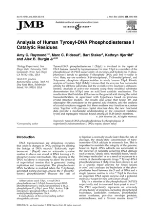
Figure 5. hTdp1 active-site overlays. a, Stereoview of active-site residues from five hTdp1 crystal structure models is
diagrammed. Structures from protein crystallized in the absence of a substrate analog are shown in yellow (PDB: 1JY1,
1QZQ), structures containing a transition state analog are shown in green (PDB: 1MU7, 1MU9, 1NOP). The Figure also
shows a portion of the DNA, vanadate, and TopoI-derived peptide (i.e. substrate analog) used to obtain 1NOP.
b, Conformation of H493, K495, N516 in structures (1JY1, 1QZQ) from protein crystallized in the absence of a substrate
analog is shown. The proposed interaction from the Nz
of K495 to the N12
of H493 is shown with a continuous line. The
average distance from the Nz
of K495 to the Og
of N516 is shown with a broken line. c, Conformation of H493, K495,
N516 in structures from protein crystallized with a substrate analog (1MU7, 1MU9 and 1NOP) is diagrammed. The
average distance from the Nz
of K495 to the N12
of H493 is shown with a broken line. The proposed hydrogen bonds
between K495, N516 and the substrate analog are shown with continuous lines.
Functional Analysis of Tdp1 Catalytic Residues 901
8. because the enzyme–substrate complex formation,
rather than enzymatic chemistry, is limiting.
Another explanation for the discrepancy between
yeast and hTdp1 is that hTdp1 is one component
of a multi-enzyme complex and additional proteins
may be involved in recognizing 30
-phoshotyrosyl
DNA in vivo; hTdp1 has been shown to associate
with XRCC1.31
The kinetic data cannot directly
address this possibility; however, the results do
show that additional proteins are not required for
efficient hTdp1-mediated cleavage in vitro.
As mentioned above, the precise in vivo substrate
of hTdp1 is not known. The substrate-binding
pocket of hTdp1 can accommodate a fairly large
topoisomerase I peptide fragment,18
and hTdp1
can cleave a 12 amino acid residue trypsin-resistant
fragment of topoisomerase I linked to the 30
-end of
DNA.12
It is therefore attractive to speculate that
hTdp1 makes additional contacts to the topo-
isomerase I-derived peptide that are necessary for
recognition or cleavage by hTdp1. However, the
only feature that is shared by the three efficiently
cleaved substrates in this study is a single phenyl
ring, demonstrating that a single phenyl ring is
sufficient for Tdp1-mediated recognition and
cleavage in vitro. Although a detailed comparison
of these substrates with larger substrates is
necessary to draw any conclusions, the efficiency
with which the 4-methyl and 4-nitro substrates are
cleaved by hTdp1 in vitro raises the possibility
that the topoisomerase I is degraded to a single
tyrosine residue before cleavage by hTdp1 in vivo.
Another possibility that could be entertained is
that hTdp1 may have a general role in the repair
of many different small residues linked to the
30
-end of DNA. For example, it has been shown
that yTdp1 can cleave 30
-phosphoglycolate linkages
in vitro.30
Previous crystal structure models have shown
that histidine 263 (H263) is positioned for in-line
attack of the 30
-phosphotyrosyl bond and histidine
493 (H493) is appropriately positioned to protonate
the tyrosine leaving group.18,19
It was therefore
proposed that H263 is the nucleophile and H493
was the general acid; however, structural data
alone cannot assign these roles, since the models
represent the most stable conformation in the
crystallization condition and may not be the
catalytically active conformation. We have shown
that when histidine 263 is mutated to alanine
(H263A), the enzyme is catalytically inactive on
any substrate,18,19
whereas the H493A mutant can
convert 4-nitro DNA to 30
-phosphate product, but
the 4-methyl DNA remains intact. While only the
4-nitro DNA was converted to product, the H493A
mutant formed covalent intermediate with both
substrates. The H493A covalent intermediate
formed with both substrates is the result of the
transesterification step in the reaction, which pro-
ceeds prior to the general acid activity required in
the hydrolysis step. H493A is able to convert the
4-nitro DNA to product because the lower pKa of
the phenoxide oxygen atom relieves the mutant
enzyme of the responsibility for protonating the
substrate leaving group and the reaction proceeds
even in the absence of a general acid. The functional
data are therefore consistent with H263 serving as
the nucleophile and demonstrates that H493 is the
general acid. More importantly, the data provide the
first direct evidence that members of the phosho-
lipase D (PLD) superfamily of enzymes use a general
acid/base catalytic mechanism.
These results also allowed us to identify
additional active-site residues that are involved in
protonating the tyrosine leaving group. We
examined six conserved residues that have been
proposed to participate in catalysis: K265, N283,
Q294, K495, N516, and E413. No conclusions can
be drawn from the analysis of K265A and E538A
because both mutants failed to cleave the 4-methyl
or the 4-nitro substrates. This is expected, since it
has been proposed that E538 coordinates the H263
(nucleophile), and K265 coordinates the non-
bridging oxygen atom of the phosphotyrosyl
bond.18,19
In addition, no conclusions can be
drawn from the analysis of N283A and Q294A,
because these mutants were able to cleave both
substrates during the course of the reaction. Struc-
tural studies show that N283 is one of several resi-
dues (N516, K495 and K265) that can coordinate
non-bridging oxygen atoms of the scissile phos-
phodiester. It is therefore possible that these other
residues supply enough redundant function that
stabilization by N283 is not necessary for the first
transesterification reaction. The observation that
Q294A is able to cleave both substrates is more sur-
prising, because it has been proposed that Q294
coordinates the H493 general acid;18,19
if H493
requires coordination, the Q294A mutant should
cleave the 4-nitro substrate, but should not cleave
the 4-methyl substrate. Although there may be a
modest reduction of activity using with the
4-methyl substrate (Figure 4), the simplest
explanation of this data is that the coordination of
H493 by Q294 alone is not critical for hTdp1
activity in vitro.
The most definitive conclusions come from the
analysis of K495A and N516A, since these mutants
are able to cleave the 4-nitro substrate, but are
unable to cleave the 4-methyl substrate. These
results demonstrate that K495 and N516 are
directly or indirectly involved in protonating the
50
-leaving group during the wild-type reaction. An
overlay of five different crystal structures of
hTdp1 shows that K495 lies adjacent to both N516
and the general acid H493. In two structures,
obtained in the absence of a transition state mimic,
the Nz
of K495 is close to the N12
of H493 (3.5 A˚ )
and is not in position to make a hydrogen bond
contact to the Od1
of N516. Whereas in three struc-
tures obtained in the presence of different tran-
sition-state mimics, the Nz
of K495 has moved
away from the N12
of H493 and is now in position
to make a hydrogen bond contact to the Od1
of
N516 (3.2 A˚ ). It is important to note that although
the two conformations of K495 only differ by
902 Functional Analysis of Tdp1 Catalytic Residues
9. 1–2 A˚ and the resolution of five crystal structures
varies between 2.0 A˚ and 2.4 A˚ , the five structures
were refined independently and represent three
different crystal forms obtained in two different
laboratories.
One mechanism that explains the functional and
structural data is that K495 participates in a
“proton relay” and transfers a proton to the
general acid H493 prior to substrate binding.
Upon substrate binding, the lysine residue moves
away from the general acid and makes a hydrogen
bond contact to the Od1
of N516. According to this
model, K495 participates directly in the general
acid reaction by protonating H493, which in turn
transfers a proton to the tyrosine leaving group.
N516 participates indirectly in the general acid
reaction by contacting and stabilizing K495; the
hydrogen bond contact between N516 and K495
made upon substrate binding, and presumably the
hydrogen bond contacts to the scissile phospho-
diester, allows H493 to protonate the tyrosine
leaving group.
Another possible explanation for the roles of
K495 and N516 is that K495 is sterically important
for positioning the H493 general acid. When K495
is mutated, or when K495 cannot be appropriately
positioned through a hydrogen bond contact to
N516, H493 is destabilized and cannot act as a
general acid. We disfavor this explanation, since
other residues that have been proposed to help
position the general acid do not significantly
inhibit the single turn-over reaction; for example,
Q294A mutants are able to cleave both 4-methyl
and 4-nitro substrates (Figure 4).
K265 and K493 are the only active-site residues
that are observed in two distinct conformations,
depending upon the presence or absence of a tran-
sition-state mimic, and lysine has been shown to
act as a general acid during phosphoryl transfer
reactions catalyzed by the type 1B topoisomerase/
tyrosine recombinase superfamily of enzymes.32,33
It is therefore interesting to speculate that the
individual lysine residues within each of the two
conserved HKD motifs (K265 and K493) have simi-
lar roles during Tdp1 catalysis. For example, if
K493 transfers a proton to H493 during the first
transesterification reaction as we propose above, it
is possible that K265 transfers a proton to H263
during the second hydrolysis reaction. In other
words, K265 would function as the general acid
for the second hydrolysis step. This may explain
why an increased steady-state concentration of the
Tdp1–DNA covalent adduct (30
-phosphohistidine)
was observed in the single-turnover reactions
when K265A was used. Specifically, if K265A was
able to cleave a small portion of the more reactive
4-nitro substrate, but unable to promote the
general acid reaction allowing hydrolysis of the
30
-phosphohistidine intermediate, very little
30
-phosphate product would be observed
(Figure 4a, lane 7) and the hTdp1–DNA covalent
complex would accumulate (Figure 4c, lane 7). If
E538 is important for coordinating H263 during
the hydrolysis reaction, then this would also
explain the accumulation of the 30
-phospho-
histidine intermediate when E538A was used in
the single turnover reactions (Figure 4a, lane 19;
Figure 4c, lane 19). Clearly, developing an assay to
isolate the hydrolysis reaction that does not
depend upon the initial transesterification reaction
is necessary to address these possibilities.
In conclusion, we have shown that single-strand
DNA oligonucleotide substrates containing a
single 30
-phospho-phenyl ring are efficient sub-
strates for hTdp1 in vitro, and the rate of sub-
strate–enzyme association, rather than catalysis, is
rate limiting. The efficiency of these substrates
suggests that a single phenyl ring is sufficient for
catalysis and potential contacts between hTdp1
and topoisomerase I peptide fragments are not
necessary for efficient cleavage. The ability of
H493A to efficiently cleave 4-nitro substrates pro-
vides evidence that hTdp1 uses a general acid/
base catalytic mechanism. Because all PLD family
share similar structures,23,34,35
it suggests that all
PLD members share this same general acid/base
catalytic mechanism. Finally, the ability of the
4-nitro substrate to relieve the need for additional
active site residues (K495 and N516) shows that
these residues participate directly or indirectly in
protonating the tyrosine leaving group. On the
basis of the functional and structural data, we pro-
pose that N516 participates in the general acid
reaction by coordinating K495, and that K495
transfers a proton to H493, which in turn transfers
a proton to the tyrosine leaving group.
Materials and Methods
Synthesis of 30
-derivatized oligonucleotide
substrates
The 30
-(4-nitro)phenyl phosphate oligonucleotides
were prepared as described.36
The 30
-phosphotyrosine
oligonucleotides were purchased from Midland Certified
Reagent Company (Midland, TX). The 30
-(4-methyl)-
phenyl oligonucleotides were prepared by post-synthetic
condensation. Oligonucleotides (6 £ 1 mmol) were syn-
thesized using 30
-PO4 CPG (Glen Research, Sterling, VA)
and standard DNA phosphoramidites on an ABI 392
automated DNA synthesizer. The resin was incubated
in concentrated ammonium hydroxide at 65 8C for 12
hours, and the 50
-OH containing DNA was ethanol-
precipitated. The resulting DNA pellet was resuspended
in 1 ml of 2 mM MgCl2, 100 mM Mes (pH 5.5), and
aliquots of 1 ml 99% para-cresol (Aldrich, Milwaukee,
WI) and 0.048 g of 1-[3-(dimethylamino)propyl]-3-
ethylcarbodiimide hydrochloride (EDC) (Aldrich) were
added sequentially. The resulting two immiscible layers
were mixed vigorously at 65 8C to form an emulsion.
After 20–24 hours, the aqueous layer was extracted
three times with 2 ml each of ethyl acetate and the DNA
was recovered from the aqueous phase by ethanol-
precipitation. This EDC-derivatization protocol was
repeated a total of three times. Typically, ,5–10%
of the 30
-PO4 (starting material) was converted to
30
-cresol DNA, which was purified by anion-exchange
Functional Analysis of Tdp1 Catalytic Residues 903
10. chromatography on a Hitachi analytical HPLC instru-
ment using a DNAPac PA-100 4 mm £ 250 mm column
(Dionex, Sunnyvale, CA) at 1 ml/minute, 5% to 60%
(w/v) 2 M sodium chloride gradient (buffered with
20 mM sodium phosphate, pH 7.0) over 15 minutes.
Peak fractions (typically 0.2 minute longer retention
time than underivatized DNA) were pooled, precipi-
tated, resuspended in water and stored at 220 8C. The
sequence of all oligonucleotides was 50
-CGTTGAAGCC
TGCTTTY, where Y is tyrosine or the tyrosine analog.
Preparation of hTdp1
The hTdp1 D 1-148 cDNA was PCR-amplified from
IMAGE clones 3900062 and 740522 (ResGen Invitrogen
Corp. Carlsbad CA). The cDNA was amplified in two
halves: DNA encoding G149 through L379 was amplified
from IMAGE clone 3900062, and DNA encoding K380
through S608 was amplified from IMAGE clone 740522.
These two halves were ligated via a naturally occurring
HindIII site embedded in the sequence coding for L379
and K380. The full-length gene and the N-terminal
deletion (D 1-148) were cloned into a modified pBAD
(Invitrogen) expression vector. The final expressed pro-
tein contains an N-terminal 6 £ His tag and a seven
amino acid epitope tag (EEYMPTE). Mutant and wild-
type hTdp1 D 1-148 constructs were sequence verified
and transfected into TOP10 cells (Invitrogen), and
grown at 30 8C. Protein expression was induced by add-
ing arabinose to 0.2% (w/v) at mid-log phase, and cells
were harvested after four hours, concentrated and then
lysed in 100 mM NaCl2, 20 mM Tris–HCl (pH 9), 2 mM
BME, and 0.1 mg/ml of lysozyme by nitrogen cavitation.
Lysate was clarified by centrifugation and filtration, and
loaded onto HiTRAP nickel chelating columns
(Amersham, Piscataway, NJ). Protein was purified at
1 ml/minute in 100 mM NaCl2, 20 mM Tris–HCl (pH 8)
and eluted in a gradient of 24% to 100% (w/v) 500 mM
imidazole. Peak fractions were pooled and loaded onto
a 20 ml affinity column (monoclonal antibody which
recognizes the EEYMPTE epitope tag) equilibrated in
500 mM NaCl2, 20 mM Tris–HCl (pH 8). The column
was then loaded with 20 ml of 5 mg/ml of peptide (EEY
MPTE) in the same equilibration buffer and incubated
for 30 minutes at 4 8C. hTdp1 was eluted from the
column with 1 mg/ml of peptide and fractions were ana-
lyzed by SDS-PAGE. Pure hTdp1 was dialyzed exhaus-
tively in 250 mM NaCl2, 15 mM Tris–HCl (pH 8.2),
3 mM DTT at 4 8C. Following purification, all versions
of hTdp1 D 1-148 migrated as a single band of the pre-
dicted molecular mass on SDS-PAGE, and the identity
of wild-type and mutant hTdp1 was confirmed by
MALDI TOF mass spectroscopy. All residue numbering
corresponds to full-length protein.
Kinetic determinations
hTdp1 activity assays were performed in 50 mM Tris–
HCl (pH 8.0), 5 mM MgCl2, 80 mM KCl, 2 mM EDTA,
1 mM DTT, and 40 mg/ml of BSA. Enzyme stocks were
diluted in 50 mM Tris–HCl (pH 8.0), 100 mM NaCl,
5 mM DTT, 10% (v/v) glycerol, 500 mg/ml of BSA and
diluted tenfold for each reaction. Cleavage substrates
were 50
-radiolabeled with [a-32
P]ATP (Perkin Elmer,
Boston, MA) using phage T4 polynucleotide kinase
(New England Biolabs, Beverly, MA) at 37 8C for
15 minutes, followed by ten minutes at 90 8C to denature
the kinase.
Unless otherwise noted, all reactions were performed
at 37 8C in 96 well V-bottom reaction plates and
quenched by the addition of an equal volume of 8 M
urea, 0.05% (w/v) SDS, 30% glycerol, 0.25% (v/v)
bromophenol blue, and resolved on 20% (w/v) poly-
acrylamide sequencing gels. The concentration of 30
-
phosphate DNA was determined by measuring the frac-
tion of substrate converted to product by densitometry
analysis of the gel image. Initial velocities were deter-
mined by plotting the concentration of 30
-phosphate
DNA as a function of time; only concentrations repre-
senting less than 20% of initial substrate concentrations
were used, all lines extrapolated to zero product at the
start of the reaction, and at least five time-points were
used to determine the slope of the line (velocity).
Finally, all velocity measurements were performed in
triplicate on three different days. Apparent KM and Vmax
values were determined by fitting the initial velocity
versus substrate concentration to the Michaelis equation,
v ¼ ðVmax½SŠÞ=ðKM þ ½SŠÞ: Very similar values were
obtained from linear regression analysis of Eadie–
Hofstee plots of the same data (data not shown).
hTdp1 crystallization
Pure protein was concentrated to 6.5 mg/ml and
screened at 4 8C against 10–20% (w/v) PEG 8000,
100 mM Ches (pH 9.4–10), 8 mM spermine. Crystals
were observed in 16%, 18% and 20% PEG 8000 for all
Ches pH ranges within one week, with fully formed
crystals observed after one month. Crystals were cryo-
protected by soaking in 30% glycerol, 20% PEG 8000,
100 mM Ches (pH 10), 8 mM spermine for 30 seconds
and flash-cooled in liquid nitrogen prior to diffraction.
Data were collected at 100 K at COM-CAT, Sector 32,
Advanced Photon Source, Argonne National Laboratory.
The structure was solved by molecular replacement
with AmoRe37
by using the published D1-148 hTdp1
structure (PDB: 1JY1).23
The structure was rebuilt and
refined by CNX with simulated annealing38
and iterative
model adjustments by XTALVIEW (see Table 2).39
Protein Data Bank accession numbers
Coordinates have been deposited in the Protein Data
Bank with accession number 1QZQ.
Acknowledgements
We thank Zachary Halloran for technical
assistance in growing Tdp1-expressing cells and
S. Lovell at COM-CAT (Sector 32) Advanced
Photon Source, Argonne National Laboratory for
assistance in data collection. We thank Anca Segall,
Lance Stewart and Mark Gurney for critical
reading of the manuscript. ACR was supported
by Predoctoral Fellowship 1F31 GM66372-01 from
the National Institutes of Health. This research
was funded in part by the Cooperative Planning
of the SDSU/UCSD Cancer Partnership (grant
CCA92079-01A2) to ACR.
904 Functional Analysis of Tdp1 Catalytic Residues
11. References
1. Wang, J. C. (2002). Cellular roles of DNA topo-
isomerases: a molecular perspective. Nature Rev.
Mol. Cell. Biol. 3, 430–440.
2. Champoux, J. J. (2001). DNA topoisomerases: struc-
ture, function, and mechanism. Annu. Rev. Biochem.
70, 369–413.
3. Pourquier, P., Pilon, A. A., Kohlhagen, G.,
Mazumder, A., Sharma, A. & Pommier, Y. (1997).
Trapping of mammalian topoisomerase I and
recombinations induced by damaged DNA contain-
ing nicks or gaps. Importance of DNA end phos-
phorylation and camptothecin effects. J. Biol. Chem.
272, 26441–26447.
4. Pourquier, P., Ueng, L. M., Kohlhagen, G.,
Mazumder, A., Gupta, M., Kohn, K. W. & Pommier,
Y. (1997). Effects of uracil incorporation, DNA mis-
matches, and abasic sites on cleavage and religation
activities of mammalian topoisomerase I. J. Biol.
Chem. 272, 7792–7796.
5. Lesher, D. T., Pommier, Y., Stewart, L. & Redinbo,
M. R. (2002). 8-Oxoguanine rearranges the active
site of human topoisomerase I. Proc. Natl Acad. Sci.
USA, 99, 12102–12107.
6. Chrencik, J. E., Burgin, A. B., Pommier, Y., Stewart, L.
& Redinbo, M. R. (2003). Structural impact of the
leukemia drug 1-beta-D-arabinofuranosylcytosine
(Ara-C) on the covalent human topoisomerase
I-DNA complex. J. Biol. Chem. 278, 12461–12466.
7. Meng, L. H., Liao, Z. Y. & Pommier, Y. (2003). Non-
camptothecin DNA topoisomerase I inhibitors in
cancer therapy. Curr. Top. Med. Chem. 3, 305–320.
8. Pizzolato, J. F. & Saltz, L. B. (2003). The campto-
thecins. Lancet, 361, 2235–2242.
9. Pouliot, J. J., Yao, K. C., Robertson, C. A. & Nash,
H. A. (1999). Yeast gene for a Tyr-DNA phospho-
diesterase that repairs topoisomerase I complexes.
Science, 286, 552–555.
10. Yang, S. W., Burgin, A. B., Jr, Huizenga, B. N.,
Robertson, C. A., Yao, K. C. & Nash, H. A. (1996).
A eukaryotic enzyme that can disjoin dead-end
covalent complexes between DNA and type I
topoisomerases. Proc. Natl Acad. Sci. USA, 93,
11534–11539.
11. Connelly, J. C. & Leach, D. R. (2004). Repair of DNA
covalently linked to protein. Mol. Cell, 13, 307–316.
12. Interthal, H., Pouliot, J. J. & Champoux, J. J. (2001).
The tyrosyl-DNA phosphodiesterase Tdp1 is a mem-
ber of the phospholipase D superfamily. Proc. Natl
Acad. Sci. USA, 98, 12009–12014.
13. Koonin, E. V. (1996). A duplicated catalytic motif in a
new superfamily of phosphohydrolases and phos-
pholipid synthases that includes poxvirus envelope
proteins. Trends Biochem. Sci. 21, 242–243.
14. Ponting, C. P. & Kerr, I. D. (1996). A novel family of
phospholipase D homologues that includes phos-
pholipid synthases and putative endonucleases:
identification of duplicated repeats and potential
active site residues. Protein Sci. 5, 914–922.
15. Waite, M. (1999). The PLD superfamily: insights into
catalysis. Biochim. Biophys. Acta, 1439, 187–197.
16. Gottlin, E. B., Rudolph, A. E., Zhao, Y., Matthews,
H. R. & Dixon, J. E. (1998). Catalytic mechanism of
the phospholipase D superfamily proceeds via a
covalent phosphohistidine intermediate. Proc. Natl
Acad. Sci. USA, 95, 9202–9207.
17. Iwasaki, Y., Horiike, S., Matsushima, K. & Yamane, T.
(1999). Location of the catalytic nucleophile of
phospholipase D of Streptomyces antibioticus in the
C-terminal half domain. Eur. J. Biochem. 264,
577–581.
18. Davies, D. R., Interthal, H., Champoux, J. J. & Hol,
W. G. (2003). Crystal structure of a transition state
mimic for tdp1 assembled from vanadate, DNA,
and a topoisomerase I-derived peptide. Chem. Biol.
10, 139–147.
19. Davies, D. R., Interthal, H., Champoux, J. J. & Hol,
W. G. (2002). Insights into substrate binding and
catalytic mechanism of human tyrosyl-DNA phos-
phodiesterase (Tdp1) from vanadate and tungstate-
inhibited structures. J. Mol. Biol. 324, 917–932.
20. Debethune, L., Kohlhagen, G., Grandas, A. &
Pommier, Y. (2002). Processing of nucleopeptides
mimicking the topoisomerase I-DNA covalent
complex by tyrosyl-DNA phosphodiesterase. Nucl.
Acids Res. 30, 1198–1204.
21. Cheng, T. J., Rey, P. G., Poon, T. & Kan, C. C. (2002).
Kinetic studies of human tyrosyl-DNA phospho-
diesterase, an enzyme in the topoisomerase I DNA
repair pathway. Eur. J. Biochem. 269, 3697–3704.
22. Redinbo, M. R., Champoux, J. J. & Hol, W. G. (2000).
Novel insights into catalytic mechanism from a
crystal structure of human topoisomerase I in
complex with DNA. Biochemistry, 39, 6832–6840.
23. Davies, D. R., Interthal, H., Champoux, J. J. & Hol,
W. G. (2002). The crystal structure of human tyrosyl-
DNA phosphodiesterase, Tdp1. Structure (Camb), 10,
237–248.
24. Sung, T. C., Roper, R. L., Zhang, Y., Rudge, S. A.,
Temel, R., Hammond, S. M. et al. (1997). Mutagenesis
of phospholipase D defines a superfamily including
a trans-Golgi viral protein required for poxvirus
pathogenicity. EMBO J. 16, 4519–4530.
25. Yang, H. & Roberts, M. F. (2003). Phosphohydrolase
and transphosphatidylation reactions of two
Streptomyces phospholipase D enzymes: covalent
versus noncovalent catalysis. Protein Sci. 12,
2087–2098.
26. Pouliot, J. J., Robertson, C. A. & Nash, H. A. (2001).
Pathways for repair of topoisomerase I covalent
complexes in Saccharomyces cerevisiae. Genes Cells, 6,
677–687.
27. Vance, J. R. & Wilson, T. E. (2002). Yeast Tdp1 and
Rad1-Rad10 function as redundant pathways for
repairing Top1 replicative damage. Proc. Natl Acad.
Sci. USA, 99, 13669–13674.
28. Caldecott, K. W. (2003). DNA single-strand break
repair and spinocerebellar ataxia. Cell, 112, 7–10.
29. Takashima, H., Boerkoel, C. F., John, J., Saifi, G. M.,
Salih, M. A., Armstrong, D. et al. (2002). Mutation of
TDP1, encoding a topoisomerase I-dependent DNA
damage repair enzyme, in spinocerebellar ataxia
with axonal neuropathy. Nature Genet. 32, 267–272.
30. Inamdar, K. V., Pouliot, J. J., Zhou, T., Lees-Miller,
S. P., Rasouli-Nia, A. & Povirk, L. F. (2002). Conver-
sion of phosphoglycolate to phosphate termini on 30
overhangs of DNA double strand breaks by the
human tyrosyl-DNA phosphodiesterase hTdp1.
J. Biol. Chem. 277, 27162–27168.
31. Plo, I., Liao, Z. Y., Barcelo, J. M., Kohlhagen, G.,
Caldecott, K. W., Weinfeld, M. & Pommier, Y. (2003).
Association of XRCC1 and tyrosyl DNA phospho-
diesterase (Tdp1) for the repair of topoisomerase
I-mediated DNA lesions. DNA Repair (Amst), 2,
1087–1100.
32. Krogh, B. O. & Shuman, S. (2000). Catalytic
Functional Analysis of Tdp1 Catalytic Residues 905
12. mechanism of DNA topoisomerase IB. Mol. Cell, 5,
1035–1041.
33. Krogh, B. O. & Shuman, S. (2002). Proton relay
mechanism of general acid catalysis by DNA topo-
isomerase IB. J. Biol. Chem. 277, 5711–5714.
34. Leiros, I., Secundo, F., Zambonelli, C., Servi, S. &
Hough, E. (2000). The first crystal structure of a
phospholipase D. Struct. Fold. Des. 8, 655–667.
35. Stuckey, J. A. & Dixon, J. E. (1999). Crystal structure
of a phospholipase D family member. Nature Struct.
Biol. 6, 278–284.
36. Woodfield, G., Cheng, C., Shuman, S. & Burgin, A. B.
(2000). Vaccinia topoisomerase and Cre recombinase
catalyze direct ligation of activated DNA substrates
containing a 30
-para-nitrophenyl phosphate ester.
Nucl. Acids Res. 28, 3323–3331.
37. Navaza, J. (2001). Implementation of molecular
replacement in AMoRe. Acta Crystallog. sect. D, 57,
1367–1372.
38. Brunger, A. T., Adams, P. D., Clore, G. M., DeLano,
W. L., Gros, P., Grosse-Kunstleve, R. W. et al. (1998).
Crystallography & NMR system: a new software
suite for macromolecular structure determination.
Acta Crystallog. sect. D, 54, 905–921.
39. McRee, D. E. (1999). XtalView/Xfit–a versatile
program for manipulating atomic coordinates and
electron density. J. Struct. Biol. 125, 156–165.
Edited by D. E. Draper
(Received 19 January 2004; received in revised form 28 February 2004; accepted 2 March 2004)
906 Functional Analysis of Tdp1 Catalytic Residues