A New Look At Brain Tumor Removal | MD Buyline
•Als DOCX, PDF herunterladen•
1 gefällt mir•260 views

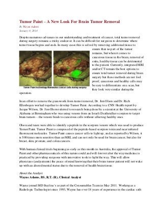
Despite numerous advances in our understanding and treatment of cancer, total tumor removal during surgery remains a tricky endeavor. It can be difficult for surgeons to determine where tumor tissue begins and ends. In many cases this is solved by removing additional tissue to ensure that no part of the tumor remains, but when it comes to cancerous tissue in the brain, removal of extra, healthy tissue can be detrimental to the patient.

Empfohlen
Weitere ähnliche Inhalte
Andere mochten auch
Mehr von MD Buyline
Mehr von MD Buyline (20)
Kürzlich hochgeladen
Kürzlich hochgeladen (20)
A New Look At Brain Tumor Removal | MD Buyline
- 1. Tumor Paint – A New Look For Brain Tumor Removal By Wayne Adams January 9, 2014 Despite numerous advances in our understanding and treatment of cancer, total tumor removal during surgery remains a tricky endeavor. It can be difficult for surgeons to determine where tumor tissue begins and ends. In many cases this is solved by removing additional tissue to ensure that no part of the tumor remains, but when it comes to cancerous tissue in the brain, removal of extra, healthy tissue can be detrimental to the patient. Currently, integrated MRI and/or CT remain the best options to ensure total tumor removal during brain surgery but these methods are not fool proof; cancerous and healthy cells may be easy to differentiate on a scan, but they look very similar during the operation. In an effort to remove the guesswork from tumor removal, Dr. Jim Olson and Dr. Rich Ellenbogen worked together to develop Tumor Paint. According to a CNN Health report by Jacque Wilson, Dr. Jim Olson referred to research being done by a scientist at the University of Alabama at Birmingham who was using venom from an Israeli Deathstalker scorpion to target brain tumors – the venom binds to cancerous cells without affecting healthy ones. Olson and team were able to identify a peptide in the scorpion venom which was used to produce Tumor Paint. Tumor Paint is composed of the peptide-based scorpion toxin and near infrared fluorescent molecules. Tumor Paint causes cancer cells to light up, and as reported by Wilson, it is 500 times more sensitive than an MRI, and can not only be used for brain cancer, but also for breast, skin, prostate, and colon cancers. With human clinical trials beginning as early as this month in Australia, the approval of Tumor Paint and other pharmaceuticals of this nature could and will forever alter the way medicine is practiced by providing surgeons with innovative tools to light the way. This will allow physicians (and patients) the peace of mind knowing that their brain tumor patient will not wake up with an altered mental status due to the removal of health brain tissue. About the Analyst Wayne Adams, BS, R.T. (R), Clinical Analyst Wayne joined MD Buyline’s as part of the Consumables Team in May 2011. Working as a Radiologic Technologist since 1993, Wayne has over 18 years of experience in the cardiac cath
- 2. lab/peripheral vascular field. He graduated from the University of Louisiana at Monroe with a BS in Radiologic Technology. He is registered with the American Registry of Radiologic Technologists and a member of the American Society of Radiologic Technologists. For more information on how MD Buyline helps hospitals and health systems through medical device research and analysis, visit http://www.mdbuyline.com