Clonal b cells in patients with hepatitis c virus–associated mixed

This study examined clonal B cells from patients with hepatitis C virus (HCV) associated mixed cryoglobulinemia (MC). Despite being fundamentally involved in MC pathogenesis, transcriptional profiling revealed these B cells have features of anergy and apoptosis rather than neoplastic transformation. Phenotypically, a large proportion of the patients' clonal B cells were CD21low memory B cells, which typically exhibit decreased activation and increased anergy. Functionally, the CD21low subset showed impaired calcium signaling and failed to efficiently differentiate into antibody-secreting plasmablasts, suggesting anergy. The down-regulation of activation pathways in many of the patients' expanded B cells may attenuate overstimulation from persistent

Recommended
Recommended
More Related Content
What's hot
What's hot (19)
Viewers also liked
Viewers also liked (20)
Similar to Clonal b cells in patients with hepatitis c virus–associated mixed
Similar to Clonal b cells in patients with hepatitis c virus–associated mixed (20)
More from Clinical Immunology Laboratory, HMRUO, Oran.
More from Clinical Immunology Laboratory, HMRUO, Oran. (20)
Recently uploaded
Recently uploaded (20)
Clonal b cells in patients with hepatitis c virus–associated mixed
- 1. IMMUNOBIOLOGY Clonal B cells in patients with hepatitis C virus–associated mixed cryoglobulinemia contain an expanded anergic CD21low B-cell subset Edgar D. Charles,1,2 Claudia Brunetti,1,3 Svetlana Marukian,1 Kimberly D. Ritola,1 Andrew H. Talal,2 Kristen Marks,2 Ira M. Jacobson,2 Charles M. Rice,1 and Lynn B. Dustin1 1Center for the Study of Hepatitis C, Laboratory of Virology and Infectious Disease, Rockefeller University, New York, NY; 2Department of Medicine, Division of Gastroenterology and Hepatology, Weill Medical College of Cornell University, New York, NY; and 3University of Bari, Bari, Italy Hepatitis C virus (HCV) is associated with the B-cell lymphoproliferative disorders mixed cryoglobulinemia (MC) and non- Hodgkin lymphoma. We have previously reported that HCV؉MC؉ patients have clonal expansions of hypermutated, rheu- matoid factor–bearing marginal zone-like IgM؉CD27؉ peripheral B cells using the VH1-69 gene. Here we coupled transcrip- tional profiling with immunophenotypic and functional studies to ascertain these cells’ role in MC pathogenesis. Despite their fundamental role in MC disease, these B cells have overall transcriptional features of anergy and apoptosis instead of neoplastic transformation. Highly up- regulated genes include SOX5, CD11C, galectin-1, and FGR, similar to a previ- ously described FCRL4؉ memory B-cell subset and to an “exhausted,” anergic CD21low memory B-cell subset in HIV؉ patients. Moreover, HCV؉MC؉ patients’ clonal peripheral B cells are enriched with CD21low, CD11c؉, FCRL4high, IL-4Rlow memory B cells. In contrast to the func- tional, rheumatoid factor–secreting CD27؉CD21high subset, the CD27؉CD21low subpopulation exhibits decreased cal- cium mobilization and does not efficiently differentiate into rheumatoid factor– secreting plasmablasts, suggesting that a large proportion of HCV؉MC؉ patients’ clonally expanded peripheral B cells is prone to anergy and/or apoptosis. Down- regulation of multiple activation path- ways may represent a homeostatic mechanism attenuating otherwise uncon- trolled stimulation of circulating HCV- containing immune complexes. This study was registered at www.clinicaltrials.gov as #NCT00435201. (Blood. 2011;117(20): 5425-5437) Introduction Hepatitis C virus (HCV) chronically infects approximately 170 million people worldwide and is the leading indicator for liver transplantation in the United States. Although hepatocytes are the primary target for HCV infection, the B-cell lymphoprolifera- tive disorder mixed cryoglobulinemia (MC) affects up to 50% of HCV patients.1 MC is characterized by the aberrant production of monoclonal rheumatoid factor (RF)–containing immune complexes that deposit on vascular endothelium of organs, such as skin, kidneys, and peripheral nerves, eliciting a complement C1q-mediated vasculitis.2 HCV has also been associated with B-cell non-Hodgkin lymphoma (NHL),3 most frequently of low-grade marginal zone or mucosa-associated lymphoid tissue subtypes, although associations with higher-grade NHL have been reported. HCV-induced B-cell dysregulation probably represents a con- tinuum from the relatively benign clonal B-cell expansion of MC to overt NHL. The continued presence of HCV is necessary for abnormal B-cell lymphoproliferation, as eradication of HCV typically results in resolution of both HCV-related MC and NHL.4 Clonal B-cell populations are present in the liver and peripheral blood of HCVϩMCϩ patients5; such B cells demonstrate biased usage of the RF-encoding VH1-69 and V3-20 gene segments,6 as do B cells isolated from lymph nodes of HCV-NHL patients.7 It remains unclear why B cells undergo clonal proliferation during chronic HCV infection. It is probable that HCV-induced B-cell lymphoproliferation is not the result of direct B-cell infection or transformation, but rather, an indirect process arising from chronic antigenic stimulation of a limited pool of preexisting autoreactive B cells. We have proposed that persistently high levels of HCV-containing immune complexes stimulate the proliferation of RF-bearing B cells,6 but the precise antigen(s) and stimulatory mechanisms have remained elusive. We have previously shown that HCVϩMCϩ patients’ clonal B cells are predominantly IgM memory B cells expressing modestly hypermu- tated immunoglobulin genes; phylogenetic analysis supports a process of antigen-directed affinity maturation. However, many of these clonal cells have decreased expression of CD21, the CR2 complement receptor.6 Because CD21 augments B-cell receptor (BCR)-mediated signaling as part of the B-cell coreceptor com- plex, its down-regulation may confer a state of relative anergy to these cells, as has been demonstrated among CD21low naive B cells from patients with chronic variable immunodeficiency and rheuma- toid arthritis.8 To better understand how HCV elicits the expansion of autore- active B-cell clones, we have performed transcriptional, immuno- phenotypic, and functional analyses on HCVϩMCϩ patients’clonal B cells. Contrary to expectations, these cells have a global transcriptional profile suggestive of anergy and apoptosis, and a large proportion of them have immunophenotypic features of anergy. Taken together, our data suggest that, although HCVϩMCϩ Submitted October 11, 2010; accepted March 6, 2011. Prepublished online as Blood First Edition paper, March 18, 2011; DOI 10.1182/blood-2010-10-312942. The online version of this article contains a data supplement. The publication costs of this article were defrayed in part by page charge payment. Therefore, and solely to indicate this fact, this article is hereby marked ‘‘advertisement’’ in accordance with 18 USC section 1734. © 2011 by The American Society of Hematology 5425BLOOD, 19 MAY 2011 ⅐ VOLUME 117, NUMBER 20 For personal use only.on March 7, 2015.by guestwww.bloodjournal.orgFrom
- 2. patients clearly have expanded peripheral B cells capable of differentiating into RF-secreting plasmablasts, these cells do not have transcriptional features of neoplastic transformation, and a significant proportion of this clonal population may be refractory to ongoing antigenic stimulation. Methods Patients The studies were approved by the Institutional Review Boards at the Rockefeller University and New York Presbyterian Hospitals. Donors gave written informed consent according to the Declaration of Helsinki before enrollment. We enrolled HCV AbϪ, HCV Abϩ/HCV RNAϩ, and HCV Abϩ/HCV RNAϪ volunteers. No subjects received interferon or immuno- suppressive therapy within 6 months of enrollment. Blood was obtained by peripheral blood draw and leukapheresis. Peripheral blood mononuclear cells (PBMCs) were prepared as previously described.6 Clinical tests HCV RNA was quantified clinically by the Roche Amplicor assay (Version 2.0; Roche Diagnostics); results are standardized to international units. Liver biopsies were evaluated by pathologists according to the Scheuer system. These tests, in addition to serum alanine aminotransferase measure- ments, were performed as part routine medical care. Testing for MC was performed as previously described.6 IgM؉؉CD27؉ B-cell isolation IgMϩϩ B cells were isolated from PBMCs by negative selection to minimize transcriptional changes effected by BCR signaling. All steps were performed at 4°C. B cells were immunomagnetically isolated using a B Cell Isolation Kit (Miltenyi Biotec). These were incubated with phycoerythrin- conjugated anti-IgG, anti-IgA, and anti-, then with anti–phycoerythrin- conjugated microbeads, and the negative fraction was magnetically puri- fied. The CD27ϩ fraction was immunomagnetically isolated using anti– CD27-conjugated microbeads. RNA extraction, cDNA synthesis, amplification, and labeling RNA was extracted from 5000 to 10 000 cells using the RNeasy Plus Micro Kit (QIAGEN) with on-column DNase digestion. RNA integrity and concentration were determined using Lab on a Chip Pico. Samples with RNA integrity numbers Ͼ 9.0 were used for downstream processing. A total of 2 ng RNA was reverse-transcribed with random hexamers as primers and amplified using the WT-Ovation Pico Kit (Nugen), and 5 g cDNA labeled using uracyl-N-glycosylase (Epicentre Biotechnologies) and biotinylated aldehyde-reactive probe. Microarray procedures Human V3 BeadChips (Illumina) were hybridized with 1.5 g cDNA. Chips were scanned on an Illumina Beadstation and analyzed with Illumina BeadStudio software (Version 3.2). Datasets were analyzed using Gene- Spring GX Version 11.1 (Agilent Technologies). Raw signal values were log-transformed, chips were normalized to the 50th percentile, and genes normalized to the median signal. This dataset was filtered to include genes with signals above background. Welch t test (P ϭ .05, Benjamini-Hochberg false discovery rate ϭ 0.05) was used to test for differences in genes between groups. The resulting set was filtered to include genes that were 2-fold up- or down-regulated. Hierarchical clustering was performed using the weighted pairwise group method with centroid average, using the Pearson correlation as the distance metric. Statistics were calculated using GeneSpring GX and Prism (GraphPad Software). Quantitative RT-PCR RNA was prepared from isolated B cells, as described under “RNA extraction, cDNA synthesis, amplification, and labeling.” Random-primed cDNA was synthesized using Superscript III (Invitrogen). Primers (supple- mental Table 1, available on the Blood Web site; see the Supplemental Materials link at the top of the online article) were constructed using the PrimerBank Database (www.pga.mgh.harvard.edu/primerbank) and were designed to span exon-intron borders to reduce the possibility of genomic DNA amplification. SYBR Green PCR Master Mix (Applied Biosystems) was used for quantitative reverse-transcribed polymerase chain reaction (RT-PCR). We normalized all samples to RPS11 and to the target gene in Universal Human Reference RNA (Stratagene). Fold change expression was calculated using the 2Ϫ⌬⌬Ct method. Groups were compared using the Kruskal-Wallis test; when Kruskal-Wallis P Ͻ .05, the HCVϩMCϩ and HCVϩMCϪ groups were compared using the Dunn post-test. Flow cytometry Cells were stained with monoclonal antibodies (mAbs) in phosphate- buffered saline supplemented with 2% (weight/volume) bovine serum albumin (Fraction V; Fisher Biotech) and 0.02% NaN3. All antibodies and reagents were from BD Biosciences, except for G6 (provided by R. Jefferis), F(abЈ)2 anti-FCRL4 biotin (provided by G. Erhardt and M. Cooper), and Ki-67 fluorescein isothiocyanate (Invitrogen). Conjugation of G6 to biotin and AlexaFluor-594 was performed using commercial kits (Pierce Chemi- cal, Invitrogen). Analysis was performed within 1 hour on a BD LSRII flow cytometer (BD Biosciences). G6؉ B-cell subset isolation PBMCs were stained with anti-CD20, anti-CD27, anti-CD21, G6 mAbs, and 4,6-diamidino-2-phenylindole (to exclude dead cells). Live CD27ϩ/ϪCD21high/low G6ϩ, CD20ϩ B cells were bulk-sorted on a BD FACSAria II (BD Biosciences). For assessment of postsort viability, sorted samples were restained with 4,6-diamidino-2-phenylindole and were reanalyzed by flow cytometry. Postsort analysis confirmed more than 85% viability and more than 99% purity of sorted populations. Cell cycle analysis B cells were negatively isolated from PBMCs using EasySep Human B Cell Enrichment Kit (StemCell Technologies) and incubated with biotinylated G6 mAb. G6ϩ and G6Ϫ B-cell subsets were purified using streptavidin- conjugated immunomagnetic beads. Cells were fixed in 80% ethanol. After incubation with FITC-labeled Ki-67, cells were resuspended in PBS containing 10 mg/mL RNase A and incubated at 37°C. Propidium iodide 20 g/mL was added before flow cytometry. Electron microscopy Cells were fixed in 2% glutaraldehyde, incubated in 1% osmium, dehy- drated in a graded alcohol series, embedded in spur resin, and then treated with 2% uranyl acetate and Reynold lead citrate. Transmission electron microscopy was performed at ϫ2000 and ϫ10 000 magnification. Calcium mobilization assay A total of 2 ϫ 106 B cells were incubated with 1M Indo-1 for 30 minutes. Cells were then labeled with anti-CD19, IgG, CD27, and CD21 mAbs and suspended in HBSS with Ca2ϩ and 1% bovine serum albumin. Emission at 405 and 495 nm was measured to obtain a baseline, and then for 5 minutes. After addition of 10 g/mL goat F(abЈ)2 anti-IgM, 405/495 nm emission ratios of IgGϪ B-cell subsets were analyzed with FlowJo software Version 9.2 (TreeStar). Annexin V apoptosis assay A total of 2 ϫ 106 PBMCs were incubated with 1 g/mL anti-CD95 or mouse IgG1 and 2 g/mL Protein G (Invitrogen) in RPMI/10% fetal calf serum at 37°C for 0 and 6 hours. Cells were washed with phosphate- buffered saline and resuspended in 0.01M N-2-hydroxyethylpiperazine-NЈ- 2-ethanesulfonic acid (pH 7.4), 0.14M NaCl, 2.5mM CaCl2, and 2% fetal 5426 CHARLES et al BLOOD, 19 MAY 2011 ⅐ VOLUME 117, NUMBER 20 For personal use only.on March 7, 2015.by guestwww.bloodjournal.orgFrom
- 3. calf serum. After incubation with annexin V–phycoerythrin and 7-amino- actinomycin at room temperature, cells were stained with anti-CD20 FITC anti-CD21–allophycocyanin, and G6-biotin at room temperature. After staining cells with streptavidin-Cy7-allophycocyanin, flow cytometry was performed. Immunoglobulin secretion assays Cells (50 000/well in 96-well round-bottom plates) were cultured for 6 days in RPMI supplemented with 10% fetal calf serum, 2mM L-glutamine, 100 U/mL penicillin/streptomycin, and 0.25 g/mL amphotericin B, with the addition of 6 U/mL IL-2 (R&D Systems), 200 ng/mL IL-10 (R&D Systems), and 1 g/mL flag-tagged CD40L with 2 g/mL mouse IgG1 anti-flag Ab (Alexis Biochemicals). For ELISPOT, cells were washed with RPMI, placed on MultiScreen filter plates (Millipore), coated with goat F(abЈ)2 anti–human IgM (Jackson ImmunoResearch Laboratories), and incubated at 37°C for 6 hours. Plates were then incubated with horseradish peroxidase-labeled anti–human IgM, and the assays were developed with 3-amino-9-ethylcarbazole (Sigma- Aldrich). Spots were counted using an ImmunoSpot Analysis Instrument (Cellular Technology). For ELISA, cell culture supernatants were added to MaxiSorb plates (Nunc) coated with anti-IgM (Bethyl Laboratories), or for RF assay, human IgG1 (Sigma-Aldrich), and incubated at room temperature for 1 hour. Plates were then incubated with HRP-labeled goat-anti–human IgM, and the assays were developed with TMB (BioFX Laboratories). After stopping reactions with 1N H2SO4, A450 was measured on a FLUOstar Omega microplate reader (BMG Laboratories). Accession numbers Microarray data are accessible through NCBI Gene Expression Omnibus accession number GSE18084 (www.ncbi.nlm.nih.gov/geo/query/ acc.cgi?acc ϭ GSE18084). Results Characteristics of study subjects Five HCV AbϪ volunteers, 7 sustained virologic responders (SVRs), and 27 persons with chronic HCV infection were enrolled for gene transcriptional analyses. Three additional HCVϩMCϪ and 2 additional HCVϩMCϩ subjects were enrolled only for immuno- phenotypic and functional analyses (Table 1). Sixteen HCVϩ subjects were MCϩ, and all 16 had evidence of clonal B-cell populations, as demonstrated by complementarity-determining region 3 PCR.6 Four of these subjects had lymphadenopathy; lymph node biopsies performed by their physicians confirmed low-grade B-NHL (subjects 1116, 1308, 1716, and ECH 529). Fifteen of 16 patients had evidence of clonal IgM gammopathy by serum immunofixation electrophoresis and were classified as being IgM MCϩ. Subject ECH 529 had evidence of clonal IgAgammopa- thy by immunofixation electrophoresis. In addition, cervical lymph node biopsy in this patient revealed abnormal numbers of IgAϩϩ B cells. Plasma from 10 of 12 HCV RNAϪ and 25 of 27 HCV RNAϩ patients had detectable anti-Epstein-Barr virus nuclear antigen 1 IgG, indicating previous exposure to Epstein-Barr virus. HCVϩ IgM MCϩ subjects had significantly expanded populations of IgMϩϩ peripheral B cells, although overall B-cell numbers were not increased, consistent with our earlier report6 (supplemen- tal Figure 1). IgM؉؉CD27؉ B cells from HCV؉MC؉ patients have a distinct transcriptional profile A total of 69 unique genes (33 up-regulated, 36 down-regulated) were found to be more than 2-fold differentially expressed in IgMϩϩCD27ϩ B cells from IgM MCϩ, compared with IgM MCϪ, subjects (Figure 1). Notably, the transcriptional profile of 2 HCV RNAϩMCϪ patients (ECH 516 and ECH 522) shared several features with that of the MCϩ population. Several of the differentially expressed genes were grouped accord- ing to broad function (Table 2). The overall transcriptional pattern was suggestive not of oncogenesis, but of dampened activation and aug- mented proapoptotic pathways. Unsurprisingly, several IFN-induced genes were up-regulated: growth interferon-inducible protein X (PY- HIN1), myeloid nuclear differentiation antigen (MNDA), and 2Ј, 5Ј- oligoadenylate synthetase 1 (OAS1). Several genes associated with B-cell anergy were up-regulated: galectin-1 (LGALS1), lymphocyte transmembrane adapter 1 (LAX1), and CD200 receptor 1 (CD200R1), an inhibitory receptor highly expressed on memory B cells and plasmablasts. Significantly up-regulated proapoptotic genes included: galectin-1, the interferon-response gene, PYHIN1, death-associated protein kinase 2 (DAPK2), and MNDA. The prosurvival gene, T-cell lymphoma 1A(TCL1A), was markedly down-regulated. In addition to having an overall transcriptional program sugges- tive of B-cell anergy and apoptosis, HCVϩMCϩ patients’ IgMϩϩCD27ϩ B cells demonstrated differential regulation of several genes previously reported increased in patients with NHL. Up-regulated genes included: SRY-box 5 (SOX5), ␣-X integrin (ITGAX, CD11C), and MNDA. Down-regulated genes included: L-selectin (SELL), LIM only 2 (LMO2), forkhead box 1 (FOXP1), and TCL1A. Also down-regulated was IL-4 receptor (IL-4R), polymorphisms of which have been associated with diffuse large B-cell lymphoma25 and which may be down-regulated in mantle cell lymphoma.9 In addition, BTB and CNC homology 1, basic leucine zipper transcription factor 2 (BACH2) was down-regulated; both BACH2 and IL-4R are reported to be down-regulated in B cells from patients with Waldenstro¨m macroglobulinemia.10 Because many of the up-regulated genes (eg, CD11C, CD84, CD200R1, and bone morphogenetic protein receptor 1A[BMPR1A]) are known to be expressed in activated and/or memory B cells,11,26-28 we hypothesized that their up-regulation reflected a particular stage of differentiation of HCVϩMCϩ patients’ clonally expanded B cells. Several of the most significantly up-regulated genes (SOX5, Gardner-Rasheed feline sarcoma viral oncogene ho- molog [FGR], and CD11C) have previously been found to be highly up-regulated in FCRL4-expressing tonsillar B cells, a recently described memory B-cell subset thought to play an important role in mucosal defense.29 Despite this transcriptional similarity, our microarray data did not reveal differences in FCRL4 transcript between MCϩ and MCϪ patients’ IgMϩϩCD27ϩ peripheral B cells. However, quantitative RT- PCR of unamplified cDNA confirmed the up-regulated expres- sion of SOX5, FGR, and CD11C in HCVϩMCϩ patients’ expanded IgMϩϩCD27ϩ B cells (Figure 2; supplemental Table 2). In addition, quantitative RT-PCR confirmed the up- regulation of galectin-1 and OAS1 and the down-regulation of IL-4R and TCL1A, and it detected no significant difference in FCRL4 expression. We did not detect Epstein-Barr virus nuclear antigen 2 or latent membrane protein 1 transcripts by quantita- tive RT-PCR (data not shown). A significant proportion of clonal cells from IgM MC؉ subjects are CD21low, CD11c؉, FCRL4high, IL-4Rlow memory B cells We and others have previously shown that clonally expanded IgMϩϩCD27ϩ B cells from IgM MCϩ patients preferentially use the VH1–69 gene segment.6,30 We used the G6 mAb, which recognizes the complementarity-determining region 2 of Ig VH1- B-CELLANERGY IN MIXED CRYOGLOBULINEMIA 5427BLOOD, 19 MAY 2011 ⅐ VOLUME 117, NUMBER 20 For personal use only.on March 7, 2015.by guestwww.bloodjournal.orgFrom
- 4. 69,31 to more precisely immunophenotype HCVϩMCϩ patients’ clonally expanded B cells. We have previously confirmed the specificity of G6 for VH1-69 by RT-PCR.32 These G6ϩ B cells from HCVϩMCϩ patients are frequently CD21low and IgMϩϩ (supple- mental Figure 2). They are also morphologically normal, nonprolif- erating, and predominantly CD20high, CD10Ϫ, CD21low, CD27ϩ, CD11cϩ, FCRL4high, and IL-4Rlow (Figure 3). We immunopheno- typed B cells from 11 MCϪ (SVR ϭ 2, HCV AbϪ ϭ 3, HCV RNAϩ ϭ 6) and 9 HCV RNAϩMCϩ subjects (Figure 4). As expected, we found that MCϩ patients had a significant (P Ͻ .005) expansion of G6ϩ B cells (median, 25.9% of B cells) compared with HCV RNAϩMCϪ patients (median, 5.1%), SVR (median, 3.0%), and HCV AbϪ (median, 3.8%) patients. In 3 MCϩ patients (LDU 125, 110, and 1432), more than 50% of total peripheral B cells were G6ϩ. When we examined G6ϩ and G6Ϫ B cells from each person, we confirmed that G6ϩ, compared with G6Ϫ, B cells from HCVϩMCϩ patients were predominantly CD21low, CD11cϩ, FCRL4high, and IL-4Rlow. Interestingly, G6ϩ, compared with G6Ϫ, B cells from MCϪ persons were also disproportionately CD21low. However, they did not have significantly increased expression Table 1. Characteristics of the study patients Condition/subject no. Age, y Sex Ethnicity HCV RNA GT Stage (0-4) Treatment history Clonal CDR3 HCV Ab- LDU 099 35 Male White NA NA NA NA No LDU 128 40 Male Black NA NA NA NA No ECH 503 35 Male Asian American NA NA NA NA No ECH 527 50 Female White/Hispanic NA NA NA NA No ECH 528 51 Female White NA NA NA NA No SVR 543 48 Female White Ͻ 50 1 4 pIFN/RBV 2001 No 731 52 Female White Ͻ 50 2b 2 pIFN/RBV 2003 No 856 61 Female White Ͻ 50 1a 2 pIFN/RBV 2003 No 1154 56 Female Asian American Ͻ 50 1a 2 pIFN/RBV 2003 No 1197 38 Female White Ͻ 50 2a 1 pIFN/RBV 2003 No ECH 521 43 Male White Ͻ 50 1 ND pIFN/RBV 2005 No ECH 542 38 Female Black Ͻ 50 1 ND pIFN/RBV 2007 No HCV؉, IgM MC؊ 1235 44 Female White 3.85 ϫ 106 2b ND Naive No 1330 54 Female White/Hispanic Ͼ 7 ϫ 105 1a 2 Naive No 1419 48 Female White 0.39 ϫ 106 1 3 Naive No 1864 58 Male White 0.02 ϫ 106 4 3 Naive No LDU 107 50 Male White/Hispanic Ͼ 7 ϫ 105 2 2 Naive No ECH 507 54 Male White/Hispanic Ͼ 7 ϫ 105 1 ND Naive No ECH 512* 62 Male White Ͼ 7 ϫ 105 1 ND Naive No ECH 516 57 Male White/Hispanic 2.4 ϫ 106 1b 2 Naive No ECH 519 52 Female White/Hispanic Ͼ 7 ϫ 105 1 ND Naive No ECH 520 57 Male Black Ͼ 5 ϫ 106 1b 2 Naive No ECH 522 50 Female White/Hispanic Ͼ 7 ϫ 105 1 ND Naive No ECH 529†‡ 52 Female White Ͼ 7 ϫ 105 3 4 Naive Yes ECH 530 49 Male White/Hispanic Ͼ 7 ϫ 105 1 ND Naive No ECH 536* 50 Male White/Hispanic 0.2 ϫ 106 1 1 Naive No ECH 537* 54 Male Black 0.9 ϫ 106 1a ND Naive No HCV؉, IgM MC؉ 110 53 Male White/Hispanic 1.70 ϫ 106 1 3 Naive Yes 880 54 Female White 3 ϫ 106 1b 2 Naive Yes 1116‡ 37 Male White 3 ϫ 104 2b 2 Naive Yes 1308‡ 55 Female White 0.72 ϫ 106 1 1 Naive Yes 1403 44 Female White/Hispanic 0.50 ϫ 106 1 2 Naive Yes 1432 69 Female White 0.03 ϫ 106 1 4 Naive Yes 1540 37 Male White/Hispanic 0.28 ϫ 106 1 ND Naive Yes 1716‡ 30 Female White 1.45 ϫ 106 1a ND Naive Yes 1931 56 Female White 2.2 ϫ 106 1b 2 Naive Yes 92200 57 Female White 2 ϫ 106 2 2 Naive Yes LDU 125 60 Male White 0.2 ϫ 106 3 ND Naive Yes ECH 531 56 Male White Ͼ 7 ϫ 105 1 ND Naive Yes ECH 532 51 Female Black Ͼ 7 ϫ 105 1 ND Naive Yes ECH 533 63 Male White 3.8 ϫ 106 1 0 pIFN/RBV 2007 Yes ECH 535 57 Male White 0.74 ϫ 106 1 ND Naive Yes ECH 546* 52 Male White Ͼ 7 ϫ 105 1 3 pIFN/RBV 2008 ND ECH 559* 58 Female White/Hispanic 1.27 ϫ 106 1 4 pIFN/RBV 2001 ND GT indicates genotype; CDR3, Ig complementarity determining region 3; pIFN/RBV, pegylated interferon/ribavirin; NA, not applicable; and ND, not done. *Subjects used for immunophenotypic and functional assays only. †Subject with ϩ IgA MC. ‡Subjects with marginal zone B-cell NHL (documented on lymph node biopsy). 5428 CHARLES et al BLOOD, 19 MAY 2011 ⅐ VOLUME 117, NUMBER 20 For personal use only.on March 7, 2015.by guestwww.bloodjournal.orgFrom
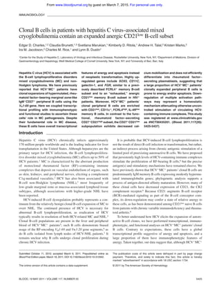
- 5. of CD11c or FCRL4, nor did they have decreased expression of IL-4R. Reflective of having G6ϩ B-cell expansions, HCVϩMCϩ pa- tients had increased percentages of total B cells that were CD21low, CD11cϩ, FCRL4high, and IL-4Rlow. These markers were signifi- cantly correlated with the percentage of peripheral B cells that were G6ϩ. We did not detect any significant differences in any of these cell surface markers among the total B cells of SVR, HCV AbϪ, and HCV RNAϩ patients, probably reflective of the low proportion of G6ϩ B cells in these MCϪ persons. FCRL4ϩ memory B cells from normal tonsils and HIV-viremic blood are reported to be CD20highCD11cϩCD95ϩCD21low.29,33 In healthy tonsils, these FCRL4ϩ B cells have been variously reported to be CD27Ϫ or CD27ϩ memory B cells.34,35 Although we detected increased FCRL4 expression on G6ϩ, compared with G6Ϫ, B cells, we did not detect significant differences in FCRL4 expression 110 1931 1308 1432 1716 1116 922000 LDU125 1540 ECH533 1403 880 ECH535 ECH532 ECH531 ECH516 ECH522 1154 LDU107 856 ECH528 ECH521 1864 ECH503 ECH527 ECH529 ECH507 ECH520 ECH530 ECH519 1197 543 731 1330 1419 1235 ECH542 LDU128 LDU099 IgM MC + IgM MC - Expression 6 01 SOX5 AEBP1 SLC7A7 PYHIN1 SLC11A1 ITGAX (CD11c) PNMA2 TESC LGALS1 (Galectin-1) PPAP2B FGR HOXB7 FLJ40869 OAS1 FCGR2A MNDA MIDN STS-1 LOC649199 RASGRF1 N/A N/A CD200R1 CD84 DAPK2 LAX1 TFEC LRRN1 OAS1 SRGN C13orf15 TUBB2A TUBB2A ENC1 DUSP5 CD68 TMEM44 N/A ADARB1 TSPAN13 SELL P2RY14 N/A TCL1A IL4R C3orf37 LMO2 SPRY1 ZNF135 LARGE SMAD3 N/A EPB41L2 VPREB3 N/A PPAPDC1B LIX1 SSBP2 MGC39372 GCNT1 TAPT1 CRIM1 CR1 CR1 C1orf162 LOC647450 LOC652493 LOC652694 N/A PELI2 CD1A NT5E IL13RA1 N/A LOC652113 N/A SERPINE2 FOXP1 BACH2 FLJ14213 IL15 Figure 1. Transcriptome analysis of IgM؉؉CD27؉ B cells. The transcripts differentially expressed by IgMϩϩCD27ϩ B cells from HCVϩMCϩ and MCϪ patients are displayed. Up- and down-regulated tran- scripts are indicated in red and blue, respectively. The magnitude of expression is indicated by the color bar. B-CELLANERGY IN MIXED CRYOGLOBULINEMIA 5429BLOOD, 19 MAY 2011 ⅐ VOLUME 117, NUMBER 20 For personal use only.on March 7, 2015.by guestwww.bloodjournal.orgFrom
- 6. among CD27ϩ, CD27Ϫ, CD21high, or CD21low B-cell subsets (supplemental Figure 3). In an attempt to reconcile the findings of unchanged FCRL4 transcript levels and increased FCRL4 protein expression in MCϩ compared with MCϪ B cells, we performed quantitative RT-PCR on RNAisolated from the bulk-sorted FCRL4ϩ and FCRL4Ϫ G6ϩ B cells from subject LDU 125. However, we failed to detect any difference in FCRL4 mRNA between these 2 groups (data not shown). CD21low B cells from healthy, HCV؉MC؊, and HCV؉MC؉ persons are anergic to BCR-mediated stimulation CD21low memory B cells have been described in HIV-viremic patients as being relatively anergic, “exhausted” B cells.33 We sought to determine whether IgGϪ, CD21low B cells had attenuated signaling on BCR ligation with anti–human IgM. Data for one healthy subject with an elevated (22%) CD21low B-cell frequency (ECH 503), 2 HCVϩMCϪ (ECH 530, ECH 537) volunteers and 5 HCVϩMCϩ (ECH 546, ECH 559, 110, LDU 125, and 1931) volunteers are shown in Figure 5. In all but one (110), the CD27ϩCD21low subset had decreased Ca2ϩ mobilization; the reason for this outlier is unclear. Furthermore, in all subjects, the CD27ϪCD21low B-cell subset had diminished amplitude of Ca2ϩ mobilization; ECH 546, for unknown reasons, had an intermediate response to BCR triggering. Finally, in ECH 559 and 1931, CD27ϩCD21high cells had low responses to BCR stimulation; IgA may possibly be a prominent non-IgG BCR in their CD27ϩCD21high subsets. Overall, these data suggest that CD21low B cells, whether from healthy, HCVϩMCϪ, or HCVϩMCϩ persons, are anergic to BCR-mediated stimulation. G6؉ B cells are preferentially prone to death and apoptosis Our microarray data suggested that HCVϩMCϩ patients’ IgMϩϩCD27ϩ B cells up-regulate several genes associated with apoptosis. We tested whether these cells were prone to apoptosis in vitro by incubating them with either media, anti-CD95 mAb, or isotype control IgG (Figure 6). After 6 hours of incubation, PBMCs were stained with annexin V (to measure apoptosis), 7-amino- actinomycin D (7-AAD; to measure cell death), anti-CD20, and-CD21, and G6 Abs. After incubation with media alone, isotype control, or anti-CD95, G6ϩ, compared with G6Ϫ, B cells had significantly higher binding of annexin V, either alone or in combination with 7-AAD. The percentage of annexin Vϩ/7-AADϪ cells was highest in the anti-CD95-stimulated G6ϩ B-cell subset. However, whereas G6Ϫ cells demonstrated a 2.1-fold increase in annexin V binding on stimulation with anti-CD95, compared with isotype control, G6ϩ cells had a 1.4-fold increase (Figure 6A). This Table 2. Functional grouping of differentially expressed genes Functional group/gene Fold difference P Significance Interferon response PYHIN1 (IFIX), (growth interferon-inducible protein X) 2.8 .000052 Interferon-inducible HIN-200 gene family member17 MNDA (myeloid nuclear differentiation antigen) 2.2 .0092 HIN-200 gene family member17,19 OAS1 (2Ј, 5Ј oligoadenylate cyclase) 2.1 .000052 IFN-inducible gene which activates RNaseL B-cell anergy LGALS1 (galectin-1) 3.6 .00038 Negatively regulates B-cell proliferation upon BCR ligation14; up-regulated in anergic murine B cells12 CD200R1 (CD200 receptor 1) 2.4 .0034 Inhibitory receptor highly expressed on memory B cells and plasmablasts; definitive role in B cell activation not established11 LAX1 (lymphocyte transmembrane adapter 1) 2.1 .032 Dampens B-cell response to BCR engagement16 B-cell apoptosis LGALS1 3.6 .00038 Expressed in IgMϩ memory cells, reported to enhance apoptosis via inhibition of Akt phosphorylation and up-regulation of Bim13 PYHIN1 2.8 .000052 As an HIN-200 gene family member, contains an N-terminal PAAD/DAPIN/Pyrin domain that mediates binding with proteins involved in apoptotic NF-B and caspase signaling17 DAPK2 (death-associated protein kinase 2) 2.4 .043 Calcium/calmodulin-dependent protein kinase, which induces apoptosis15 MNDA 2.2 .0092 As an HIN-200 gene family member, contains an N-terminal PAAD/DAPIN/Pyrin domain that mediates binding with proteins involved in apoptotic NF-B and caspase signaling17 B-cell survival TCL1A (T-cell lymphoma antigen 1A) 0.34 .000075 Reported to enhance B-cell survival by induction of Mcl-113 B-cell lymphomagenesis SOX5 (SRY-box 5) 3.6 .00062 Reported to be up-regulated in splenic follicular lymphoma20 ITGAX, (CD11C), (integrin, ␣ X) 2.9 .00066 High expression associated with splenic marginal zone lymphoma21 MNDA 2.2 .0092 Expressed in marginal zone B cells, up-regulated in marginal zone lymphoma22 BACH2 0.46 .022 Down-regulated in Waldenström macroglobulinemia10 FOXP1 0.44 .00072 Up-regulation of FOXP1 in mucosa-associated lymphoid tissue lymphoma may be associated with neoplastic transformation23 TCL1A (T-cell lymphoma 1A) 0.34 .00075 Up-regulated in splenic marginal zone lymphoma18 SELL (L-selectin) 0.34 .00031 Up-regulated in splenic marginal zone lymphoma18 IL-4R (IL-4 receptor) 0.31 .00017 Down-regulated in mantle cell lymphoma9 and Waldenström macroglobulinemia10 LMO2 (LIM domain only 2) 0.31 .0010 Up-regulation corresponds to increased survival in diffuse large B-cell lymphoma24 IFN indicates interferon; PAAD/DAPIN, Pyrin, AIM (Absent in Melanoma), ASC (Apoptosis-associated-speck-like protein containing a caspase recruitment domain [CARD]) and death-domain (DD)-like Domain in Apoptosis and Interferon response; and NFB, nuclear factor-B. *Bonferroni corrected. 5430 CHARLES et al BLOOD, 19 MAY 2011 ⅐ VOLUME 117, NUMBER 20 For personal use only.on March 7, 2015.by guestwww.bloodjournal.orgFrom
- 7. indicates that G6ϩ B cells’ propensity for apoptosis is not fully dependent on CD95, although CD95 was upregulated on several HCVϩMCϩ patients’ G6ϩ cells. A larger percentage of apoptotic annexin Vϩ7-AADϪ cells had lower CD21 expression compared with annexin VϪ7-AADϪ cells (Figure 6B). This was true for both G6ϩ and G6Ϫ populations. Surface CD20 expression was similar among all subsets, except for G6Ϫ annexin Vϩ7-AADϩ cells. Importantly, 6 hours of incubation with anti-CD95 did not signifi- cantly alter the percentages of CD20ϩ B cells that were G6ϩ (37 vs 29% at 0 and 6 hours, respectively), nor did it cause down- regulation of CD21 on G6ϩ or G6Ϫ B cells (Figure 6C), indicating that cell surface CD20, CD21, and G6 expression was stable and not significantly affected by apoptosis or cell death. Taken together, these data suggest that, in the absence of survival signals, G6ϩ B cells, and in particular, the CD21low G6ϩ subset, are prone to spontaneous cell death and apoptosis. CD27؉CD21high, but not CD27؉CD21low, G6؉ B cells from HCV؉MC؉ patients efficiently differentiate into antibody-secreting plasmablasts To determine whether CD27ϩCD21low B cells could proliferate and differentiate in response to stimulatory signals, we stimulated sorted CD27ϩCD21high, CD27ϩCD21low, CD27ϪCD21high, and CD27ϪCD21low G6ϩ B cells from 4 HCVϩMCϩ subjects. For a fifth patient, CD27ϩCD21low, CD27ϩCD21high, CD27Ϫ G6ϩ B cells and total G6Ϫ B cells were sorted. We used 2 independent samples to test whether our cell sorting procedure caused early cell death; in all subsets, more than 85% of cells were viable, as measured by 4,6-diamidino-2-phenylindole staining (supplemental Figure 4). Cells were cultured for 6 days with CD40L, IL-2, and IL-10 and analyzed for differentiation toward antibody-secreting cells. Using IgM ELISPOT, we found that the CD27ϩCD21high, com- pared with CD27ϩCD21low and CD27Ϫ G6ϩ B-cell subsets, were more efficient at differentiating to IgM-secreting plasmablasts on CD40L/IL-2/IL-10 stimulation (Figure 7A). To confirm these findings, we quantitated IgM in the cell culture supernatants using ELISA. The CD27ϩCD21high cell culture supernatants all had higher levels of IgM compared with their CD27ϩCD21low and CD27Ϫ counterparts (Figure 7B). Class switch did not account for the decreased IgM in these subsets, as we consistently detected Ͻ 150 ng/mL IgG in cell culture supernatants. We confirmed that the IgM produced by the stimulated G6ϩ B-cell subsets had RF activity by demonstrating that they could bind human IgG1 (Figure 7C). As a control, we tested patient LDU 125’s G6Ϫ B-cell supernatant. Although IgM was present at a total concentration of approximately 0.5 g/mL, we detected no signifi- cant RF activity. Taken together, these results suggest that differen- tiation to pathogenic RF-secreting plasmablasts on CD40L/IL-2/ IL-10 stimulation is less efficient in the CD27ϩCD21low, compared with the CD27ϩCD21high, G6ϩ B-cell subset. Discussion We previously reported that HCVϩMCϩ patients have clonal expansions of IgMϩϩCD27ϩ peripheral B cells.6 Here we have transcriptionally profiled IgMϩϩCD27ϩ B cells isolated from HCVϩMCϩ patients, and we have identified a set of 69 genes that are differentially expressed. This set is notable for interferon response genes, B-cell activation markers, transcription factors, and glycoprotein synthesis genes. Although several genes previ- ously implicated in B-cell neoplasia (eg, SOX5, CD11C, and MNDA) are up-regulated among these cells, others (LMO2, IL-4R, SELL, TCL1A, FOXP1, and BACH2) are down-regulated. Thus, although IgMϩϩCD27ϩ B cells are responsible for MC pathogen- esis, their overall gene expression pattern suggests that there exists a subpopulation that is biased toward anergy and/or apoptosis. The most highly up-regulated gene was the -galactoside– binding lectin galectin-1, which is intriguing given its known pleiotropic roles in innate and adaptive immune responses. Galec- tin-1 (and CD11C) transcripts have been identified as being up-regulated in anergic murine B cells.12 Moreover, galectin-1 has been shown to be overexpressed in IgMϩCD27ϩ B cells, is further HCVAb- HCV+ MC- HCV+ MC+ SVR HCVAb- HCV+ MC- HCV+ MC+ SVR HCVAb- HCV+ MC- HCV+ MC+ SVR HCVAb- HCV+ MC- HCV+ MC+ SVR HCVAb- HCV+ MC- HCV+ MC+ SVR HCVAb- HCV+ MC- HCV+ MC+ SVR HCVAb- HCV+ MC- HCV+ MC+ SVR HCVAb- HCV+ MC- HCV+ MC+ SVR p < 0.05 p < 0.001 p < 0.001 p = n.s. OAS1 FCRL4IL4R TCL1A LGALS1 SOX5 FGR CD11C p < 0.001p < 0.001 0 1 2 3 4 5 6 7 0.0 2.5 5.0 7.5 10.0 12.5 0 10000 20000 30000 40000 0 10 20 30 p < 0.01 0 1 2 3 4 5 6 0 5 10 15 0 1000 2000 3000 4000 0.0 0.5 1.0 1.5 2.0 p < 0.0001 Figure 2. Relative expression of selected genes in IgM؉؉CD27؉ B cells determined by quantitative RT-PCR. Values are normalized to RPS11 for cDNA content and to a universal standard RNA. Comparisons between groups were made using the Kruskal-Wallis test, and P values for HCVϩMCϪ compared with HCVϩMCϩ subjects were computed using Dunn post test. N.S. indicates not significant (P Ͼ .05). B-CELLANERGY IN MIXED CRYOGLOBULINEMIA 5431BLOOD, 19 MAY 2011 ⅐ VOLUME 117, NUMBER 20 For personal use only.on March 7, 2015.by guestwww.bloodjournal.orgFrom
- 8. induced on BCR ligation, has been associated with Bim-mediated apoptosis,13 and may negatively regulate B-cell proliferation and BCR-mediated tyrosine phosphorylation.14 Also up-regulated were CD200R1, a potentially inhibitory immune receptor that is up- regulated in human tonsillar memory B cells and plasmablasts,11 and DAPK2, a calcium/calmodulin-dependent serine/threonine kinase responsible for IFN-␥, TNF-␣, and Fas ligand–induced apoptosis.15 Similarly up-regulated was LAX1, a negative regulator of BCR-mediated calcium flux, Akt activation, and cell survival and proliferation.16 PYHIN1 (IFIX) and MNDA, 2 members of the IFN-inducible HIN-200 gene family implicated in apoptosis,17 were also up-regulated. One of the most down-regulated genes was TCL1A, a proto-oncogene up-regulated in splenic marginal zone lymphoma cells.18 TCL1A is induced in naive B cells on BCR ligation, and this may protect B cells from apoptosis.13 The down-regulation of IL-4R in HCVϩMCϩ patients’ clonally ex- panded B cells is additionally supportive of a proapoptotic state, given that IL-4 is an effective antiapoptotic cytokine for B cells and CD10 G6 [log10] [log10] CD21 CD27 FCRL4 IL4R CD20+ Propidium Iodide Ki-67 CD20+ G6+ CD20+ G6- 0 50K 100K 150K 200K 250K 0 20 40 60 80 100 FSC-A %ofMax G6 CD20 Lymphocytes HCV + MC + HCV + MC - G6+ G6- A B G [log10] [log10] Propidium Iodide Ki-67 C D E F CD20+ 0 50K 100K 150K 200K 250K 0 102 10 3 104 10 5 0.79 00.45 98.8 0 50K 100K 150K 200K 250K 0 102 10 3 104 105 0.88 0.0150.7 98.4 G6 CD11c Figure 3. Phenotypic analysis of HCV؉MC؉ patients’ clonal B cells. PBMCs from one HCVϪMCϪ (ECH 542, red) and one HCVϩMCϩ patient (LDU 125, blue) representative of 6 samples each are shown. (A) Staining of PBMCs with anti-CD20 and G6 mAbs. (B) Forward light scatter analysis of G6ϩ and G6Ϫ B cells from the HCVϩMCϩ person. Scanning electron micrographs of immunomagnetically sorted G6ϩ and G6Ϫ B cells from the HCVϩMCϩ person. (C-D) Scale bars on the low and high power magnifications represent 5 m and 1 m, respectively. Propidium iodide and Ki-67 staining of immunomagnetically sorted G6ϩ and G6Ϫ B cells from the HCVϩMCϩ person for Ki-67 and propidium iodide (E-F). Staining for cell surface CD10, CD21, CD27, CD11c, FCRL4, and IL-4R (G). Electron micrographs (original magnifica- tion ϫ10 000) were taken with a FEI Tecnai G2 Spirit BioTWIN Transmission Electron Microscope equipped with a Gatan ES500W Erlangshen CCD camera and DigitalMicrograph Software. 5432 CHARLES et al BLOOD, 19 MAY 2011 ⅐ VOLUME 117, NUMBER 20 For personal use only.on March 7, 2015.by guestwww.bloodjournal.orgFrom
- 9. that IL-4R engagement can abrogate Fas-mediated B cell apoptosis.36 We found that HCVϩMCϩ patients’IgMϩϩCD27ϩ B cells had up-regulation of CD11C, SOX5, and FGR, similar to what was recently reported in an FCRL4ϩ CD27Ϫ memory tonsillar B-cell subset. FCRL4 is an orphan receptor whose intracellular domain contains 3 immunoreceptor tyrosine-based inhibitory motifs that can inhibit BCR-mediated signaling.37 The primary location of FCRL4ϩ cells in the marginal zone equivalents of tonsillar and Peyer patch epithelia and in mucosa-associated lymphoid tissue lymphomas34 suggests that these cells may play a role in mucosal defense against pathogens. FCRL4ϩ B cells are reported to be large CD20high, CD11cϩ, CXCR3ϩ, CD95ϩ, CD21low proliferating B cells.29,33 Although we did not detect increased FCRL4 transcript in clonally expanded G6ϩ peripheral B cells, they shared several features with FCRL4ϩ cells, as they expressed FCRL4 protein and were predominantly CD20high, CD21low, CD11cϩ B cells. However, in contrast to the concordance between FCRL4 mRNA transcript and FCRL4 protein expression that has been reported in healthy persons’ tonsillar B cells,35 FCRL4 protein, but not mRNA, was elevated in HCVϩMCϩ patients’ peripheral B cells. We speculate that an increased level of FCRL4 mRNA is not necessary to maintain the relatively increased surface FCRL4 protein expression on these cells. Further research is necessary to investigate whether ** ** G6+ G6- G6+ G6- MC- MC+ G6+ G6- G6+ G6- MC- MC+ G6+ G6- G6+ G6- MC- MC+ G6+ G6- G6+ G6- MC- MC+ G6+ G6- G6+ G6- MC- MC+ G6+ G6- G6+ G6- MC- MC+ % G6+ (of CD20+ ) FCRL4MFI r = 0.7386 p < 0.0005 %CD21high/int % G6+ (of CD20+ ) r = -0.9167 p < 0.0001 * % G6+ (of CD20+ ) %CD11c+ r = 0.5240 p < 0.05 ** r = 0.2422 p = n.s. CXCR3MFI % G6+ (of CD20+ ) r = 0.5233 p < 0.05 % G6+ (of CD20+ ) CD95MFI % G6+ (of CD20+ ) %IL4R+ r = -0.4602 p < 0.05 n.s. * ** ** 0 25 50 75 100 0 25 50 75 100 0 25 50 75 100 0 25 50 75 100 0 25 50 75 100 0 25 50 75 0 1000 2000 3000 0 25 50 75 100 0 1000 2000 0 25 50 75 100 0 1000 2000 3000 0 25 50 75 100 0 25 50 75 100 0 25 50 75 0 25 50 75 100 0 1000 2000 0 1000 2000 3000 0 1000 2000 3000 4000 0 1000 2000 3000 4000 ** Figure 4. Immunophenotypic profiles of MC؉ and MC؊ patients’ G6؉, G6؊, and total B cells. Data are flow cytometric analyses of PBMCs from HCVϩMCϩ (n ϭ 9) and MCϪ (n ϭ 11; SVR ϭ 2, HCV AbϪ ϭ 2, HCV RNAϩ ϭ 6) subjects. Cell surface marker expression in each subject’s G6ϩ and G6Ϫ B cells is shown in the column graphs. The scatterplots represent expression of cell surface markers versus the proportion of total B cells that are G6ϩ. In the scatterplots: Œ represents MCϩ patients; ‚, HCV RNAϩ patients; and छ, HCV RNAϪ patients. MFI indicates geometric mean fluorescence intensity. The P values for the column graphs were determined using the Kruskal-Wallis and Dunn multiple comparison tests. *P Ͻ .05. **P Ͻ .01. n.s. indicates not significant (P Ͼ .05). Black bars represent medians. For the scatterplots, R was calculated using the Spearman correlation coefficient. B-CELLANERGY IN MIXED CRYOGLOBULINEMIA 5433BLOOD, 19 MAY 2011 ⅐ VOLUME 117, NUMBER 20 For personal use only.on March 7, 2015.by guestwww.bloodjournal.orgFrom
- 10. HCVϩMCϩ patients’ mucosal-associated and peripheral B cells share common transcriptional, immunophenotypic, and functional characteristics. Decreased CD21 expression is observed on apoptotic38 B cells. Increased CD21low B cells are seen in systemic lupus erythemato- sis,39 chronic variable immunodeficiency,8,40 and rheumatoid arthri- tis,8 and increases of CD21low, CD27Ϫ, FCRL4ϩ B cells have been described in HIV-viremic33 and Plasmodium falciparum–exposed persons.41 Interestingly, the expanded B cells in chronic variable immunodeficiency patients are activated, have up-regulated SOX5 expression, and diminished calcium signaling and proliferative responses to BCR or CD40L stimulation.8,40 In HIV-viremic patients, they have up-regulated CD95 expression and are prone to apoptosis.42 Moreover, these cells may be functionally “ex- hausted,” as they demonstrate less ability to proliferate on BCR ligation with or without T-cell help and they have decreased ability to differentiate into antibody-secreting cells on stimulation with Staphylococcus aureus Cowan and CpG.33 Given these facts, and because CD21, as part of the B-cell coreceptor complex, augments BCR-mediated signaling, we sus- pected that HCVϩMCϩ patients’ expanded CD21low G6ϩ B-cell subset represented a relatively anergic B-cell population. In addition, because transcriptional profiling revealed the up- regulation of several proapoptotic genes, we hypothesized that these B cells were prone to apoptosis. Indeed, we found that G6ϩ, compared with G6Ϫ, B cells were more prone to apoptosis and cell death and that these cells were skewed toward the CD21low subset. Our data are consistent with earlier reports suggesting that HCVϩ patients’ memory B cells are prone to apoptosis and that this may serve as a feedback inhibition mechanism to prevent exaggerated autoreactive B-cell responses.43,44 In addition, CD21low B-cell subpopulations from healthy, HCVϩMCϪ, or HCVϩMCϩ persons were relatively hyporesponsive to BCR stimulation, as measured by Ca2ϩ mobilization. Thus, the frequent down-regulation of CD21 among G6ϩ B cells suggests that these cells are relatively anergic. Moreover, the G6ϩCD21low subpopulation was only weakly in- duced to differentiate to IgM-secreting plasmablasts on stimulation with CD40L, IL-2, and IL-10, suggesting that these cells in vivo may be refractory to stimulation by T cell–mediated signals in the context of BCR engagement. Whether other stimuli (eg, TLR agonists or IFN-␣) can induce these cells to differentiate remains an open question. We surmise that the down-regulation of CD21 is a homeostatic control mechanism that attenuates autoreactive RFϩ B-cell responses to chronic HCV-containing immune complex stimulation. To summarize, we have found that HCVϩMCϩ patients’ clonally expanded peripheral B cells have global transcriptional features not of proto-oncogenesis, but rather of stimulatory hypore- sponsiveness and anergy. Immunophenotypically, they resemble activated memory B cells and share several features with previ- ously described CD21low and FCRL4ϩ B-cell populations. Al- though we have shown that the overall clonal population is capable of differentiating into RF-secreting plasmablasts, the expanded CD21low fraction is prone to anergy. Together, our results suggest overall attenuation mechanisms present in these autoreactive-prone B cells that limit their pathogenic expansion and differentiation in IgG - B Cells 0 100 200 300 0 100 200 300 0 100 200 300 ECH 530 HCV + MC - 0 100 200 300 14.8 18.4 58.48.4 100 200 300 35.3 448.39 12.4 62 18.76.69 12.6 20.7 21.233.7 24.4 25.2 16.714.3 43.9 ECH 503 healthy 8.01 28.2 49.414.4 100 200 300 15.8 54.9 20.88.46 100 200 300100 200 300 ECH 503 11.3 14.5 66.37.88 50 100 150 200 250 14.5 10.7 62.911.8 CD21 CD27 TIme (seconds) Ratio: bound/unbound 4 1 3 2 Ionomycin ECH 537 HCV + MC - ECH 559 HCV + MC + ECH 546 HCV + MC + 110 HCV + MC + 1931 HCV + MC + LDU 125 HCV + MC + Figure 5. CD27؉CD21low, compared with CD27؉CD21high, B cells have attenuated Ca2؉ responses after BCR cross-linking. Analyses of B cells from one healthy volunteer (ECH 503), 2 HCVϩMCϪ patients (ECH 530 and ECH 537), and 5 HCVϩMCϩ patients (ECH 546, ECH 559, 110, LDU 125, and 1931) are shown. PBMCs from 1931 and LDU 125 were collected 4 and 10 months, respectively, after the cells collected for the microarray and primary immunophenotyping experiments. (Left panels) CD27 and CD21 staining of IgGϪ B cells. Indo-1-AM-loaded cells were stained with anti-CD19, anti-IgG, anti-CD27, and anti-CD21, warmed to 37°C, and, after establishing a baseline for 30 seconds, stimulated with 10 g/mL goat F(abЈ)2 anti–human IgM. Kinetic graphs represent ratios of bound/unbound Indo-1 over time for CD27ϩCD21high, CD27ϩCD21low, CD27ϪCD21high, and CD27ϪCD21low B-cell populations. Single arrows indicate injection of F(abЈ)2 anti–human IgM; and double arrow, injection of 10 g/mLionomycin. 5434 CHARLES et al BLOOD, 19 MAY 2011 ⅐ VOLUME 117, NUMBER 20 For personal use only.on March 7, 2015.by guestwww.bloodjournal.orgFrom
- 11. vivo. Several clinical lines of evidence suggest that these B-cell attenuation mechanisms may serve to limit disease activity in vivo. First, the incidence of clinically apparent MC during chronic HCV infection is rather low (1 per 1000 person-years in US veterans3), even in the setting of decades of infection. Second, clinical signs and symptoms of MC frequently wax and wane, with no apparent correlation with fluctuations in HCV RNA or liver inflammation.45 Third, even among patients with MC, progression to overt B-NHL is a relatively rare phenom- enon (6.6 per 1000 person-years in an Italian cohort46). Most importantly, these attenuation mechanisms may, in part, explain the relatively ineffective anti-HCV humoral response. It has been shown that VH169 partially encoded Abs are enriched in anti-influenza47,48 and anti-HIV49 responses. One of the most potent cross-neutralizing anti-HCV mAbs, CBH5, is partially encoded by VH169.50 VH169 encodes a distinct hydrophobic complementarity- determining region-H2 loop, and it has been speculated that this confers antiviral activity by binding to hydrophobic viral targets.47 As autoreactive RF is partially encoded by VH169, it is tempting to speculate that homeostatic mechanisms that are up-regulated to attenuate self-reactivity have the unintended consequence of abro- gating effective VH169-mediated anti-HCV responses. It remains unclear why only some HCV-infected persons develop MC and why these attenuation mechanisms fail to prevent the development of NHL in some HCVϩMCϩ patients. It must be emphasized that we have studied patients’ peripheral B cells; it remains to be seen whether B cells at other anatomic sites, such as the liver or perihepatic lymph nodes, display similar features of attenuation. Further patient-oriented research will be necessary to answer these central questions, as well as to determine whether the 0h 6h 7AAD Annexin V G6+ G6- A CD21 CD20 Annexin V+ , 7AAD+ Annexin V- , 7AAD- Annexin V+ , 7AAD- B α-CD95Media ms IgG1 α-CD95: 6h G6+ %ofMax CD21 6 h 0 h C 13.2 0.86 0.3285.6 4.06 0.087 0.3295.5 1.22 24.4 34.340.2 1.54 6.4 10.581.6 0.82 28.1 47.623.5 0.99 8.16 1773.9 0.55 20.2 64.314.9 0.12 4.98 36.158.8 G6- Lymphocytes CD20+ B Cells CD20 G6 α-CD95: 0h, 6h G6+ B Cells G6- B Cells %ofMax%ofMax Figure 6. G6؉ B cells are prone to apoptosis and cell death, and the apoptotic cells are disproportionately CD21low. PBMCs were incubated for 6 hours in the presence of media alone, mouse IgG1 (isotype control), or 1 g/mL anti-CD95 with 2 g/mL protein G. Cells were then stained with annexin V, 7-AAD, anti-CD20, anti-CD21, and G6 mAbs and were analyzed by flow cytometry. Analyses of G6ϩ and G6Ϫ CD20ϩ B cells are shown. (A) Annexin V and 7-AAD staining of cells at baseline and after 6 hours of stimulation. (B) Analysis of surface CD21 and CD20 expression on cells incubated for 6 hours with ␣-CD95/protein G; annexin Vϩ7-AADϩ, annexin Vϩ7-AADϪ, and annexin VϪ7-AADϪ subsets. (C) Cell surface anti-CD20 (of total lymphocytes), G6 (of total B cells), and anti-CD21 (of G6ϩ and G6Ϫ B cells) staining at baseline, compared with 6 hours, of stimulation. PBMCs from LDU 125 were collected 10 months after the cells collected for the microarray and primary immunophenotyping experiments. Data are representative of 3 independent experiments. B-CELLANERGY IN MIXED CRYOGLOBULINEMIA 5435BLOOD, 19 MAY 2011 ⅐ VOLUME 117, NUMBER 20 For personal use only.on March 7, 2015.by guestwww.bloodjournal.orgFrom
- 12. autoregulatory mechanisms described here depend on anatomic context, are specific for HCVϩMCϩ patients or are common to other autoimmune diseases. Acknowledgments The authors thank the patient volunteers for their generosity; Roy Jefferis for G6 monoclonal antibody; Go¨tz Ehrhardt for F(abЈ)2 anti-FCRL4 antibody; Donna Brassil, Veronica Whalen, and Rhonda Kost of the Rockefeller University Center for Clinical and Transla- tional Science for assistance with subject enrollment and study management; Natasha Levenkova for statistical advice; and Santa Maria Di Vittorio for administrative assistance. This study was supported in part by the National Institutes of Health/National Institute of Allergy and Infectious Diseases (R01AI60561, L.B.D.; and K08AI075031, E.D.C.), the Irma T. Hirschl/Monique Weill-Caulier Trust (L.B.D.), Center for Transla- tional Science Award (Pilot Grant CCL3001018) (E.D.C.), and Center for Translational Science Award (grant UL1 RR024143, to Rockefeller University), from the National Center for Research Resources, a component of National Institutes of Health. Sorting on the FACSAria II was made possible by support from the Empire State Stem Cell Fund (New York State Department of Health contract C023046). Opinions expressed here are solely those of the authors and do not necessarily reflect those of the Empire State Stem Cell Fund, the New York State Department of Health, or the state of New York. Authorship Contribution: E.D.C., C.B., and L.B.D. devised and conducted the experiments; E.D.C., K.M., A.H.T., and I.M.J. provided patient referrals; E.D.C., L.B.D., C.M.R., S.M., K.D.R., K.M., A.H.T., and I.M.J. interpreted results; and E.D.C. and L.B.D. wrote the paper. Conflict-of-interest disclosure: The authors declare no compet- ing financial interests. Correspondence: Lynn B. Dustin, Center for the Study of Hepatitis C, Laboratory of Virology and Infectious Disease, Rockefeller University, Box 64, 1230 York Ave, New York, NY 10065; e-mail: dustinl@rockefeller.edu. References 1. Agnello V, Chung RT, Kaplan LM. A role for hepa- titis C virus infection in type II cryoglobulinemia. N Engl J Med. 1992;327(21):1490-1495. 2. Sansonno D, Dammacco F. Hepatitis C virus, cryo- globulinaemia, and vasculitis: immune complex rela- tions. Lancet Infect Dis. 2005;5(4):227-236. 3. Giordano TP, Henderson L, Landgren O, et al. Risk of non-Hodgkin lymphoma and lymphoprolif- erative precursor diseases in US veterans with hepatitis C virus. JAMA. 2007;297(18):2010-2017. 4. Hermine O, Lefrere F, Bronowicki JP, et al. Re- gression of splenic lymphoma with villous lymphocytes after treatment of hepatitis C virus infection. N Engl J Med. 2002;347(2):89-94. 5. Charles ED, Dustin LB. Hepatitis C virus-induced cryoglobulinemia. Kidney Int. 2009;76(8):818- 824. 6. Charles ED, Green RM, Marukian S, et al. Clonal expansion of immunoglobulin MϩCD27ϩ B cells in HCV-associated mixed cryoglobulinemia. Blood. 2008;111(3):1344-1356. A B ECH 546 ECH 559 1931 110 LDU 125 21lo 21hi 27+ 21lo 21hi 27 - 21lo 21hi 27+ 21lo 21hi 27 - 21lo 21hi 27+ 21lo 21hi 27 - 21lo 21hi 27+ 21lo 21hi 27 - 21lo 21hi 27+ 21lo 21hi 27 - 21lo 21hi 27+ 21lo 21hi 27 - 21lo 21hi 27+ 21lo 21hi 27 - 21lo 21hi 27+ 21lo 21hi 27 - 27 - IgM-ASC/100,000Cells IgM-ASC/100,000Cells IgM-ASC/100,000Cells IgM-ASC/100,000Cells IgM-ASC/100,000Cells IgM(μg/ml) IgM(μg/ml) IgM(μg/ml) IgM(μg/ml) IgM(ng/ml) 0 50 100 150 200 250 300 350 0 5 10 15 20 0 5 10 15 20 0 5 10 15 20 25 0 1000 2000 3000 4000 5000 6000 0 2500 5000 7500 10000 12500 0 500 1000 1500 2000 0 25 50 75 100 0 1 2 3 0 1 2 3 0 1 2 3 0 1 2 3 C A450 A450 A450 A450 A450 1:101 1:102 1:103 1:104 1:105 Dilution 1:101 1:102 1:103 1:104 1:105 Dilution 1:101 1:102 1:103 1:104 1:105 Dilution 1:101 1:102 1:103 1:104 1:105 Dilution 1:101 1:102 1:103 1:104 1:105 Dilution CD27+ CD21lo CD27+ CD21hi CD27 - CD21lo CD27 - CD21hi 0.0 0.5 1.0 1.5 2.0 2.5 21lo 21hi 27+ G6 - 21lo 21hi 27+ 27 - 0 250 500 750 1000 1250 0 1 2 3 CD27+ CD21lo CD27+ CD21hi CD27 - G6 - X Figure 7. CD27؉CD21low, compared with CD27؊ and CD27؉CD21high, G6؉ B cells from HCV؉MC؉ patients demonstrate decreased differentiation to IgM RF-secreting plasmablasts on CD40L/IL-2/IL-10 stimulation. Analyses of 5 HCVϩMCϩ patients’ B cells are shown. B cells from LDU 125 were collected 10 months after the cells collected for the microarray and primary immunophenotyping experiments. CD27ϩCD21low, CD27ϩCD21high, CD27ϪCD21low, and CD27ϩCD21high G6ϩ B cells were bulk-sorted and plated in a 96-well dish. For patient LDU 125, CD27ϩCD21low, CD27ϩCD21high, CD27Ϫ G6ϩ B cells and total G6Ϫ B cells were sorted. After 6 days of incubation in media supplemented with CD40L, IL-2, and IL-10, cells and supernatants were collected. (A) IgM enzyme-linked immunospot of stimulated B cells. (B) IgM ELISA of cell culture supernatants. (C) IgM RF ELISA of cell culture supernatants. 5436 CHARLES et al BLOOD, 19 MAY 2011 ⅐ VOLUME 117, NUMBER 20 For personal use only.on March 7, 2015.by guestwww.bloodjournal.orgFrom
- 13. 7. Ivanovski M, Silvestri F, Pozzato G, et al. Somatic hypermutation, clonal diversity, and preferential expression of the VH 51p1/VL kv325 immuno- globulin gene combination in hepatitis C virus- associated immunocytomas. Blood. 1998;91(7): 2433-2442. 8. Isnardi I, Ng YS, Menard L, et al. Complement receptor 2/CD21Ϫ human naive B cells contain mostly autoreactive unresponsive clones. Blood. 2010;115(24):5026-5036. 9. Ek S, Hogerkorp CM, Dictor M, Ehinger M, Borrebaeck CA. Mantle cell lymphomas express a distinct genetic signature affecting lymphocyte trafficking and growth regulation as compared with subpopulations of normal human B cells. Cancer Res. 2002;62(15):4398-4405. 10. Gutierrez NC, Ocio EM, de Las Rivas J, et al. Gene expression profiling of B lymphocytes and plasma cells from Waldenstrom’s macroglobu- linemia: comparison with expression patterns of the same cell counterparts from chronic lympho- cytic leukemia, multiple myeloma and normal in- dividuals. Leukemia. 2007;21(3):541-549. 11. Rijkers ES, de Ruiter T, Baridi A, Veninga H, Hoek RM, Meyaard L. The inhibitory CD200R is differentially expressed on human and mouse T and B lymphocytes. Mol Immunol. 2008;45(4): 1126-1135. 12. Clark AG, Chen S, Zhang H, et al. Multifunctional regulators of cell growth are differentially ex- pressed in anergic murine B cells. Mol Immunol. 2007;44(6):1274-1285. 13. Tabrizi SJ, Niiro H, Masui M, et al. T cell leukemia/ lymphoma 1 and galectin-1 regulate survival/cell death pathways in human naive and IgMϩ memory B cells through altering balances in Bcl-2 family pro- teins. J Immunol. 2009;182(3):1490-1499. 14. Yu X, Siegel R, Roeder RG. Interaction of the B cell-specific transcriptional coactivator OCA-B and galectin-1 and a possible role in regulating BCR-mediated B cell proliferation. J Biol Chem. 2006;281(22):15505-15516. 15. Cohen O, Feinstein E, Kimchi A. DAP-kinase is a Ca2ϩ/calmodulin-dependent, cytoskeletal- associated protein kinase, with cell death- inducing functions that depend on its catalytic activity. EMBO J. 1997;16(5):998-1008. 16. Zhu M, Granillo O, Wen R, et al. Negative regula- tion of lymphocyte activation by the adaptor pro- tein LAX. J Immunol. 2005;174(9):5612-5619. 17. Ludlow LE, Johnstone RW, Clarke CJ. The HIN- 200 family: more than interferon-inducible genes? Exp Cell Res. 2005;308(1):1-17. 18. Ruiz-Ballesteros E, Mollejo M, Rodriguez A, et al. Splenic marginal zone lymphoma: proposal of new diagnostic and prognostic markers identified after tissue and cDNA microarray analysis. Blood. 2005;106(5):1831-1838. 19. Briggs RC, Briggs JA, Ozer J, et al. The human myeloid cell nuclear differentiation antigen gene is one of at least two related interferon-inducible genes located on chromosome 1q that are ex- pressed specifically in hematopoietic cells. Blood. 1994;83(8):2153-2162. 20. Storlazzi CT, Albano F, Lo Cunsolo C, et al. Up- regulation of the SOX5 by promoter swapping with the P2RY8 gene in primary splenic follicular lymphoma. Leukemia. 2007;21(10):2221-2225. 21. Kost CB, Holden JT, Mann KP. Marginal zone B-cell lymphoma: a retrospective immunopheno- typic analysis. Cytometry B Clin Cytom. 2008; 74(5):282-286. 22. Kanellis G, Roncador G, Arribas A, et al. Identifi- cation of MNDA as a new marker for nodal mar- ginal zone lymphoma. Leukemia. 2009;23(10): 1847-1857. 23. Sagaert X, de Paepe P, Libbrecht L, et al. Fork- head box protein P1 expression in mucosa- associated lymphoid tissue lymphomas predicts poor prognosis and transformation to diffuse large B-cell lymphoma. J Clin Oncol. 2006; 24(16):2490-2497. 24. Lossos IS, Czerwinski DK, Alizadeh AA, et al. Prediction of survival in diffuse large-B-cell lym- phoma based on the expression of six genes. N Engl J Med. 2004;350(18):1828-1837. 25. Habermann TM, Wang SS, Maurer MJ, et al. Host immune gene polymorphisms in combination with clinical and demographic factors predict late sur- vival in diffuse large B-cell lymphoma patients in the pre-rituximab era. Blood. 2008;112(7):2694- 2702. 26. Postigo AA, Corbi AL, Sanchez-Madrid F, de Landazuri MO. Regulated expression and function of CD11c/CD18 integrin on human B lymphocytes: relation between attachment to fi- brinogen and triggering of proliferation through CD11c/CD18. J Exp Med. 1991;174(6):1313- 1322. 27. Tangye SG, van de Weerdt BC, Avery DT, Hodgkin PD. CD84 is up-regulated on a major population of human memory B cells and recruits the SH2 domain containing proteins SAP and EAT-2. Eur J Immunol. 2002;32(6):1640-1649. 28. Tomayko MM, Anderson SM, Brayton CE, et al. Systematic comparison of gene expression be- tween murine memory and naive B cells demon- strates that memory B cells have unique signaling capabilities. J Immunol. 2008;181(1):27-38. 29. Ehrhardt GR, Hijikata A, Kitamura H, Ohara O, Wang JY, Cooper MD. Discriminating gene ex- pression profiles of memory B cell subpopula- tions. J Exp Med. 2008;205(8):1807-1817. 30. Carbonari M, Caprini E, Tedesco T, et al. Hepatitis C virus drives the unconstrained monoclonal ex- pansion of VH1–69-expressing memory B cells in type II cryoglobulinemia: a model of infection- driven lymphomagenesis. J Immunol. 2005; 174(10):6532-6539. 31. Potter KN, Li Y, Mageed RA, Jefferis R, Capra JD. Molecular characterization of the VH1- specific variable region determinants recognized by anti-idiotypic monoclonal antibodies G6 and G8. Scand J Immunol. 1999;50(1):14-20. 32. Charles ED, Orloff MI, Dustin LB.Aflow cytometry- based strategy to identify and express IgM from VH1–69ϩ clonal peripheral B cells. J Immunol Meth- ods. 2011;363(2):210-220. 33. Moir S, Ho J, Malaspina A, et al. Evidence for HIV-associated B cell exhaustion in a dysfunc- tional memory B cell compartment in HIV-infected viremic individuals. J Exp Med. 2008;205(8): 1797-1805. 34. Falini B, Tiacci E, Pucciarini A, et al. Expression of the IRTA1 receptor identifies intraepithelial and subepithelial marginal zone B cells of the mu- cosa-associated lymphoid tissue (MALT). Blood. 2003;102(10):3684-3692. 35. Ehrhardt GR, Hsu JT, Gartland L, et al. Expres- sion of the immunoregulatory molecule FcRH4 defines a distinctive tissue-based population of memory B cells. J Exp Med. 2005;202(6):783- 791. 36. Rothstein TL. Inducible resistance to Fas- mediated apoptosis in B cells. Cell Res. 2000; 10(4):245-266. 37. Ehrhardt GR, Davis RS, Hsu JT, Leu CM, Ehrhardt A, Cooper MD. The inhibitory potential of Fc receptor homolog 4 on memory B cells. Proc Natl Acad Sci U S A. 2003;100(23):13489- 13494. 38. Segundo C, Medina F, Rodriguez C, Martinez- Palencia R, Leyva-Cobian F, Brieva JA. Surface molecule loss and bleb formation by human ger- minal center B cells undergoing apoptosis: role of apoptotic blebs in monocyte chemotaxis. Blood. 1999;94(3):1012-1020. 39. Wehr C, Eibel H, Masilamani M, et al. A new CD21low B-cell population in the peripheral blood of patients with SLE. Clin Immunol. 2004;113(2): 161-171. 40. Rakhmanov M, Keller B, Gutenberger S, et al. Circulating CD21low B cells in common variable immunodeficiency resemble tissue homing, in- nate-like B cells. Proc Natl Acad Sci U S A. 2009; 106(32):13451-13456. 41. Weiss GE, Crompton PD, Li S, et al. Atypical memory B cells are greatly expanded in individu- als living in a malaria-endemic area. J Immunol. 2009;183(3):2176-2182. 42. Moir S, Malaspina A, Pickeral OK, et al. De- creased survival of B cells of HIV-viremic patients mediated by altered expression of receptors of the TNF superfamily. J Exp Med. 2004;200(7): 587-599. 43. Racanelli V, Frassanito MA, Leone P, et al. Anti- body production and in vitro behavior of CD27- defined B-cell subsets: persistent hepatitis C vi- rus infection changes the rules. J Virol. 2006; 80(8):3923-3934. 44. De Re V, Pavan A, Sansonno S, Sansonno D, Racanelli V. Clonal CD27ϩ CD19ϩ B cell expan- sion through inhibition of FC gammaIIR in HCV(ϩ) cryoglobulinemic patients. Ann N Y Acad Sci. 2009;1173:326-333. 45. Cacoub P, Hausfater P, Musset L, Piette JC. Mixed cryoglobulinemia in hepatitis C patients: GERMIVIC. Ann Med Interne (Paris). 2000; 151(1):20-29. 46. Monti G, Pioltelli P, Saccardo F, et al. Incidence and characteristics of non-Hodgkin lymphomas in a multicenter case file of patients with hepatitis C virus-related symptomatic mixed cryoglobuline- mias. Arch Intern Med. 2005;165(1):101-105. 47. Sui J, Hwang WC, Perez S, et al. Structural and functional bases for broad-spectrum neutraliza- tion of avian and human influenza A viruses. Nat Struct Mol Biol. 2009;16(3):265-273. 48. Corti D, Suguitan AL Jr, Pinna D, et al. Hetero- subtypic neutralizing antibodies are produced by individuals immunized with a seasonal influenza vaccine. J Clin Invest. 2010;120(5):1663-1673. 49. Huang CC, Venturi M, Majeed S, et al. Structural basis of tyrosine sulfation and VH-gene usage in antibodies that recognize the HIV type 1 corecep- tor-binding site on gp120. Proc Natl Acad Sci U S A. 2004;101(9):2706-2711. 50. Chan CH, Hadlock KG, Foung SK, Levy S. V(H)1–69 gene is preferentially used by hepatitis C virus-associated B cell lymphomas and by nor- mal B cells responding to the E2 viral antigen. Blood. 2001;97(4):1023-1026. B-CELLANERGY IN MIXED CRYOGLOBULINEMIA 5437BLOOD, 19 MAY 2011 ⅐ VOLUME 117, NUMBER 20 For personal use only.on March 7, 2015.by guestwww.bloodjournal.orgFrom
- 14. online March 18, 2011 originally publisheddoi:10.1182/blood-2010-10-312942 2011 117: 5425-5437 Marks, Ira M. Jacobson, Charles M. Rice and Lynn B. Dustin Edgar D. Charles, Claudia Brunetti, Svetlana Marukian, Kimberly D. Ritola, Andrew H. Talal, Kristen B-cell subsetlowcryoglobulinemia contain an expanded anergic CD21 associated mixed−Clonal B cells in patients with hepatitis C virus http://www.bloodjournal.org/content/117/20/5425.full.html Updated information and services can be found at: (5282 articles)Immunobiology Articles on similar topics can be found in the following Blood collections http://www.bloodjournal.org/site/misc/rights.xhtml#repub_requests Information about reproducing this article in parts or in its entirety may be found online at: http://www.bloodjournal.org/site/misc/rights.xhtml#reprints Information about ordering reprints may be found online at: http://www.bloodjournal.org/site/subscriptions/index.xhtml Information about subscriptions and ASH membership may be found online at: Copyright 2011 by The American Society of Hematology; all rights reserved. of Hematology, 2021 L St, NW, Suite 900, Washington DC 20036. Blood (print ISSN 0006-4971, online ISSN 1528-0020), is published weekly by the American Society For personal use only.on March 7, 2015.by guestwww.bloodjournal.orgFrom