1. Materials & Methods
Microanalysis of the Vasculature of the Canine Middle Ear using
Microcomputed Tomography for Improved Anatomical Understanding
Keith A. Jarrett and Cathryn K. Stevens-Sparks
Department of Comparative Biomedical Sciences, School of Veterinary Medicine, Louisiana State University, Baton Rouge, LA 70803
Introduction
Clinical Significance: Hearing loss in dogs and
cats following dental or ear procedures performed
under anesthesia has recently been reported.
Vascular occlusion due to jaw manipulation may be
a possible cause of acute onset deafness in the dog.
Background: Structures adjacent to the
temporomandibular joint (TMJ) are of interest
because changes in jaw orientation could disrupt
vessels in this area. Current descriptions of the
anatomy of the vasculature supplying the canine ear
are either incomplete or inconsistent. An anatomical
variation of the canine rostral tympanic artery (RTA)
has been previously described. The RTA was shown
to be a branch of a parent vessel that branched from
the maxillary artery caudal to the retroarticular
process of the temporal bone (Fig. 1). The RTA
extends medially and curves around the caudal
aspect of the retroarticular process of the temporal
bone and enters a small foramen medial to the
mandibular fossa (Fig. 2). The parent vessel, which
gives off three other branches in addition to the
rostral tympanic artery, is equivalent to the
temporomandibular ramus, as described by Evans
(Fig. 3). There were limitations in following the
complete course of this vessel due to its minimal
size (0.4 mm) and its bony course.
Purpose: The goal of this research is to provide
more accurate anatomical descriptions of relevant
canine juxta-articular (TMJ) vessels using micro-
computed tomography (micro-CT).
Hypothesis: The hypothesis of this study is that
high-resolution micro-CT combined with computer-
aided 3-D reconstruction will allow a more complete
analysis of the course of the RTA.
*
1
2
3
MB
MA
Figure 1. A parent branch (asterisk) arose from the maxillary artery (MA)
caudal to the joint capsule (JC) of the TMJ and gave rise to three small
branches: One branch (1) extended dorsally and caudally toward the
retroarticular foramen. A second branch (2) supplied the temporomandibular
joint capsule. A third branch (3) extended medially along the caudal aspect of
the retroarticular process. A fourth muscular branch (MB) was also observed.
Figure 2. (A) Oblique lateral view of the ventral surface of the canine skull. (B)
Magnified view of the region boxed in (A). The rostral tympanic artery (C)
curved around the caudal aspect of the retroarticular process (RAP) and
entered a foramen (arrow, B) immediately medial to the mandibular fossa (MF)
of the temporal bone. RTA = rostral tympanic artery, TB = tympanic bulla.
Perfuse dog
with saline.
Decapitate and
inject common
carotid aa. with
Batson’s #17 or
Microfil.
Freeze and then
section frozen head
into manageable
blocks.
Core Microfil
injected specimens
and submit core for
microCT scanning.
Reconstruct a 3-D
image using Avizo
computer software.
Suspend Batson’s
injected blocks in
water-filled bucket
for maceration.
Results
Figure 5. (A) 3-D reconstruction of the external carotid (EC) and maxillary (MA)
arteries near the ear base and TMJ in lateral view. Associated arterial branches
include: CA = caudal auricular a., CDT = caudal deep temporal a., FA = facial
a., IA = inferior alveolar a., MM = middle meningeal a., PB = parent branch, and
ST = superficial temporal artery. (B) 3-D reconstruction of the TMJ and
associated vessels with surface generation and surface selected.
Caudoventrolateral view.
Acknowledgements
We wish to thank Dr. Margaret McNulty, Dr.
George Strain, Dr. Dominique Homberger, Dr.
Amanda Cozic, Dr. Hermann Bragulla, Dr. Martha
Littlefield, and Caitlin Thorn.
This project was supported by Merial through
Louisiana State University, School of Veterinary
Medicine’s 2014 Summer Scholars Research
Program.
Discussion
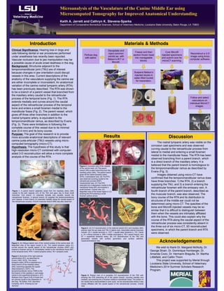
Figure 4. Corrosion cast of the vessels
of the canine head. (A) The parent
branch (PB) is seen branching from the
maxillary artery (MA). The parent branch
gives off the rostral tympanic artery
(RTA). Craniolateral view. (B) The RTA
was observed coursing caudal to the
retroarticular process (RAP).
Caudoventral view. (C) The RTA
extended into a depression medial to the
mandibular fossa (MF) and entered a
small foramen. Cranial view. (D)
Magnified view of the boxed region in (C)
showing the RTA entering a small
foramen (yellow arrow).
B
RAP
RTA
The rostral tympanic artery was visible on the
corrosion cast specimens and was observed
curving caudal to the retroarticular process from
lateral to medial and entering a small foramen
medial to the mandibular fossa. The RTA has been
observed branching from a parent branch, which
is a direct branch of the maxillary artery. It is
believed that the parent branch is homologous to
the temporomandibular ramus as described by
Evans (Fig. 3).
Images obtained using micro-CT have
confirmed that the temporomandibular ramus does
have three branches: 1) the RTA, 2) a branch
supplying the TMJ, and 3) a branch entering the
retroarticular foramen with the emissary vein. A
fourth branch of the parent branch, described as
the muscular branch, was also observed. The
bony course of the RTA and its distribution to
structures of the middle ear could not be
determined using micro-CT. The opacities of the
bone and Microfil injected vessels may be so
similar that it is difficult to distinguish between
them when the vessels are intimately affiliated
with the bone. This could also explain why the
course of the RTA along the caudal aspect of the
retroarticular process was consistently absent in
the three out of six micro-CT, 3D reconstructed
specimens, in which the parent branch and RTA
were observed.
Follow and select
vasculature on
individual MicroCT
images.
Figure 6. Rostral view of a complete 3-D reconstruction of the TMJ with
showing the RTA branching off of the parent branch, coursing rostrally and
medially, and entering into a foramen medial to mandibular fossa. The portion of
the vessel that is demonstrated in pink could not be reconstructed where it was
closely affiliated with the caudal aspect of the retroarticular process. Cranial
view.
PB
ST
CA
MA
EC
IA
CDT
MM
FA
BA
C
MF
A
B
C
TB
RAP
RAP
RTA
MF
A
RTA
MA
PB
MF
Figure 3. Branches of the right external
carotid artery (EC), as described by
Evans (1993, 2010). The rostral
tympanic artery (RTA) is depicted as a
direct branch of the maxillary artery
(MA), branching opposite the caudal
deep temporal a. (CDT). The
temporomandibular ramus (TMR) is the
first branch of the maxillary a. and arises
rostral to the superficial temporal a. (ST).
Redrawn from Figure 5-45 in Evans and
de Lahunta Guide to the Dissection of
the Dog, 7th Edition. W.B. Saunders
Company, 2010. Drawing by Lee
Aymond.
CA
ST
TMR
RTA
IA
CDT
MM
FA
EC
MA
MA