Hand out service plan มหาราช นำเสนอ สสจ. 29 เมษายน 2559
Kdigoaki 120826211850-phpapp02
1. http://www.kidney-international.org chapter 2.1
& 2012 KDIGO
Section 2: AKI Definition
Kidney International Supplements (2012) 2, 19–36; doi:10.1038/kisup.2011.32
Chapter 2.1: Definition and classification of AKI
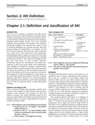
INTRODUCTION Table 2 | Staging of AKI
AKI is one of a number of conditions that affect kidney Stage Serum creatinine Urine output
structure and function. AKI is defined by an abrupt decrease
1 1.5–1.9 times baseline o0.5 ml/kg/h for
in kidney function that includes, but is not limited to, ARF. It
OR 6–12 hours
is a broad clinical syndrome encompassing various etiologies, X0.3 mg/dl (X26.5 mmol/l) increase
including specific kidney diseases (e.g., acute interstitial
2 2.0–2.9 times baseline o0.5 ml/kg/h for
nephritis, acute glomerular and vasculitic renal diseases); X12 hours
non-specific conditions (e.g, ischemia, toxic injury); as well
3 3.0 times baseline o0.3 ml/kg/h for
as extrarenal pathology (e.g., prerenal azotemia, and acute OR X24 hours
postrenal obstructive nephropathy)—see Chapters 2.2 and Increase in serum creatinine to OR
2.3 for further discussion. More than one of these conditions X4.0 mg/dl (X353.6 mmol/l) Anuria for X12 hours
OR
may coexist in the same patient and, more importantly, Initiation of renal replacement therapy
epidemiological evidence supports the notion that even mild, OR, In patients o18 years, decrease in
reversible AKI has important clinical consequences, including eGFR to o35 ml/min per 1.73 m2
increased risk of death.2,5 Thus, AKI can be thought of more
like acute lung injury or acute coronary syndrome.
Furthermore, because the manifestations and clinical con- 2.1.2: AKI is staged for severity according to the following
sequences of AKI can be quite similar (even indistinguish- criteria (Table 2). (Not Graded)
able) regardless of whether the etiology is predominantly 2.1.3: The cause of AKI should be determined whenever
within the kidney or predominantly from outside stresses on possible. (Not Graded)
the kidney, the syndrome of AKI encompasses both direct
injury to the kidney as well as acute impairment of function. RATIONALE
Since treatments of AKI are dependent to a large degree on Conditions affecting kidney structure and function can be
the underlying etiology, this guideline will focus on specific considered acute or chronic, depending on their duration.
diagnostic approaches. However, since general therapeutic AKI is one of a number of acute kidney diseases and
and monitoring recommendations can be made regarding all disorders (AKD), and can occur with or without other acute
forms of AKI, our approach will be to begin with general or chronic kidney diseases and disorders (Figure 2). Whereas
measures. CKD has a well-established conceptual model and definition
that has been useful in clinical medicine, research, and public
Definition and staging of AKI health,42–44 the definition for AKI is evolving, and the
AKI is common, harmful, and potentially treatable. Even concept of AKD is relatively new. An operational definition
a minor acute reduction in kidney function has an adverse of AKD for use in the diagnostic approach to alterations
prognosis. Early detection and treatment of AKI may in kidney function and structure is included in Chapter 2.5,
improve outcomes. Two similar definitions based on SCr with further description in Appendix B.
and urine output (RIFLE and AKIN) have been proposed and The conceptual model of AKI (Figure 3) is analogous to
validated. There is a need for a single definition for practice, the conceptual model of CKD, and is also applicable to
research, and public health. AKD.42,45 Circles on the horizontal axis depict stages in the
development (left to right) and recovery (right to left) of
2.1.1: AKI is defined as any of the following (Not Graded): AKI. AKI (in red) is defined as reduction in kidney function,
K Increase in SCr by X0.3 mg/dl (X26.5 lmol/l) including decreased GFR and kidney failure. The criteria for
within 48 hours; or the diagnosis of AKI and the stage of severity of AKI are
K Increase in SCr to X1.5 times baseline, which based on changes in SCr and urine output as depicted in the
is known or presumed to have occurred within triangle above the circles. Kidney failure is a stage of AKI
the prior 7 days; or highlighted here because of its clinical importance. Kidney
K Urine volume o0.5 ml/kg/h for 6 hours. failure is defined as a GFR o15 ml/min per 1.73 m2 body
Kidney International Supplements (2012) 2, 19–36 19
2. chapter 2.1
surface area, or requirement for RRT, although it is Existing evidence supports the validity of both RIFLE and
recognized that RRT may be required earlier in the evolution AKIN criteria to identify groups of hospitalized patients with
of AKI. Further description is included in Chapter 2.5 and increased risk of death and/or need for RRT.2,5,25,28–30
Appendix A. Epidemiological studies, many multicentered, collectively
It is widely accepted that GFR is the most useful overall enrolling more than 500 000 subjects have been used to
index of kidney function in health and disease, and changes establish RIFLE and/or AKIN criteria as valid methods to
in SCr and urine output are surrogates for changes in GFR. In diagnose and stage AKI. Recently, Joannidis et al.29 directly
clinical practice, an abrupt decline in GFR is assessed from an compared RIFLE criteria with and without the AKIN
increase in SCr or oliguria. Recognizing the limitations of the modification. While AKI classified by either criteria were
use of a decrease in kidney function for the early detection associated with a similarly increased hospital mortality, the
and accurate estimation of renal injury (see below), there is a two criteria identified somewhat different patients. The
broad consensus that, while more sensitive and specific original RIFLE criteria failed to detect 9% of cases that were
biomarkers are needed, changes in SCr and/or urine output detected by AKIN criteria. However, the AKIN criteria missed
form the basis of all diagnostic criteria for AKI. The first 26.9% of cases detected by RIFLE. Examination of the cases
international interdisciplinary consensus criteria for diag- missed by either criteria (Table 4) shows that cases identified
nosis of AKI were the RIFLE criteria32 proposed by the by AKIN but missed by RIFLE were almost exclusively Stage 1
ADQI. Modifications to these criteria have been proposed in (90.7%), while cases missed by AKIN but identified by RIFLE
order to better account for pediatric populations (pRIFLE)32 included 30% with RIFLE-I and 18% RIFLE-F; furthermore,
and for small changes in SCr not captured by RIFLE (AKIN these cases had hospital mortality similar to cases identified
criteria).23 Recommendations 2.1.1 and 2.1.2 represent the by both criteria (37% for I and 41% for F). However, cases
combination of RIFLE and AKIN criteria (Table 3). missed by RIFLE but identified as Stage 1 by AKIN also had
hospital mortality rates nearly twice that of patients who had
no evidence of AKI by either criteria (25% vs. 13%). These
data provide strong rationale for use of both RIFLE and
AKIN criteria to identify patients with AKI.
AKD AKI CKD Staging of AKI (Recommendation 2.1.2) is appropriate
because, with increased stage of AKI, the risk for death and
need for RRT increases.2,5,25,28–31 Furthermore, there is now
Figure 2 | Overview of AKI, CKD, and AKD. Overlapping ovals accumulating evidence of long-term risk of subsequent
show the relationships among AKI, AKD, and CKD. AKI is a subset development of cardiovascular disease or CKD and mortality,
of AKD. Both AKI and AKD without AKI can be superimposed even after apparent resolution of AKI.47–49
upon CKD. Individuals without AKI, AKD, or CKD have no known
kidney disease (NKD), not shown here. AKD, acute kidney diseases
For staging purposes, patients should be staged accord-
and disorders; AKI, acute kidney injury; CKD, chronic kidney ing to the criteria that give them the highest stage. Thus
disease. when creatinine and urine output map to different stages,
Stages defined by Complications
creatinine and
urine output
are surrogates
GFR
Increased Kidney
Normal Damage ↓ GFR Death
risk failure
Damage
Antecedents Markers such
Intermediate Stage as NGAL, KIM-1,
AKI and IL-18 are
Outcomes surrogates
Figure 3 | Conceptual model for AKI. Red circles represent stages of AKI. Yellow circles represent potential antecedents of AKI, and the
pink circle represents an intermediate stage (not yet defined). Thick arrows between circles represent risk factors associated with the
initiation and progression of disease that can be affected or detected by interventions. Purple circles represent outcomes of AKI.
‘‘Complications’’ refers to all complications of AKI, including efforts at prevention and treatment, and complications in other organ systems.
AKI, acute kidney injury; GFR, glomerular filtration rate. Adapted from Murray PT, Devarajan P, Levey AS, et al. A framework and key research
questions in AKI diagnosis and staging in different environments. Clin J Am Soc Nephrol 2008; 3: 864–868 with permission from American
Society of Nephrology45 conveyed through Copyright Clearance Center, Inc.; accessed http://cjasn.asnjournals.org/content/3/3/864.full
20 Kidney International Supplements (2012) 2, 19–36
3. chapter 2.1
Table 3 | Comparison of RIFLE and AKIN criteria for diagnosis and classification of AKI
AKI staging Urine output RIFLE
Serum creatinine (common to both) Class Serum creatinine or GFR
Stage 1 Increase of more than or equal to 0.3 mg/dl Less than 0.5 ml/kg/h for Risk Increase in serum creatinine  1.5 or GFR
(X26.5 mmol/l) or increase to more than or equal to more than 6 hours decrease 425%
150% to 200% (1.5- to 2-fold) from baseline
Stage 2 Increased to more than 200% to 300% Less than 0.5 ml/kg per hour Injury Serum creatinine  2 or GFR decreased
(42- to 3-fold) from baseline for more than 12 hours 450%
Stage 3 Increased to more than 300% (43-fold) Less than 0.3 ml/kg/h for Failure Serum creatinine  3, or serum creatinine
from baseline, or more than or equal to 4.0 mg/dl 24 hours or anuria for 44 mg/dl (4354 mmol/l) with an acute
(X354 mmol/l) with an acute increase of at least 12 hours rise 40.5 mg/dl (444 mmol/l) or GFR
0.5 mg/dl (44 mmol/l) or on RRT decreased 475%
Loss Persistent acute renal failure=complete
loss of kidney function 44 weeks
End-stage kidney ESRD 43 months
disease
Note: For conversion of creatinine expressed in SI units to mg/dl, divide by 88.4. For both AKIN stage and RIFLE criteria, only one criterion (creatinine rise or urine output
decline) needs to be fulfilled. Class is based on the worst of either GFR or urine output criteria. GFR decrease is calculated from the increase in serum creatinine above
baseline. For AKIN, the increase in creatinine must occur in o48 hours. For RIFLE, AKI should be both abrupt (within 1–7 days) and sustained (more than 24 hours). When
baseline creatinine is elevated, an abrupt rise of at least 0.5 mg/dl (44 mmol/l) to 44 mg/dl (4354 mmol/l) is sufficient for RIFLE class Failure (modified from Mehta et al.23 and
the report of the Acute Dialysis Quality Initiative consortium22).
AKI, acute kidney injury; AKIN, Acute Kidney Injury Network; ESRD, end-stage renal disease; GFR, glomerular filtration rate; RIFLE, risk, injury, failure, loss, and end stage; RRT,
renal replacement therapy. Reprinted from Endre ZH. Acute kidney injury: definitions and new paradigms. Adv Chronic Kidney Dis 2008; 15: 213–221 with permission from
National Kidney Foundation46; accessed http://www.ackdjournal.org/article/S1548-5595(08)00049-9/fulltext
Table 4 | Cross-tabulation of patients classified by RIFLE vs. AKIN
RIFLE
AKIN Non-AKI Risk Injury Failure Total (AKIN)
Non-AKI n* 8759 (12.9%) 781 (27.7%) 452 (37.4%) 271 (41.3%) 10 263 (15.9%)
Stage1 n* 457 (25.2%) 282 (33.0%) 243 (44.0%) 95 (60.0%) 1077 (34.5%)
Stage 2 n* 36 (30.6%) 21 (47.6%) 885 (25.9%) 91 (54.9) 1033 (29.0%)
Stage 3 n* 11 (18.2%) 8 (12.5%) 16 (62.5%) 1948 (41.3) 1983 (41.2%)
Total (RIFLE) n* 9263 (13.6%) 1092 (29.2%) 1596 (32.3%) 2405 (42.6%) 14 356 (21.7%)
*Number of patients classified into the respective stages of AKI by AKIN or RIFLE are cross-tabulated against each other. Hospital mortality of each group is given in
parentheses. Shaded fields denote patients assigned to the same degree of AKI by both classification systems.
AKI, acute kidney injury; AKIN, Acute Kidney Injury Network; RIFLE, risk, injury, failure, loss, and end stage. With kind permission from Springer Science+Business Media:
Intensive Care Med. Acute kidney injury in critically ill patients classified by AKIN versus RIFLE using the SAPS 3 database. 35 (2009): 1692–1702. Joannidis M, Metnitz B,
Bauer P et al.29; accessed http://www.springerlink.com/content/r177337030550120/
the patient is staged according to the highest (worst) stage. the definition of AKI in Recommendation 2.1.1). This is
The changes in GFR that were published with the original problematic for smaller pediatric patients, including infants
RIFLE criteria do not correspond precisely to changes in SCr. and children with low muscle mass who may not be able to
As SCr is measured and GFR can only be estimated, achieve a SCr of 4.0 mg/dl (354 mmol/l). Thus, the pediatric-
creatinine criteria should be used along with urine output modified RIFLE AKI criteria32 were developed using a change
for the diagnosis (and staging) of AKI. One additional change in estimated creatinine clearance (eCrCl) based on the
in the criteria was made for the sake of clarity and simplicity. Schwartz formula. In pRIFLE, patients automatically reach
For patients reaching Stage 3 by SCr 44.0 mg/dl Stage 3 if they develop an eCrCl o35 ml/min per 1.73 m2.
(4354 mmol/l), rather than require an acute increase of However, with this automatic pRIFLE threshold, the SCr
X0.5 mg/dl (X44 mmol/l) over an unspecified time period, we change based AKI definition (recommendation 2.1.1) is
instead require that the patient first achieve the creatinine- applicable to pediatric patients, including an increase of
based change specified in the definition (either X0.3 mg/dl 0.3 mg/dl (26.5 mmol/l) SCr.32
[X26.5 mmol/l] within a 48-hour time window or an increase There are important limitations to these recommenda-
of X1.5 times baseline). This change brings the definition and tions, including imprecise determination of risk (see Chapter
staging criteria to greater parity and simplifies the criteria. 2.2) and incomplete epidemiology of AKI, especially outside
Recommendation 2.1.2 is based on the RIFLE and AKIN the ICU. Clinical judgment is required in order to determine
criteria that were developed for average-sized adults. The if patients seeming to meet criteria do, in fact, have disease, as
creatinine change–based definitions include an auto- well as to determine if patients are likely to have AKI even if
matic Stage 3 classification for patients who develop SCr incomplete clinical data are available to apply the diagnostic
44.0 mg/dl (4354 mmol/l) (provided that they first satisfy criteria. The application of the diagnostic and staging criteria
Kidney International Supplements (2012) 2, 19–36 21
4. chapter 2.1
Table 5 | Causes of AKI and diagnostic tests some patients with specific kidney diseases (e.g., glome-
Selected causes of AKI requiring rulonephritis) for which a specific treatment is available. As
immediate diagnosis and specific such, it is always necessary to search for the underlying cause
therapies Recommended diagnostic tests of AKI (see Chapter 2.3).
Decreased kidney perfusion Volume status and urinary
diagnostic indices Research Recommendations
Acute glomerulonephritis, vasculitis, Urine sediment examination, K The role of biomarkers other than SCr in the early
interstitial nephritis, thrombotic serologic testing and
microangiopathy hematologic testing
diagnosis, differential diagnosis, and prognosis of AKI
Urinary tract obstruction Kidney ultrasound patients should be explored. Some important areas in
AKI, acute kidney injury. which to focus include:
J Early detection where the gold standard is AKI by
clinical diagnosis after the fact and the biomarker is
compared to existing markers (SCr and urine
is discussed in greater detail, along with specific examples in output) at the time of presentation.
Chapter 2.4. J Prognosis where a biomarker is used to predict risk
The use of urine output criteria for diagnosis and staging for AKI or risk for progression of AKI.
has been less well validated and in individual patients J Prognosis where a biomarker is used to predict
the need for clinical judgment regarding the effects of drugs recovery after AKI vs. death or need for long-term RRT.
(e.g., angiotensin-converting enzyme inhibitors [ACE-I]), K The influence of urinary output criteria on AKI staging
fluid balance, and other factors must be included. For very needs to be further investigated. Influence of fluid
obese patients, urine output criteria for AKI may include balance, percent volume overload, diuretic use, and
some patients with normal urine output. However, these differing weights (actual, ideal body weight, lean body
recommendations serve as the starting point for further mass) should be considered. Also, it is currently not
evaluation, possibly involving subspecialists, for a group of known how urine volume criteria should be applied (e.g.,
patients recognized to be at increased risk. average vs. persistent reduction for the period specified).
Finally, it is axiomatic that patients always be managed K The influence of SCr or eGFR criteria on AKI staging
according to the cause of their disease, and thus it is needs to be further investigated. The use of different
important to determine the cause of AKI whenever possible. relative and absolute SCr increments or eGFR decrements
In particular, patients with decreased kidney perfusion, acute at different time points and with differently ascertained
glomerulonephritis, vasculitis, interstitial nephritis, throm- baseline values requires further exploration and valida-
botic microangiopathy, and urinary tract obstruction require tion in various populations.
immediate diagnosis and specific therapeutic intervention, in
addition to the general recommendations for AKI in the SUPPLEMENTARY MATERIAL
remainder of this guideline (Table 5). Appendix A: Background.
Appendix B: Diagnostic Approach to Alterations in Kidney Function
It is recognized that it is frequently not possible to deter-
and Structure.
mine the cause, and often the exact cause does not dictate a Supplementary material is linked to the online version of the paper at
specific therapy. However, the syndrome of AKI includes http://www.kdigo.org/clinical_practice_guidelines/AKI.php
22 Kidney International Supplements (2012) 2, 19–36
5. http://www.kidney-international.org chapter 2.2
& 2012 KDIGO
Chapter 2.2: Risk assessment
The kidney is a fairly robust organ that can tolerate exposure to Table 6 | Causes of AKI: exposures and susceptibilities for
several insults without suffering significant structural or non-specific AKI
functional change. For this reason, any acute change in kidney
Exposures Susceptibilities
function often indicates severe systemic derangement and
predicts a poor prognosis. Risk for AKI is increased by exposure Sepsis Dehydration or volume depletion
Critical illness Advanced age
to factors that cause AKI or the presence of factors that increase Circulatory shock Female gender
susceptibility to AKI. Factors that determine susceptibility of the Burns Black race
kidneys to injury include dehydration, certain demographic Trauma CKD
characteristics and genetic predispositions, acute and chronic Cardiac surgery (especially Chronic diseases (heart, lung, liver)
with CPB)
comorbidities, and treatments. It is the interaction between Major noncardiac surgery Diabetes mellitus
susceptibility and the type and extent of exposure to insults that Nephrotoxic drugs Cancer
determines the risk of occurrence of AKI. Radiocontrast agents Anemia
Understanding individual ‘‘risk factors’’ may help in Poisonous plants and animals
preventing AKI. This is particularly gratifying in the hospital CKD, chronic kidney disease; CPB, cardiopulmonary bypass.
setting, where the patient’s susceptibility can be assessed
before certain exposures as surgery or administration of However, the chances of developing AKI after exposure to the
potentially nephrotoxic agents. Accordingly, some suscept- same insult differ among different individuals. This is
ibility factors may be modified, and contemplated exposures attributed to a number of susceptibility factors which vary
avoided or tailored to reduce the risk of AKI. widely from individual to individual. Our understanding of
Risk assessment in community-acquired AKI is different susceptibility factors (Table 6) is based on many observa-
from hospital-acquired AKI, for two main reasons: i) Available tional studies that address different settings with regards to
evidence on risk factors is largely derived from hospital data and the type, severity, duration, and multiplicity of insults. While
extrapolation to the community setting is questionable. ii) The this heterogeneity provides insight into some susceptibility
opportunity to intervene, prior to exposure, is quite limited. factors that are common across various populations, the
Most patients are seen only after having suffered an exposure generalizability of results from one particular setting to the
(trauma, infection, poisonous plant, or animal). However, there next is uncertain.
is still room to assess such patients, albeit after exposure, in The course and outcome of AKI are modified by other
order to identify those who are more likely to develop AKI, factors, but since these are manifested within the context of
thereby requiring closer monitoring and general supportive actual disease, they must be categorized as ‘‘prognostic’’
measures. It may also be helpful to identify such patients in rather than ‘‘risk’’ factors, hence being discussed separately in
order to avoid additional injury. A more complete discussion of Appendix D. Lastly, the fact that some 30% of patients who
the approach to identification and management of risk for AKI recover from AKI remain at increased risk of CKD,
is provided in Appendices C and D. cardiovascular disease, and death calls for the identification
of the risk factors that can identify such patients in the hopes
2.2.1: We recommend that patients be stratified for risk of AKI of providing them with timely preventive measures.50–52
according to their susceptibilities and exposures. (1B) Finally, it is important to screen patients who have
2.2.2: Manage patients according to their susceptibilities and undergone an exposure (e.g., sepsis, trauma) and to continue
exposures to reduce the risk of AKI (see relevant monitor high-risk patients until the risk has subsided. Exact
guideline sections). (Not Graded) intervals for checking SCr and in which individuals to
2.2.3: Test patients at increased risk for AKI with measure- monitor urine output remain matters of clinical judgment;
ments of SCr and urine output to detect AKI. (Not however, as a general rule, high risk in-patients should have
Graded) Individualize frequency and duration of SCr measured at least daily and more frequently after an
monitoring based on patient risk and clinical course. exposure, and critically ill patients should have urine output
(Not Graded) monitoring. This will necessitate urinary bladder catheteriza-
tion in many cases, and the risks of infection should also be
considered in the monitoring plan.
RATIONALE A recent clinical practice assessment in the UK concluded
There are many types of exposures that may cause AKI that only 50% of patients with AKI were considered to have
(Table 6) and these are discussed in detail in Appendix C. received a ‘‘good’’ overall standard of care. This figure fell to
Kidney International Supplements (2012) 2, 19–36 23
6. chapter 2.2
just over 30% if AKI developed during a hospital admission K Studies are needed to develop and validate scoring systems
rather than being diagnosed before admission.53 The authors for AKI risk prediction in various settings, in addition to
also felt that there was an unacceptable delay in recognizing cardiac surgery and exposure to radiocontrast material.
AKI in 43% of those that developed the condition after K Genome-wide association studies are needed to deter-
admission, and that in a fifth of such patients its develop- mine risk of AKI in different hospital settings and with
ment was predictable and avoidable. Their recommendations respect to long-term outcomes.
were simple: risk assessment for AKI as part of the initial K Studies are needed on risk factors for the development of,
evaluation of emergency admissions, along with appropriate recovery from, and long-term outcomes of community-
serum biochemistry on admission and at frequent intervals acquired AKI, including sepsis, trauma, tropical infec-
thereafter.53 tions, snake bites, and ingestion of toxic plants, etc.
RESEARCH RECOMMENDATIONS SUPPLEMENTARY MATERIAL
K Better delineation of risk for hospital- and community- Appendix C: Risk Determination.
Appendix D: Evaluation and General Management Guidelines for
acquired AKI is needed.
Patients with AKI.
K Better delineation of the effects of age on the risk for AKI Supplementary material is linked to the online version of the paper at
is needed. http://www.kdigo.org/clinical_practice_guidelines/AKI.php
24 Kidney International Supplements (2012) 2, 19–36
7. http://www.kidney-international.org chapter 2.3
& 2012 KDIGO
Chapter 2.3: Evaluation and general management of
patients with and at risk for AKI
Given that AKI is associated with significant morbidity and with AKI include both reducing kidney injury and complica-
mortality, and because no specific treatment is available to tions related to decreased kidney function.
reverse AKI, early recognition and management is para-
mount. Indeed, recognition of patients at risk for AKI, or 2.3.1: Evaluate patients with AKI promptly to determine
with possible AKI but prior to clinical manifestations, is the cause, with special attention to reversible
likely to result in better outcomes than treating only causes. (Not Graded)
established AKI. Chapter 2.2 introduced the approach to 2.3.2: Monitor patients with AKI with measurements of
risk assessment with further detail provided in Appendix C. SCr and urine output to stage the severity,
This chapter will concern itself with the evaluation according to Recommendation 2.1.2. (Not Graded)
and general management of patients with, or even at risk 2.3.3: Manage patients with AKI according to the stage
for, AKI. Further detail is provided in Appendix D. We (see Figure 4) and cause. (Not Graded)
highlight the importance of beginning management at the 2.3.4: Evaluate patients 3 months after AKI for resolu-
earliest point in the development of AKI—in patients with tion, new onset, or worsening of pre-existing CKD.
suspected AKI or even in those at increased risk who have (Not Graded)
been exposed to the various factors discussed in Chapters 2.2 K If patients have CKD, manage these patients as
and Appendix C. detailed in the KDOQI CKD Guideline (Guide-
Although much of the remaining chapters in this guide- lines 7–15). (Not Graded)
line pertain to management of specific aspects of AKI, there K If patients do not have CKD, consider them to be
are general management principles that are common to all at increased risk for CKD and care for them as
patients and these will be discussed here and further detailed in the KDOQI CKD Guideline 3 for
expounded upon in Appendix D. Treatment goals in patients patients at increased risk for CKD. (Not Graded)
Figure 4 | Stage-based management of AKI. Shading of boxes indicates priority of action—solid shading indicates actions that are
equally appropriate at all stages whereas graded shading indicates increasing priority as intensity increases. AKI, acute kidney injury;
ICU, intensive-care unit.
Kidney International Supplements (2012) 2, 19–36 25
8. chapter 2.3
RATIONALE The clinical evaluation of AKI includes a careful history
As emphasized in Chapter 2.2, AKI is not a disease but and physical examination. Drug history should include over-
rather a clinical syndrome with multiple etiologies. While the-counter formulations and herbal remedies or recreational
much of the literature examining epidemiology and clinical drugs. The social history should include exposure to tropical
consequences of AKI appear to treat this syndrome as a diseases (e.g., malaria), waterways or sewage systems, and
homogeneous disorder, the reality is that AKI is hetero- exposure to rodents (e.g., leptospirosis, hantavirus). Physical
geneous and often is the result of multiple insults. Figure 5 examination should include evaluation of fluid status, signs
illustrates an approach to evaluation of AKI. Further for acute and chronic heart failure, infection, and sepsis.
discussion of evaluation in clinical practice is provided in Measurement of cardiac output, preload, preload respon-
Appendix D. siveness, and intra-abdominal pressure should be considered
Figure 5 | Evaluation of AKI according to the stage and cause.
26 Kidney International Supplements (2012) 2, 19–36
9. chapter 2.3
in the appropriate clinical context. Laboratory parameters— variables like central venous pressure are not nearly as useful
including SCr, blood urea nitrogen (BUN), and electrolytes, as dynamic variables, such as pulse-pressure variation,
complete blood count and differential—should be obtained. inferior vena cava filling by ultrasound and echocardio-
Urine analysis and microscopic examination as well as graphic appearance of the heart (see also Appendix D).
urinary chemistries may be helpful in determining the Note that while the actions listed in Figure 4 provide
underlying cause of AKI. Imaging tests, especially ultrasound, an overall starting point for stage-based evaluation and
are important components of the evaluation for patients with management, they are neither complete not mandatory for
AKI. Finally, a number of biomarkers of functional change an individual patient. For example, the measurement of urine
and cellular damage are under evaluation for early diagnosis, output does not imply that the urinary bladder catheteriza-
risk assessment for, and prognosis of AKI (see Appendix D tion is mandatory for all patients, and clinicians should
for detailed discussion). balance the risks of any procedures with the benefits.
Individualize frequency and duration of monitoring based Furthermore, clinicians must individualize care decisions
on patient risk, exposure and clinical course. Stage is a predictor based on the totality of the clinical situation. However, it is
of the risk for mortality and decreased kidney function (see advisable to include AKI stage in these decisions.
Chapter 2.4). Dependent on the stage, the intensity of future The evaluation and management of patients with AKI
preventive measures and therapy should be performed. requires attention to cause and stage of AKI, as well as factors
Because the stage of AKI has clearly been shown to that relate to further injury to the kidney, or complications
correlate with short-term2,5,27,29 and even longer-term out- from decreased kidney function. Since AKI is a risk factor for
comes,31 it is advisable to tailor management to AKI stage. CKD, it is important to evaluate patients with AKI for new
Figure 4 lists a set of actions that should be considered for onset or worsening of pre-existing CKD. If patients have
patients with AKI. Note that for patients at increased risk (see CKD, manage patients as detailed in the KDOQI CKD
Chapters 2.2 and 2.4), these actions actually begin even Guideline (Guidelines 7–15). If patients do not have CKD,
before AKI is diagnosed. consider them to be at increased risk for CKD and care for
Note that management and diagnostic steps are both them as detailed in the KDOQI CKD Guideline 3 for patients
included in Figure 4. This is because response to therapy is an at increased risk for CKD.
important part of the diagnostic approach. There are few
specific tests to establish the etiology of AKI. However, a RESEARCH RECOMMENDATIONS
patient’s response to treatment (e.g., discontinuation of a K Clinical research aimed at testing early management
possible nephrotoxic agent) provides important information strategies is urgently needed. Such trials should also
as to the diagnosis. address the risks and benefits of commonly used fluid-
Nephrotoxic drugs account for some part of AKI in 20–30% management strategies, including intravenous (i.v.) fluids
of patients. Often, agents like antimicrobials (e.g., aminoglyco- and diuretics.
sides, amphotericin) and radiocontrast are used in patients that K Methods to better assess fluid status in critically ill and
are already at high risk for AKI (e.g., critically ill patients with other hospitalized patients at risk for AKI are needed.
sepsis). Thus, it is often difficult to discern exactly what K Research is needed, with follow-up beyond hospital stay,
contribution these agents have on the overall course of AKI. to better understand the clinical consequences of AKI in
Nevertheless, it seems prudent to limit exposure to these agents patients with and without underlying CKD.
whenever possible and to weigh the risk of developing or
worsening AKI against the risk associated with not using the SUPPLEMENTARY MATERIAL
agent. For example, when alternative therapies or diagnostic Appendix C: Risk Determination.
Appendix D: Evaluation and General Management Guidelines for
approaches are available they should be considered.
Patients with AKI.
In order to ensure adequate circulating blood volume, it is Supplementary material is linked to the online version of the paper at
sometimes necessary to obtain hemodynamic variables. Static http://www.kdigo.org/clinical_practice_guidelines/AKI.php
Kidney International Supplements (2012) 2, 19–36 27
10. http://www.kidney-international.org chapter 2.4
& 2012 KDIGO
Chapter 2.4: Clinical applications
This chapter provides a detailed application of the AKI diagnose patients as rapidly as possible. For example, case A
definition and staging for clinical diagnosis and management. can be diagnosed with AKI on day 2 by the first criterion,
The definitions and classification system discussed in whereas the second criterion is not satisfied until day 3
Chapter 2.1 can be used easily in many patients and requires (increase from 1.3 to 1.9). However, this is only true because
little clinical interpretation. However, in real time, clinicians the episode of AKI began prior to medical attention, and thus
do not always have a complete dataset to work with the day 1 SCr level was already increased. If creatinine
and individual patients present with unique histories. As measurements had available with 48 hours prior to day 1 and
discussed in the previous chapter, it is difficult to distinguish if this level had been at baseline (1.0 mg/dl [88.4 mmol/l]), it
AKI from CKD in many cases. In addition, as many as would have been possible to diagnose AKI on day 1 using the
two-thirds of all cases of AKI begin prior to hospitali- second criterion.
zation (community-acquired AKI). Therefore, clinicians Cases F-H do not have a baseline measurement of SCr
may be faced with patients in whom kidney function available. Elevated SCr (reduced eGFR) on day 1 of the
is already decreased and, during the hospitalization, hospitalization is consistent with either CKD or AKD
improves rather than worsens. Finally, many patients without AKI. In Case F, baseline SCr can be inferred
do not have a prior measurement of kidney function to be below the day 1 value because of the subsequent
available for comparison. This chapter provides detailed clinical course; thus, we can infer the patient has had an
examples of the application of these definitions to the clinical episode of AKI. In case G, AKI can be diagnosed by
setting. application of criterion 2, but the patient may have under-
lying CKD. Case H does not fulfill the definition for
Examples of application of AKI definitions AKI based on either criteria, and has either CKD or AKD
Table 7 illustrates a number of examples whereby patients without AKI.
presenting with possible AKI can be diagnosed. Cases A-F The example of Case A raises several important issues.
have a measurement of baseline SCr. To simplify decision- First, frequent monitoring of SCr in patients at increased risk
making, baseline estimated glomerular filtration rate (eGFR) of AKI will significantly improve diagnostic time and
exceeds 60 ml/min per 1.73 m2 in these patients, so none has accuracy. If Case A had not presented to medical attention
pre-existing CKD. Cases A-F can all be diagnosed with AKI (or if SCr had not been checked) until day 7, the case of AKI
by applying the first two criteria in Recommendation 2.1.1. (a would likely have been missed. Frequent measurement of SCr
documented increase of at least 0.3 mg/dl (426.5 mmol/l) in high-risk patients, or in patients in which AKI is suspected,
[within 48 hours or a 50% increase from presumed baseline). is therefore encouraged—see Chapter 2.3. The second issue
Note that a patient can be diagnosed with AKI by fulfilling highlighted by Case A is the importance of baseline SCr
either criterion 1 or 2 (or 3, urine output) and thus cases measurements. Had no baseline been available it would still
B,C,D, and F all fulfill the definition of AKI. Note also that have been possible to diagnose AKI on day 3 (by either using
patients may be diagnosed earlier using criterion 1 or 2. Early criterion 2 or by using criterion 1 and accepting the baseline
diagnosis may improve outcome so it is advantageous to SCr as 1.3); however, not only would this have resulted in a
Table 7 | AKI diagnosis
Serum creatinine mg/dl (lmol/l) Diagnosis AKI?
Criterion 1 Criterion 2
Case Baseline Day 1 Day 2 Day 3 Day 7 50% from baseline X0.3 mg/dl (X26.5 lmol/l) rise in p48 hours
A 1.0 (88) 1.3 (115) 1.5 (133) 2.0 (177) 1.0 (88) Yes Yes
B 1.0 (88) 1.1 (97) 1.2 (106) 1.4 (124) 1.0 (88) No Yes
C 0.4 (35) 0.5 (44) 0.6 (53) 0.7 (62) 0.4 (35) Yes No
D 1.0 (88) 1.1 (97) 1.2 (106) 1.3 (115) 1.5 (133) Yes No
E 1.0 (88) 1.3 (115) 1.5 (133) 1.8 (159) 2.2 (195) Yes Yes
F ? 3.0 (265) 2.6 (230) 2.2 (195) 1.0 (88) Yes No
G ? 1.8 (159) 2.0 (177) 2.2 (195) 1.6 (141) ? Yes
H ? 3.0 (265) 3.1 (274) 3.0 (265) 2.9 (256) ? No
28 Kidney International Supplements (2012) 2, 19–36
11. chapter 2.4
Table 8 | Overview of the approaches to determine baseline SCr in the application of RIFLE classification in previous studies
No. of pts Multi-/ Criteria % %
Study analyzed single-center used Method to determine baseline SCr recorded estimated
Bagshaw25 120123 multi cr+uo estimated by MDRD formula 0 100
Ostermann30 41972 multi cr estimated by MDRD formula 0 100
Uchino5 20126 single cr retrieved from hospital database, or estimated by MDRD formula N/A N/A
Bell54 8152 single cr+uo retrieved from hospital database, or estimated by MDRD formula N/A N/A
Hoste2 5383 single cr+uo estimated by MDRD formula, or admission creatinine value, N/A N/A
whatever was lower
Ali31 5321 multi cr retrieved from hospital database, or admission creatinine value 100 0
Cruz55 2164 multi cr+uo retrieved from hospital database, or estimated by MDRD formula 78 22
Perez-Valdivieso56 1008 single cr estimated by MDRD formula 0 100
Kuitunen57 813 single cr+uo preoperative value 100 0
Coca58 304 single cr the lowest s-creatinine value in the first 5 hospital days 100 0
Arnaoutakis59 267 single N/A N/A N/A N/A
Abosaif60 247 single cr+uo retrieved from hospital database, or admission creatinine value 100 0
Maccariello61 214 multi cr+uo retrieved from hospital database, or estimated by MDRD formula N/A N/A
Jenq62 134 single cr+uo admission creatinine value, or estimated by MDRD formula 90 10
cr, creatinine criteria; MDRD, Modification of Diet in Renal Disease; N/A, not available; pts, patients; SCr, serum creatinine; uo, urine output criteria.
Reprinted from Zavada J, Hoste E, Cartin-Ceba R et al. A comparison of three methods to estimate baseline creatinine for RIFLE classification. Nephrol Dial Transplant 2010;
25(12): 3911–3918 (Ref. 64) by permission from The European Renal Association-European Dialysis and Transplant Association; accessed http://ndt.oxfordjournals.org/content/
25/12/3911.long
Table 9 | Estimated baseline SCr
Age (years) Black males mg/dl (lmol/l) Other males mg/dl (lmol/l) Black females mg/dl (lmol/l) Other females mg/dl (lmol/l)
20–24 1.5 (133) 1.3 (115) 1.2 (106) 1.0 (88)
25–29 1.5 (133) 1.2 (106) 1.1 (97) 1.0 (88)
30–39 1.4 (124) 1.2 (106) 1.1 (97) 0.9 (80)
40–54 1.3 (115) 1.1 (97) 1.0 (88) 0.9 (80)
55–65 1.3 (115) 1.1 (97) 1.0 (88) 0.8 (71)
465 1.2 (106) 1.0 (88) 0.9 (80) 0.8 (71)
Estimated glomerular filtration rate=75 (ml/min per 1.73 m2)=186 Â (serum creatinine [SCr]) À 1.154 Â (age) À 0.203 Â (0.742 if female) Â (1.210 if black)=exp(5.228 À 1.154 Â
In [SCr]) À 0.203 Â In(age) À (0.299 if female) + (0.192 if black).
Reprinted from Bellomo R, Ronco C, Kellum JA et al. Acute renal failure - definition, outcome measures, animal models, fluid therapy and information technology needs: the
Second International Consensus Conference of the Acute Dialysis Quality Initiative (ADQI) Group. Crit Care 2004; 8: R204-212 with permission from Bellomo R et al.22; accessed
http://ccforum.com/content/8/4/R204
delay in diagnosis, it would have resulted in a delay in staging Estimating baseline SCr
(see Table 7). On day 7, it can be inferred that the patient’s Many patients will present with AKI without a reliable
baseline was no higher than 1.0 mg/dl (88 mmol/l) and thus baseline SCr on record. Baseline SCr can be estimated using
correct staging of Case A as Stage 2 (two-fold increase from the Modification of Diet in Renal Disease (MDRD) Study
the reference SCr, see below and Table 7) on day 3 could have equation assuming that baseline eGFR is 75 ml/min per 1.73
been determined in retrospect. However, if a baseline SCr was m2 (Table 9).22 This approach has been used in many, but not
available to use as the reference, the correct stage could be all, studies of AKI epidemiology using RIFLE2,5,25,30–32,54–63
determined on day 3. (see Table 8) and has recently been validated.64 Hence, most
Case B illustrates why criterion 2 can detect cases of AKI current data concerning AKI defined by RIFLE criteria are
missed by criterion 1. It also clarifies why these cases are based on estimated baseline SCr for a large proportion of
unusual. Had the SCr increased to 1.5 mg/dl (132.6 mmol/l) patients.
as opposed to peaking at 1.4 mg/dl (123.8 mmol/l), it would Table 9 shows the range of estimated SCr obtained by
have been picked up by criterion 1 as well. By contrast back-calculation for various age, sex, and race categories.
Cases C, D, and even F illustrate how criterion 2 may When the baseline SCr is unknown, an estimated SCr can be
miss cases identified by criterion 1. Note that Case F can used provided there is no evidence of CKD (see Appendix B).
only be diagnosed by inference. By day 7, it can be Fortunately, when there is a history of CKD, a baseline SCr is
inferred that the baseline was no higher than 1.0 mg/dl usually available. Unfortunately, many cases of CKD are not
(88 mmol/l) and thus it can be determined that the patient identified, and thus estimating the baseline SCr may risk
presented with AKI. However, if the baseline SCr could labeling a patient with AKI when in reality the diagnosis was
be estimated it would be possible to make this inference as unidentified CKD. As discussed further in Appendix B, it is
early as day 1. essential to evaluate a patient with presumed AKI for
Kidney International Supplements (2012) 2, 19–36 29
12. chapter 2.4
Table 10 | AKI staging
Serum creatinine mg/dl (lmol/l)
Case Baseline Day 1 Day 2 Day 3 Day 7 Reference creatinine Max AKI stage
A 1.0 (88) 1.3 (115) 1.5 (133) 2.0 (177) 1.0 (88) 1.0 (88) 2
B 1.0 (88) 1.1 (97) 1.2 (106) 1.4 (124) 1.0 (88) 1.0 (88) 1
C 0.4 (35) 0.5 (44) 0.6 (53) 0.7 (62) 0.4 (35) 0.4 (35) 1
D 1.0 (88) 1.1 (97) 1.2 (106) 1.3 (115) 1.5 (133) 1.0 (88) 1
E 1.0 (88) 1.3 (115) 1.5 (133) 1.8 (159) 2.2 (195) 1.0 (88) 2
F ? 3.0 (265) 2.6 (230) 2.2 (195) 1.0 (88) 1.0 (88) 3
G ? 1.8 (159) 2.0 (177) 2.2 (195) 1.6 (141) ? X1
H ? 3.0 (265) 3.1 (274) 3.0 (265) 2.9 (256) ? ?
AKI, acute kidney injury.
presence of CKD. Furthermore, CKD and AKI may coexist. 1.2 mg/dl (106 mmol/l) (Table 9) and his initial stage on
By using all available clinical data (laboratory, imaging, admission (day 1) would be assumed to be 2. However, once
history, and physical exam) it should be possible to arrive at his SCr recovered to 1.0 mg/dl (88 mmol/l) on day 7, it would
both an accurate diagnosis as well as an accurate estimate of be possible to restage him as having had Stage 3. Once he has
baseline SCr. Importantly, excluding some cases of hemo- recovered, there may be no difference between Stage 2 or 3 in
dilution secondary to massive fluid resuscitation (discussed terms of his care plan. On the other hand, accurately staging
below), the lowest SCr obtained during a hospitalization the severity of AKI may be important for intensity of follow-
is usually equal to or greater than the baseline. This SCr up and future risk.
should be used to diagnose (and stage) AKI. For example, if Note that Cases G and H can only be staged if the
no baseline SCr was available in Case A, diagnosis of AKI reference SCr can be inferred. Case G may be as mild as stage
could be made using the MDRD estimated SCr (Table 9). If 1 if the baseline is equal to the nadir SCr on day 7. On the
Case A were a 70-year-old white female with no evidence or other hand, if this case were a 70-year-old white female with
history of CKD, the baseline SCr would be 0.8 mg/dl no known evidence or history of CKD, the reference SCr
(71 mmol/l) and a diagnosis of AKI would be possible would be 0.8 mg/dl (71 mmol/l) based on an estimated
even on day 1 (criterion 1, X50% increase from baseline). baseline (Table 9). In this case, the severity on day 1 would
However, if the patient was a 20-year-old black male, his already be stage 2.
baseline SCr would be estimated at 1.5 mg/dl (133 mmol/l).
Since his admission SCr is lower, this is assumed to be the Urine output vs. SCr
baseline SCr until day 7 when he returns to his true baseline, Both urine output and SCr are used as measures of an acute
and this value can be taken as the baseline. These dynamic change in GFR. The theoretical advantage of urine output
changes in interpretation are not seen in epidemiologic over SCr is the speed of the response. For example, if GFR
studies, which are conducted when all the data are present, were to suddenly fall to zero, a rise in SCr would not be
but are common in clinical medicine. Note that the only detectable for several hours. On the other hand, urine output
way to diagnose AKI (by SCr criteria) in Case H is to use an would be affected immediately. Less is known about the use
estimated SCr. of urine output for diagnosis and staging compared to SCr,
since administrative databases usually do not capture urine
Examples of application of AKI stages output (and frequently it is not even measured, especially
Once a diagnosis of AKI has been made, the next step is to outside the ICU). However, studies using both SCr and urine
stage it (Recommendation 2.1.2). Like diagnosis, staging output to diagnose AKI show increased incidence, suggesting
requires reference to a baseline SCr when SCr criteria are that the use of SCr alone may miss many patients. The use
used. This baseline becomes the reference SCr for staging of urine output criteria (criterion 3) will also reduce the
purposes. Table 10 shows the maximum stage for each number of cases where criterion 1 and criterion 2 are
Case described in Table 7. Staging for Case A was already discordant (cases B,C,D, and F in Table 7), as many of these
mentioned. The maximum stage is 2 because reference SCr is cases will be picked up by urine output criteria.
1.0 mg/dl (88 mmol/l) and the maximum SCr is 2.0 mg/dl
(177 mmol/l). Had the reference SCr been 0.6 mg/dl (53 mmol/ Timeframe for diagnosis and staging
l), the maximum stage would have been 3. Case F was staged The purpose of setting a timeframe for diagnosis of AKI is to
by using the lowest SCr (1.0 mg/dl [88 mmol/l]) as the clarify the meaning of the word ‘‘acute’’. A disease process
reference. Of course, the actual baseline for this case might that results in a change in SCr over many weeks is not AKI
have been lower but this would not affect the stage, since it is (though it may still be an important clinical entity: see
already Stage 3. Note that if this patient was a 35-year-old Appendix B). For the purpose of this guideline, AKI is
white male, his MDRD estimated baseline SCr would be defined in terms of a process that results in a 50% increase in
30 Kidney International Supplements (2012) 2, 19–36
13. chapter 2.4
SCr within 1 week or a 0.3 mg/dl (26.5 mmol/l) increase Clinical judgment
within 48 hours (Recommendation 2.1.1). Importantly, there While the definitions and classification system discussed in
is no stipulation as to when the 1-week or 48-hour time Chapter 2.1 provide a framework for the clinical diagnosis of
periods can occur. It is stated unequivocally that it does not AKI, they should not be interpreted to replace or to exclude
need to be the first week or 48 hours of a hospital or ICU stay. clinical judgment. While the vast majority of cases will
Neither does the time window refer to duration of the fit both AKI diagnostic criteria as well as clinical judgment,
inciting event. For example, a patient may have a 2-week AKI is still a clinical diagnosis—not all cases of AKI will fit
course of sepsis but only develop AKI in the second week. within the proposed definition and not all cases fitting the
Importantly, the 1-week or 48-hour timeframe is for definition should be diagnosed as AKI. However, exceptions
diagnosis of AKI, not staging. A patient can be staged over should be very rare.
the entire episode of AKI such that, if a patient develops a Pseudo-AKI. As with other clinical diagnoses defined by
50% increase in SCr in 5 days but ultimately has a three-fold laboratory results (e.g., hyponatremia), the clinician must be
increase over 3 weeks, he or she would be diagnosed with AKI cautious to interpret laboratory data in the clinical context.
and ultimately staged as Stage 3. The most obvious example is with laboratory errors or errors
As with any clinical criteria, the timeframe for AKI is in reporting. Erroneous laboratory values should obviously
somewhat arbitrary. For example, a disease process that not be used to diagnose disease and suspicious lab results
results in a 50% increase in SCr over 2 weeks would not fulfill should always be repeated. Another example is when two SCr
diagnostic criteria for AKI even if it ultimately resulted in measurements are obtained by different laboratories. While
complete loss of kidney function. Similarly, a slow process the coefficient of variation for SCr is very small (o5%) by
that resulted in a steady rise in SCr over 2 weeks, and then a various clinical testing methods, variation (bias) from one
sudden increase of 0.3 mg/dl (26.5 mmol/l) in a 48-hour laboratory to the next may be considerably higher, although
period, would be classified as AKI. Such are the inevitable it is unlikely to approach 50%. Given that the SCr definition
vagaries of any disease classification. However, one scenario of AKI always uses at least two values, the variation and bias
deserves specific mention, and that is the case of the patient between each measure is further magnified—the coefficient
with an increased SCr at presentation. As already discussed, of variation for comparison of two lab tests is equal to the
the diagnosis of AKI requires a second SCr value for square root of the sum of each coefficient squared. Although
comparison. This SCr could be a second measured SCr the international standardization of SCr measurements will
obtained within 48 hours, and if it is X0.3 mg/dl largely eliminate interlaboratory bias in the future, care is
(X26.5 mmol/l) greater than the first SCr, AKI can be needed in interpreting lab values obtained from different
diagnosed. Alternatively, the second SCr can be a baseline labs. Furthermore, daily variation in SCr due to differences in
value that was obtained previously or estimated from the diet and activity may be as great as 10%. Finally, endogenous
MDRD equation (see Table 9). However, this poses two chromogens (e.g., bilirubin, ascorbic acid, uric acid) and
dilemmas. First, how far back can a baseline value be exogenous chromogens and drugs (e.g., cephalosporins,
retrieved and still expected to be ‘‘valid’’; second, how can trimethoprim, cimetidine) may interfere with the creatinine
we infer acuity when we are seeing the patient for the first assay. The cumulative effect of these various factors
time? influencing precision, bias, and biological variation may
Both of these problems will require an integrated approach the level at which it could impact the diagnosis of
approach as well as clinical judgment. In general, it is AKI. A similar problem exists with urine output. Particularly
reasonable in patients without CKD to assume that SCr outside the ICU, urine output is not often reported and urine
will be stable over several months or even years, so that collections may be inaccurate, especially in noncatheterized
a SCr obtained 6 months or even 1 year previously would patients. Finally, as discussed in Chapter 2.1, a weight-based
reasonable reflect the patient’s premorbid baseline. However, criterion for urine output will mean that some very obese
in a patient with CKD and a slow increasing SCr over several patients will fulfill the definition of AKI without any kidney
months, it may be necessary to extrapolate the baseline SCr abnormality. Clinical judgment should always be exercised in
based on prior data. In terms of inferring acuity it is most interpreting such data.
reasonable to determine the course of the disease process Atypical AKI. A complementary problem to pseudo-AKI is
thought to be causing the episode of AKI. For example, for a the situation where a case of AKI fails to meet the definition.
patient with a 5-day history of fever and cough, and chest These cases should be distinguished from conditions in which
radiograph showing an infiltrate, it would be reasonable to data are simply missing (discussed above) and refer to
infer that the clinical condition is acute. If SCr is found to be situations in which existing data are unreliable. For example,
X50% increased from baseline, this fits the definition of AKI. a patient might receive very large quantities of intravascular
Conversely, a patient presenting with an increased SCr in the fluids such that SCr is falsely lowered.65 Similarly, massive
absence of any acute disease or nephrotoxic exposure will blood transfusions will result in the SCr more closely
require evidence of an acute process before a diagnosis can be reflecting the kidney function of the blood donors than the
made. Evidence that the SCr is changing is helpful in patient. It is unusual for these cases not to result in oliguria
establishing acuity. and, thus, most patients will be diagnosed with AKI even if
Kidney International Supplements (2012) 2, 19–36 31
14. chapter 2.4
SCr is not increased. Nevertheless, the clinician should is decreased. Creatinine production may also be decreased in
be cognizant of possibility that SCr may be falsely lowered sepsis66 possibly due to decreased muscle perfusion.
by large-volume fluid resuscitation or transfusion; thus, a
normal value may not rule out AKI. Changes in creatinine SUPPLEMENTARY MATERIAL
production are also well known in conditions such as muscle Appendix B: Diagnostic Approach to Alterations in Kidney Function
and Structure.
breakdown where production increases and in muscle Supplementary material is linked to the online version of the paper at
wasting (including advanced liver disease) where production http://www.kdigo.org/clinical_practice_guidelines/AKI.php
32 Kidney International Supplements (2012) 2, 19–36
15. http://www.kidney-international.org chapter 2.5
& 2012 KDIGO
Chapter 2.5: Diagnostic approach to alterations in
kidney function and structure
Definitions of AKI, CKD and AKD GFR and SCr
AKI and CKD were defined by separate Work Groups CKD, AKD, and AKI are defined by parameters expressing
according to different criteria. The definition for each is the level of kidney function. Table 12 gives examples of each
based on alterations in kidney function or structure. AKI and condition based on GFR and different magnitudes of increase
CKD have many causes which may lead to alterations of in SCr.
kidney function and structure that do not meet the criteria To illustrate the relationship of changes in SCr to changes
for the definition of either AKI or CKD, yet patients with in eGFR, we simulated changes in eGFR that would result
these diseases and disorders may need medical attention to from changes in SCr corresponding to the KDIGO definition
restore kidney function and reverse damage to kidney of AKI in the Chronic Kidney Disease Epidemiology
structure to avoid adverse outcomes. A uniform and Collaboration cohort.67,68 Figure 6 shows the relationship
systematic nomenclature could enhance understanding and of these changes in eGFR to the definition and stages of AKI.
communication about these diseases and disorders, and lead Not all patients with AKI would meet the eGFR criteria for
to improved medical care, research, and public health. For the definition of AKD.
these reasons, the Work Group proposed an operational
definition for AKD to provide an integrated clinical approach GFR/SCr algorithm
to patients with abnormalities of kidney function and Figure 7 provides a diagnostic algorithm based on a
structure. sequential approach through three questions: i) Is GFR
Table 11 compares the definitions for AKI, CKD, and decreased or is SCr increased (according to the criteria in
AKD. We have also included an operational definition of ‘‘no Table 12)?; ii) Is SCr increasing or GFR decreasing (according
known kidney disease’’ (NKD) for those who do not meet to the criteria in Table 12)?; and iii) Does the decrease in GFR
these criteria, with the understanding that clinical judgment or increase in SCr resolve within 3 months? Based on a ‘‘yes’’
is required to determine the extent of the evaluation that is or ‘‘no’’ response to these three sequential questions, all
necessary to assess kidney function and structure. In the combinations of AKI, AKD, and CKD can be identified. In
following sections, we will elaborate on each component of this section, we review the algorithm and illustrate its use
these definitions. for classification of patients with acute and chronic kidney
disease in two previously reported cohorts.
Table 11 | Definitions of AKI, CKD, and AKD
Functional criteria Structural criteria Table 12 | Examples of AKI, CKD, and AKD based on GFR and
AKI Increase in SCr by 50% within 7 days, OR No criteria increases in SCr
Increase in SCr by 0.3 mg/dl (26.5 mmol/l) Baseline GFR Increase in GFR during
within 2 days, OR (ml/min per SCr during next
Oliguria 1.73 m2) 7 consecutive days 3 months Diagnosis
CKD GFR o60 ml/min per 1.73 m2 for Kidney damage
43 months for 43 months 460 41.5 Â NA AKI
AKD AKI, OR Kidney damage 460 o1.5 Â o60 AKD without AKI
GFR o60 ml/min per 1.73 m2 for for o3 months 460 o1.5 Â 460 NKD
o3 months, OR
Decrease in GFR by X35% or increase Baseline GFR Change in SCr GFR during
in SCr by 450% for o3 months (ml/min per during next next
NKD GFR X60 ml/min per 1.73 m2 No damage 1.73 m2) 7 days 3 months Diagnosis
Stable SCr
o60 41.5 Â NA AKI + CKD
GFR assessed from measured or estimated GFR. Estimated GFR does not reflect o60 o1.5 Â 435% decrease AKD without
measured GFR in AKI as accurately as in CKD. Kidney damage assessed by pathology, AKI + CKD
urine or blood markers, imaging, and—for CKD—presence of a kidney transplant. NKD
o60 o1.5 Â o35% decrease CKD
indicates no functional or structural criteria according to the definitions for AKI, AKD,
or CKD. Clinical judgment is required for individual patient decision-making regarding GFR assessed from measured or estimated GFR. Estimated GFR does not reflect
the extent of evaluation that is necessary to assess kidney function and structure. measured GFR in AKI as accurately as in CKD.
AKD, acute kidney diseases and disorders; AKI, acute kidney injury; CKD, chronic AKD, acute kidney diseases and disorders; AKI, acute kidney injury; CKD, chronic
kidney disease; GFR, glomerular filtration rate; NKD, no known kidney disease; kidney disease; GFR, glomerular filtration rate; NKD, no known kidney disease;
SCr, serum creatinine. SCr, serum creatinine.
Kidney International Supplements (2012) 2, 19–36 33
16. chapter 2.5
Figure 6 | Chronic Kidney Disease Epidemiology Collaboration cohort changes in eGFR and final eGFR corresponding to KDIGO
definition and stages of AKI. Panels (a) and (b) show the final eGFR and the percent changes in eGFR, respectively, corresponding to the
KDIGO definition and stages of AKI. The horizontal line in panel a and b indicates the threshold value for AKD (o60 ml/min per 1.73 m2 and
435% reduction in initial GFR, respectively). Points above the horizontal line indicate subjects who meet the SCr criteria for the definition of
AKI but do not meet eGFR criteria for the definition of AKD. AKD, acute kidney disorder/disease; AKI, acute kidney injury; eGFR, estimated
glomerular filtration rate; KDIGO, Kidney Disease: Improving Global Outcomes; SCr, serum creatinine. (Lesley Inker, personal
communication.)
GFR/S cr
1 Is GFR decreased or is serum creatinine increased ?
No Yes Yes
<3 mo or >3 mo
unknown
NKD AKD CKD
2 Is Scr increasing or GFR decreasing ?
No Yes-D Yes-I No Yes-D Yes-I No Yes-D Yes-I
AKD AKD AKD CKD+
CKD +AKD
NKD without AKI without without AKI CKD without AKI AKI
AKI AKI AKI
Yes-D, change in Scr meets AKD criteria but not AKI criteria
CKD+ AKD
CKD+
AKD without without AKI
AKI
AKI AKI
Does the decrease in GFR or increase in Scr
3
resolve within 3 months?
No Yes No Yes No Yes No Yes
CKD CKD CKD CKD CKD CKD
NKD NKD
Worse Stable Worse Stable New New
Figure 7 | GFR/SCr algorithm. See text for description. AKD, acute kidney disease/disorder; AKI, acute kidney injury; CKD, chronic kidney
disease; GFR, glomerular filtration rate; NKD, no known kidney disease; SCr, serum creatinine.
The answer to Question 1 requires ascertainment of an patients into three categories: NKD, AKD, and CKD.
index GFR/SCr as well during the prior 3 months. The index Question 2 requires repeat ascertainment of kidney function
GFR/SCr can be assigned as any of the GFR/SCr measures after the index measure. ‘‘No’’ indicates that the increase in
during the interval of observation. The answer classifies SCr or decrease in GFR after the index measure does not
34 Kidney International Supplements (2012) 2, 19–36
17. chapter 2.5
meet AKI or AKD criteria; ‘‘Yes-D’’ indicates that increase in Table 13 | Markers of kidney damage in AKD and CKD
SCr and decrease in GFR meets the AKD criteria but not AKI Markers AKD CKD
criteria; and ‘‘Yes-I’’ indicates that increase in SCr meets AKI
Pathology X X
criteria. Question 3 requires repeat ascertainment of GFR/
SCr 3 months after the index measure. ‘‘Yes’’ indicates GFR Urinary markers
460, indicating NKD. No indicates GFR o60, and based on RBC/casts X X
prior level of GFR, may indicate stable, new, or worse CKD. WBC/casts X X
RTE/casts X X
Fine and coarse granular casts X X
Oliguria as a measure of kidney function Proteinuria X X
Although urine flow rate is a poor measure of kidney
function, oliguria generally reflects a decreased GFR. If GFR Blood markers (tubular syndromes) X X
is normal (approximately 125 ml/min, corresponding to Imaging
approximately 107 ml/kg/h for a 70-kg adult), then reduction Large kidneys X X
in urine volume to o0.5 ml/kg/h would reflect reabsorption Small kidneys — X
of more than 99.5% of glomerular filtrate. Such profound Size discrepancy — X
Hydronephrosis X X
stimulation of tubular reabsorption usually accompanies Cysts X X
circulatory disturbances associated with decreased GFR. Stones X X
Oliguria is unusual in the presence of a normal GFR and is
History of kidney transplantation — X
usually associated with the non–steady state of solute balance
Kidney damage is not required for diagnosis of AKI. In the presence of AKI, findings
and rising SCr sufficient to achieve the criteria for AKI. As a of kidney damage do not indicate a separate diagnosis of AKD.
corollary, if GFR and SCr are normal and stable over an AKD, acute kidney diseases and disorders; CKD, chronic kidney disease; RBC, red
interval of 24 hours, it is generally not necessary to measure blood cells; RTE, renal tubular epithelial cells; WBC, white blood cells.
urine flow rate in order to assess kidney function.
In principle, oliguria (as defined by the criteria for AKI) Table 14 | Integrated approach to interpret measures of
can occur without a decrease in GFR. For example, low kidney function and structure for diagnosis of AKI, AKD, and
intake of fluid and solute could lead to urine volume of less CKD
than 0.5 ml/kg/h for 6 hours or 0.3 ml/kg/h for 24 hours. On Measures
the other hand, severe GFR reduction in CKD usually does Diagnosis GFR/SCr Oliguria Kidney damage Small kidneys
not lead to oliguria until after the initiation of dialysis.
AKI X X
As described in Chapter 2.1, the thresholds for urine flow AKD X X
for the definition of AKI have been derived empirically and CKD X X X X
are less well substantiated than the thresholds for increase X indicates that the measures can contribute to the diagnosis indicated.
in SCr. Urinary diagnostic indices, such as the urinary AKD, acute kidney diseases and disorders; AKI, acute kidney injury; CKD, chronic
kidney disease.
concentrations of sodium and creatinine and the fractional
reabsorption of sodium and urea, remain helpful to
distinguish among causes of AKI, but are not used in the
definition (see Appendix D). the left side and 10.9 cm on the right side. Renal size
decreased with age, almost entirely because of parenchymal
Kidney damage reduction. The lowest 10th percentiles for length of the left
Table 13 describes measures of kidney damage in AKD and and right kidney were approximately 10.5 and 10.0 cm,
CKD. Kidney damage is most commonly ascertained by respectively, at age 30 years, and 9.5 and 9.0 cm, respectively,
urinary markers and imaging studies. Most markers and at age 70 years.
abnormal images can indicate AKD or CKD, based on the
duration of abnormality. One notable exception is small Integrated approach to AKI, AKD, and CKD
kidneys, either bilateral or unilateral, indicating CKD, which Clinical evaluation is necessary for all patients with
are discussed separately below. Kidney damage is not a alterations in kidney function or structure. The expectation
criterion for AKI; however, it may be present. Renal tubular of the Work Group is that the diagnostic approach will
epithelial cells and coarse granular casts, often pigmented and usually begin with assessment of GFR and SCr. However,
described as ‘‘muddy brown’’, remain helpful in distinguish- evaluation of kidney function and structure is not complete
ing the cause of AKI, but are not part of the definition. unless markers of kidney damage—including urinalysis,
examination of the urinary sediment, and imaging studies—
Small kidneys as a marker of kidney damage have been performed. Table 14 shows a summary of the
Loss of renal cortex is considered a feature of CKD, and is diagnostic approach using measures for kidney function
often sought as a specific diagnostic sign of CKD. Kidney size and structure. Based on interpretation of each measure
is most often evaluated by ultrasound. In a study of 665 separately, the clinical diagnosis indicated by an ‘‘X’’ can be
normal volunteers,69 median renal lengths were 11.2 cm on reached.
Kidney International Supplements (2012) 2, 19–36 35
18. chapter 2.5
SPONSORSHIP contributor, copyright holder, or advertiser concerned.
KDIGO gratefully acknowledges the following sponsors that Accordingly, the publishers and the ISN, the editorial board
make our initiatives possible: Abbott, Amgen, Belo Founda- and their respective employers, office and agents accept no
tion, Coca-Cola Company, Dole Food Company, Genzyme, liability whatsoever for the consequences of any such
Hoffmann-LaRoche, JC Penney, NATCO—The Organization inaccurate or misleading data, opinion or statement. While
for Transplant Professionals, NKF—Board of Directors, every effort is made to ensure that drug doses and other
Novartis, Robert and Jane Cizik Foundation, Shire, Trans- quantities are presented accurately, readers are advised that
western Commercial Services, and Wyeth. KDIGO is new methods and techniques involving drug usage, and
supported by a consortium of sponsors and no funding is described within this Journal, should only be followed in
accepted for the development of specific guidelines. conjunction with the drug manufacturer’s own published
literature.
DISCLAIMER
While every effort is made by the publishers, editorial board,
and ISN to see that no inaccurate or misleading data, opinion SUPPLEMENTARY MATERIAL
Appendix D: Evaluation and General Management Guidelines for
or statement appears in this Journal, they wish to make
Patients with AKI.
it clear that the data and opinions appearing in the articles Supplementary material is linked to the online version of the paper at
and advertisements herein are the responsibility of the http://www.kdigo.org/clinical_practice_guidelines/AKI.php
36 Kidney International Supplements (2012) 2, 19–36