Bone Grafts /certified fixed orthodontic courses by Indian dental academy
MTS Cadaveric Fixture Poster
1. • The final manufactured product successfully met all of the design goals that
were set.
• Has the potential to attach shoulders and knees.
• Can be used to both compress and stretch limbs.
• Potential adjustments to current setup include:
1. Developing an attachable pulley system for the Achilles tendon to act as the
reaction force that is applied when the ankle joint is experiencing a load.
2. Developing a more conventional way to pot cadaver feet to avoid being offset
from the neutral position.
• Daly,P.J.,etal.,EpidemiologyofanklefracturesinRochester,Minnesota.Acta OrthopScand,1987.58(5):p.53944.
• CourtBrown,C.M.,J.McBirnie,andG.Wilson,Adultanklefracturesan
increasingproblem?ActaOrthopScand,1998.69(1):p.437.
• Weening,B.andM.Bhandari,Predictorsoffunctionaloutcomefollowing
transsyndesmoticscrewfixationofanklefractures.JOrthopTrauma,2005.19(2): p.1028.
• Ebraheim, N. A., et al. (2003). "Syndesmotic disruption in low fibular fractures associated with deltoid
ligament injury." Clin Orthop Relat Res(409): 260-267.
• Lindsjö U. Operative treatment of ankle fracture-dislocations: a follow-up study of 306/321 consecutive
cases. Clin Orthop 1985;199:28-8.
• Wright, D. G., Shrikant Madhusudan Desai, and William H. Henderson. (1964). "Action of the subtalar and
ankle-joint complex during the stance phase of walking." The Journal of Bone & Joint Surgery 46.2: 361-
464.
• Tochigi, Y., Rudert, M.J., Saltzman, C.L., Amendola, A., Brown, T.D., 2006. Contribution of articular surface
geometry to ankle stabilization. J. Bone Joint Surg. Am. 88, 2704–2713.
• Brüggemann, G. Intraarticular pressure distribution in the talocrural joint is related to lower leg muscle
forces. Clinical Biomechanics, 632-639. Retrieved July 1, 2014, from
• Latt, D. Effect of Graft Height Mismatch on Contact Pressures With Osteochondral Grafting of the Talus.
The American Journal of Sports Medicine, 2662-2669.
• An, K. Measurement of surface contact area of the ankle joint. Clinical Biomechanics, 365-370.
• Johnson, G. A kinematic model of the human ankle. Journal of Biomedical Engineering, 137-143.
Retrieved July 1, 2014, from
Determining the magnitude and distribution of the ankle joint contact area with
respect to loading level, ankle position and condition of foot stability is important
in understanding the mechanism behind degenerative arthrosis and other
abnormalities. However, the ability to evaluate ankle joint contact pressures
under these circumstances is difficult to achieve using conventional methods.
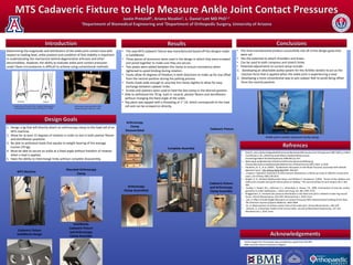
• The new MTS cadaveric fixture was manufactured based off the designs made
in SolidWorks.
• Three pieces of aluminum were used in the design in which they were screwed
and pined together to make sure they are secure.
• Two plates were added between the clamp to ensure consistency when
tightened to avoid binding during rotation.
• Tracks allow 45 degrees of freedom in both directions to make up for any offset
from the neutral position during the potting process.
• Tracks made wide enough to unscrew the clamp slightly to allow for easy
exchange between cadaver limbs.
• Screws and washers were used to hold the box clamp in the desired position.
• Able to withstand the 70 kg load in neutral, plantar flexion and dorsiflexion
without changing the fixed angle of the ankle.
• Top piece was tapped with a threading of 1”-14, which corresponds to the load
cell and can be screwed on directly.
1. Design a jig that will directly attach an arthroscopy clamp to the load cell of an
MTS machine.
2. Allow for at least 15 degrees of rotation in order to test in both plantar flexion
and dorsiflexion positions.
3. Be able to withstand loads that equate to weight bearing of the average
human (70 kg).
4. Must be able to secure an ankle at a fixed angle without freedom of rotation
when a load is applied.
5. Have the ability to interchange limbs without complete disassembly.
Frontal view of the ankle joint complex showing partial
and extended contact surfaces (Tochigi et al., 2006)
Arthroscopy clamp attached to MTS
Machine at Duke (Latt et al., 2011)
• Partial support for this project was provided by a grant from the NIH
• BME Summer Clinical Immersion Program
Arthroscopy
Clamp
Disassembled Cadaveric Fixture
Arthroscopy
Clamp Assembled
Cadaveric Fixture
and Arthroscopy
Clamp Assembly
Complete Assembly
Ankle joint contact pressure study setup
MTS Machine
Mounted Arthroscopy
Clamp
Cadaveric Fixture
SolidWorks Design
SolidWorks
Cadaveric Fixture
and Arthroscopy
Clamp Assembly