
Empfohlen
Weitere ähnliche Inhalte
Was ist angesagt?
Was ist angesagt? (20)
Andere mochten auch
Andere mochten auch (17)
Ähnlich wie 7.2 Cell Structure
Ähnlich wie 7.2 Cell Structure (20)
Mehr von JdccSeiki
Mehr von JdccSeiki (15)
Kürzlich hochgeladen
Kürzlich hochgeladen (20)
7.2 Cell Structure
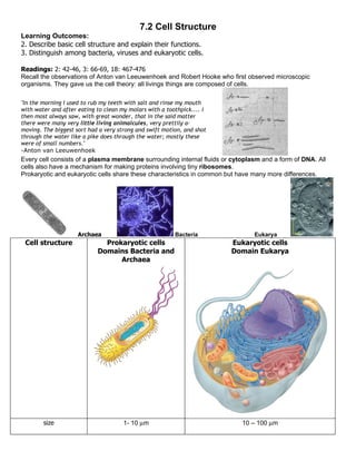
- 1. 7.2 Cell Structure Learning Outcomes: 2. Describe basic cell structure and explain their functions. 3. Distinguish among bacteria, viruses and eukaryotic cells. Readings: 2: 42-46, 3: 66-69, 18: 467-476 Recall the observations of Anton van Leeuwenhoek and Robert Hooke who first observed microscopic organisms. They gave us the cell theory: all livings things are composed of cells. "In the morning I used to rub my teeth with salt and rinse my mouth with water and after eating to clean my molars with a toothpick.... I then most always saw, with great wonder, that in the said matter there were many very little living animalcules, very prettily a- moving. The biggest sort had a very strong and swift motion, and shot through the water like a pike does through the water; mostly these were of small numbers." -Anton van Leeuwenhoek Every cell consists of a plasma membrane surrounding internal fluids or cytoplasm and a form of DNA. All cells also have a mechanism for making proteins involving tiny ribosomes. Prokaryotic and eukaryotic cells share these characteristics in common but have many more differences. Archaea Bacteria Eukarya Cell structure Prokaryotic cells Eukaryotic cells Domains Bacteria and Domain Eukarya Archaea size 1- 10 µm 10 – 100 µm
- 2. Location and in cytoplasm contained inside nucleus arrangement of arranged in a circular DNA strands are coiled around histone genetic information chromosome and small proteins and highly condensed into circular plasmids chromosomes Internal structures ribosomes √ √ Microtubules some √ for cytoskeleton Endoplasmic reticulum No √ for processing proteins Golgi apparatus No √ for further processing proteins Mitochondria No, but may have same Most have except few anaerobic protists enzymes in cytoplasm Perform energetic rxns in cytoplasm involving oxygen Chloroplasts No, if photosynthetic, Plantae, some protists perform in cytoplasm External structures 1. Flagella some some 2. Plasma membrane All cells have a plasma membrane made of phospholipids. In membranes, lipids are arranged in a double layer and only allow some small uncharged molecules to pass through (eg. CO2) (Fig 3.11a). Protein carriers/channels in membranes Water enters cells through “aquaporins” (small pores in the plasma membrane made of proteins). Other small molecules (eg. glucose) can also be transported by protein carriers through membranes. Charged molecules (eg. Na+, sodium ions) must pass through ion channels which are also composed of proteins sitting in the membrane and have specificity to only allow specific ions through. Facilitated diffusion (Fig 3.11b) means no energy is required to move some molecules as movement goes along the molecules’ concentration gradient (from an area of high concentration where there are many of these molecules to an area of low conc.) Active transport (Fig 3.12) means energy must be expended to move a molecule against its concentration gradient. Large molecules enter or exit cells through vesicles by endo or exocytosis. (endo = in exo=out)
- 3. Cell structure Prokaryotic cells Eukaryotic cells 3. Cell wall -differing compositions of Found in Plantae, Fungi, some carbohydrate in wall protists, with differing major components (eg. cellulose, chitin) Not in animal cells 4. External Outer membrane Found in Gram - bacteria (in Domain Bacteria) none Bacterial membranes and cell walls: Note: Peptidoglycan is a carbohydrate. Lipopolysaccharides are, as the name suggests, lipids with sugars (carbohydrates) linked to them. In pathogenic bacteria these lipolysaccharides often contain toxins. Prokaryotes divide by binary fission Circular chromosome is copied into two. Two copies separate Cell grows larger and divides in two with one identical chromosome in each new cell. Also shown in Fig 18.2
- 4. Bacteria can have a form of sex by sharing plasmids Note: This form of genetic transfer allows bacteria (often of different species) to share genes. E coli 0157 (pathogenic strain of E. coli that killed the people in Walkerton Ontario) make virtually the same toxin as found in Shigella dysenteriae, the organism which causes dysentery. It is thought that S. dysenteriae transferred the gene to make the toxin to E. coli through a plasmid. Viruses Fig 18.4 -consist of genetic material (DNA or RNA) and a protein coat (capsid) They may or may not have an additional covering called an envelope made of more protein. -require a living cell to make more of themselves 1. inject DNA 2. take over cell’s machinery for making DNA and proteins 3. make many copies of viral DNA and viral coat proteins assemble into viruses 4. cell lyses (breaks open) viruses are released and spread to nearby cells Viral infection ends when cells of the immune system are able to recognize and degrade viruses. Viruses can sometimes incorporate into a cell’s DNA and stay for many cell divisions/years (lysogenic cycle). Such viruses are latent. A certain environmental signal can cause virus production to suddenly begin Eg.: chickenpox virus can become active in nerve cells of adults (who once had chicken pox) causing “shingles” decades later -Viruses usually only recognize very specific cell types egs: - bacteriophage specific sp. of bacteria. -adenoviruses cells of lungs and respiratory system
- 5. Prions (Fig 18.6) A prion is a misfolded protein which in brain cells causes spongiform encephalopathy (holes in the brain tissue) The word comes from proteinaceous infectious particle. It seems to be one specific protein which, when misfolded, can cause spongiform encephalopathy. The normal form of the protein is PrPC and the misfolded disease-causing form is PrPSC. It is not known what the normal protein does, although it is found in many nerve cells. One misfolded protein can stimulate normal proteins to become misfolded themselves (Fig 18.6). This is the only protein known to have this infectious capability. Misfolded proteins build up in nerve cells causing the cells to stop functioning and die. Normally when proteins in a cell are misfolded they are either refolded by proteins called chaperones or destroyed (in lysosomes). What makes prions different from other pathogens (viruses, bacteria, eukaryotic pathogens) is that they do not carry any genetic material (DNA or RNA) with them. Previously it was thought that all pathogens needed to have their genetic material with them in order to multiply. The normal form of prions (PrPc) is found in many nerve cells and like other proteins it is coded for by a gene. QUESTION: Where is the PrP gene found? Questions to Consider: 1. Although mutations are rare they happen frequently enough for species to evolve. Because bacterial species reproduce so quickly, a mutation which provides some advantage (i.e. allows a bacterium to resist an antibiotic, become more infectious or pathogenic etc) will increase quickly in a population. Besides the quick reproduction rate of bacteria what other feature assists their quick evolution (gaining new advantageous inherited characteristics)? 2. Why could it be advantageous for a virus to remain dormant for some time rather than reproducing and causing cells to lyse rapidly? 3. Antibodies in your immune system recognize specific shapes of foreign macromolecules (proteins, carbohydrates, lipids and combinations of all three) that are different from any shape of self macromolecules (normally found in the host’s body). Based on what you know now about bacterial cells, what components of bacterial cells may be recognized by antibodies. Hint: what structures or molecules do bacteria have that are different from eukaryotic cells? (FYI: we will talk about how antibodies work in greater detail next week) 4. In order to evade the immune system some pathogens get inside the cells of the host organism. They usually do not initially kill the cells they enter. Considering what you know about how molecules enter cells, how might pathogens get inside? 5. When you get a cold, you have been infected with a virus that infects cells of your airways (bronchial passages and lungs). A virus which infects a lung cell will have the cell make many copies of the virus and then the cell bursts and the viruses spread to nearby cells. Aside from mucus, what is in a tissue when you blow your nose?