SlideShare a Scribd company logo
1 of 7
Download to read offline
Journal of Athletic Training    2002;37(3):293–299
  by the National Athletic Trainers’ Association, Inc
www.journalofathletictraining.org


Nonthermal Effects of Therapeutic
Ultrasound: The Frequency Resonance
Hypothesis
Lennart D. Johns
Quinnipiac University, Hamden, CT

Lennart D. Johns, PhD, ATC, provided conception and design; acquisition of the data; and drafting, critical revision, and final
approval of the article.
Address correspondence to Lennart D. Johns, PhD, ATC, Quinnipiac University, 275 Mount Carmel Avenue, Hamden, CT
06518. Address e-mail to Lenn.Johns@Quinnipiac.edu.


   Objective: To present the frequency resonance hypothesis,          ultrasound affects enzyme activity and possibly gene regulation
a possible mechanical mechanism by which treatment with non-          provide sufficient data to present a probable molecular mech-
thermal levels of ultrasound stimulates therapeutic effects. The      anism of ultrasound’s nonthermal therapeutic action. The fre-
review encompasses a 4-decade history but focuses on recent           quency resonance hypothesis describes 2 possible biological
reports describing the effects of nonthermal therapeutic levels       mechanisms that may alter protein function as a result of the
of ultrasound at the cellular and molecular levels.                   absorption of ultrasonic energy. First, absorption of mechanical
   Data Sources: A search of MEDLINE from 1965 through                energy by a protein may produce a transient conformational
2000 using the terms ultrasound and therapeutic ultrasound.           shift (modifying the 3-dimensional structure) and alter the pro-
   Data Synthesis: The literature provides a number of exam-          tein’s functional activity. Second, the resonance or shearing
ples in which exposure of cells to therapeutic ultrasound under       properties of the wave (or both) may dissociate a multimolecular
                                                                      complex, thereby disrupting the complex’s function. This review
nonthermal conditions modified cellular functions. Nonthermal
                                                                      focuses on recent studies that have reported cellular and mo-
levels of ultrasound are reported to modulate membrane prop-          lecular effects of therapeutic ultrasound and presents a me-
erties, alter cellular proliferation, and produce increases in pro-   chanical mechanism that may lead to a better understanding of
teins associated with inflammation and injury repair. Combined,        how the nonthermal effects of ultrasound may be therapeutic.
these data suggest that nonthermal effects of therapeutic ultra-      Moreover, a better understanding of ultrasound’s mechanical
sound can modify the inflammatory response.                            mechanism could lead to a better understanding of how and
   Conclusions: The concept of the absorption of ultrasonic en-       when ultrasound should be employed as a therapeutic modality.
ergy by enzymatic proteins leading to changes in the enzymes             Key Words: immunology, injury, signal transduction, molec-
activity is not novel. However, recent reports demonstrating that     ular mechanism, wound healing, cytokines


U
         ltrasound has become a common therapy for a number           in the field of cellular and molecular biology, specifically the
         of clinical conditions: sprained ligaments, inflamed          activation of proteins and signal-transduction pathways that
         tendons and tendon sheaths, lacerations and other soft       may result in modifications to cellular function.
tissue damage, scar tissue sensitivity and tension, varicose ul-
cers, amputations, neuromata, strained and torn muscles, in-          Thermal Effects of Ultrasound
flamed and damaged joint capsules, fasciitis, and delayed-on-
                                                                         Ultrasound is capable of producing thermal therapeutic ef-
set muscle soreness.1,2 Recent uses include the accelerated
                                                                      fects.2 In 1987, Dyson1 suggested that the tissue must reach a
healing of fractures,3–5 muscle injury,6 and thrombolysis.7–16
                                                                      temperature of 40 C to 45 C for at least 5 minutes to be ther-
   Over the past several years, research investigating the cel-       apeutic in nature. Experiments performed with nonperfused
lular and molecular effects of nonthermal levels of ultrasound        tissue demonstrated that ultrasound could increase the tissue
has accumulated. While clinicians state that ultrasound is used       temperature at a rate of 0.86 C/min (1 W/cm2, 1 MHz).17
to accomplish heating within deep tissue, there is a common,          However, the results of these experiments were difficult to
whispered belief that heating alone cannot account for the clin-      interpret because they were performed in nonperfused tissue.
ical effects, especially when ultrasound is delivered at non-         In living tissue, as the temperature increases, the normal blood
thermal settings. My purpose is to review the past 4 decades          flow to the area dissipates the heat. More recent, direct in vivo
of ultrasound research and to propose a molecular mechanism           measurement of tissue temperature during ultrasound treatment
whereby the mechanical properties of ultrasound interact with         has resolved the question of tissue heating.18–21 Draper et
the molecular and multimolecular complexes within the cell.           al,18,19 Ashton et al,20 and Chan et al21 inserted thermistors to
The frequency resonance hypothesis incorporates past research         various depths (5 cm or less) and measured the increase in
demonstrating ultrasound’s mechanical properties (absorption,         muscle temperature during a 10-minute treatment with either
cavitation, acoustical streaming) with current knowledge with-        1-MHz or 3-MHz ultrasound. The data show that treatment


                                                                                              Journal of Athletic Training        293
with 1-MHz or 3-MHz ultrasound resulted in a time- and dose-         organelles and molecules of different molecular weight exist.
dependent increase in tissue temperature.18–21 The 3-MHz fre-        While many of these structures are stationary, many are free
quency increased tissue temperature at a faster rate than the        floating and may be driven to move around more stationary
1-MHz frequency.19 More recently, Ashton et al20 and Chan            structures. This mechanical pressure applied by the wave pro-
et al21 employed similar techniques to study increases in tem-       duces unidirectional movement of fluid along and around cell
perature in the patellar tendon and the effects of coupling me-      membranes.25
dia on increases in tissue temperature. While a number of               Cavitation is defined as the physical forces of the sound
questions remain unanswered with respect to the thermal ef-          waves on microenvironmental gases within fluid. As the sound
fects of ultrasound, the purpose of my review is to focus on         waves propagate through the medium, the characteristic com-
the nonthermal effects of ultrasound. I will not include the         pression and rarefaction causes microscopic gas bubbles in the
various therapeutic applications of ultrasound that have re-         tissue fluid to contract and expand. It is generally thought that
cently been reviewed elsewhere.22                                    the rapid changes in pressure (caused by the leading and lag-
                                                                     ging edges of the sound wave), both in and around the cell,
                                                                     may cause damage to the cell. Substantial injury to the cell
Nonthermal Effects of Ultrasound                                     can occur when microscopic gas bubbles expand and then col-
   A number of experimental designs appear to have success-          lapse rapidly, causing a ‘‘microexplosion.’’ Although true mi-
fully isolated the nonthermal from the thermal effects of ul-        croexplosions, referred to as unstable cavitation, are not
trasound within cellular systems.1,2,23–25 In vivo, a portion of     thought to commonly occur at therapeutic levels of ultrasound,
the energy from the ultrasound wave is absorbed into the tissue      pulsation of gas bubbles may disrupt cellular activity, altering
structure and converted into heat energy.2,24 The amount of          the function of the cell.27
heating is determined by the frequency and intensity of the             Early studies investigating the gross effects of acoustic
ultrasound (dosage) and the type of tissue that is exposed to        streaming and cavitation on cells showed growth retardation
acoustic energy. A 1982 report demonstrated a direct relation-       of cells in vitro,28–31 increases in protein synthesis,32,33 and
ship between the absorption of ultrasound and amount of pro-         membrane alterations.34,35 Combined, these results may sug-
tein.26 More simply, as the concentration of protein increased,      gest that ultrasound first ‘‘injures’’ the cell, resulting in growth
the absorption of ultrasound increased. In normal tissue, the        retardation, and then initiates a cellular recovery response
absorption of ultrasound energy varies depending on the              characterized by an increase in protein production. These find-
amount of protein in the tissue.26 In 1980, Love and Kremkau23       ings encompass both continuous and pulsed ultrasound at ther-
demonstrated that by eliminating extracellular tissue structures     apeutic levels ranging from 0.1 to 1.7 W/cm2.28–35
(collagen, fibrin, elastin, etc) and placing only the cells in tis-
sue culture media maintained at 37 C, they could treat cells at      Attraction of Immune Cells to the Injured Area
therapeutic levels without significant increases in temperature
(less than 0.5 C over a 10-minute exposure). Our data confirm            The natural course of tissue injury can be categorized into
that cell cultures treated with either 1-MHz or 3-MHz ultra-         4 distinct phases: acute inflammation, clearance of tissue de-
sound at intensities of 0.5 W/cm2 sustained less than 0.5 C          bris, cellular proliferation, and tissue remodeling.1,36
increases over a 10-minute exposure (unpublished observation,           With the early arrival of immune cells to the injured tissue,
1998). At first it may seem that these data are contradictory         the immune system can be destructive in nature. When soft
to those of Draper et al,18,19 Ashton et al,20 and Chan et al21;     tissue is injured, platelets and mast cells are activated and re-
however, the 2 experimental protocols were significantly dif-         lease chemokines, attracting polymorphonuclear cells and
ferent. The in vivo measurements performed by Draper et              blood monocytes (macrophages). Once activated, macrophages
al,18,19 Ashton et al,20 and Chan et al21 recorded actual in-        produce a unique set of proteins that aid in the destruction of
creases within intact muscle and tendon. The tissue culture          damaged tissue and attract additional lymphocytes to the area.
protocol eliminates the extracellular structural proteins (col-      A concerted recruitment of lymphocytes is accomplished by
lagen, fibrin, elastin, etc)23,26 that are responsible for most of    the production of chemokines and the activation of adhesion
the increase in temperature observed within the intact tis-          molecules on the surface of the local capillaries. Adhesion
sues.18–21 Moreover, the tissue culture protocol makes it pos-       molecules can be viewed as docking proteins that grab circu-
sible to ‘‘eliminate’’ the thermal effects of ultrasound and to      lating lymphocytes and aid in their migration to the injured
study the mechanical effects of ultrasound in an attempt to          tissue.37,38 Reports39–42 suggest that the nonthermal effects of
identify a mechanical mechanism of action.                           ultrasound aid the immune response by inducing vasodilation
   While exposure of single cells to ultrasound does not in-         of arterioles and activation of adhesion molecules. Both va-
crease the overall temperature of the experimental system,23 it      sodilation and the activation of adhesion molecules are regu-
is difficult to determine whether larger temperature increases        lated by signal-transduction pathways,37–42 suggesting that the
occurred at the cell surface or within the microenvironments         ultrasound treatment modified cellular activity by modulating
of the cell. Theoretically, larger increases in temperature could    one or more signal-transduction pathways. In general, signal-
occur within microenvironments of the cell as a result of cav-       transduction pathways are composed of a series of enzymatic
itation.24 However, direct measurements of these types of mi-        proteins that are turned on and off by the addition and deletion
croenvironmental changes in temperature are currently not            of phosphate molecules. Phosphate modifications to a mole-
possible.                                                            cule lead to distinct changes in conformation (3-dimensional
   Therapeutic ultrasound produces a combination of nonther-         configuration) and regulate the enzymatic activity of protein.
mal effects (acoustic streaming and cavitation) that are difficult    A simple analogy for changes in 3-dimensional shape altering
to isolate. Acoustic streaming is defined as the physical forces      function is a pocketknife. When the knife is open, the blade
of the sound waves that provide a driving force capable of           is functionally available and can cut; however, when the knife
displacing ions and small molecules.24 At the cellular level,        is closed, the blade is functionally not available. Similarly,


294      Volume 37     • Number 3 • September 2002
Cellular and Molecular Effects of Nonthermal Ultrasound on Wound Healing
Increase in Protein or Cellular Function          Producing Cell Type                               Effector Function

Interleukin-1 45
                                                Osteoblasts, monocytes                 General inflammatory mediator
Interleukin-248                                 T cells                                T-cell growth
Interleukin-845,50                              Osteoblasts                            Endothelial cell migration and proliferation
Vascular endothelial growth factor45,50         Osteoblasts, monocytes                 Endothelial cell migration and proliferation
Basic fibroblast growth factor45                 Osteoblasts                            Endothelial cell migration and proliferation
Fibroblast growth factor50,53                   Monocytes                              Fibroblast growth
Collagen5,45                                    Osteoblasts, fibroblasts                Wound healing
Chloramphenicol acetyl transferase56            HeLa, NIH/3T3, C1271                   Gene expression of liposomal transfection
Increased proliferation45                       Fibroblasts                            Enhanced wound healing
Increased proliferation45                       Osteoblasts                            Enhanced wound healing
Lymphocyte adhesion41                           Endothelial cells                      Enhanced lymphocyte trafficking
Vasodilation39,40,42                            Capillary, endothelium                 Enhanced blood flow


proteins have an ‘‘active site’’ that can be either available or     ability, calcium flux, and proliferation), possibly activating sig-
not available, depending on the 3-dimensional shape of the           nal-transduction pathways that lead to gene regulation (Ta-
protein.                                                             ble).37,39–42,45,48,53,56,57 Importantly, exposure to ultrasound
                                                                     caused an increase in intracellular calcium in fibroblasts, sug-
Inflammatory Response, Injury Repair, and                             gesting that the mechanical effects disrupt the normal function
Therapeutic Ultrasound                                               of the membrane, permitting leaking of calcium into the cell.49
                                                                     After ultrasound exposure, the cells rapidly expelled the cal-
   Later in the inflammatory process, immune cells alter their
                                                                     cium and returned to a homeostatic state. Mortimer and Dy-
course of action, aiding in the clearance of tissue debris and
                                                                     son49 eliminated the effects of transient cavitation and gross
stimulating tissue remodeling. This pivotal action is directed
                                                                     heating as possible mechanisms for the resultant increases in
by cytokines. For example, the arrival of T cells in an injured
                                                                     intracellular calcium. Cells employ calcium as a cofactor in
area may enhance the immunologic response by releasing T-
                                                                     regulating the activity of enzymes, many of which are asso-
cell growth factors (interleukin [IL]-2 and IL-4) and immu-
                                                                     ciated with signal-transduction pathways. Activation of calci-
noregulatory cytokines (IL-10 and interferon- ).43 At a certain
                                                                     um-sensitive signal-transduction pathways (protein kinase C
point in the immune intervention, anti-inflammatory cytokines
                                                                     and cyclic AMP) commonly results in gene activation. The
(largely transforming growth factor- ) are either produced or
                                                                     resultant protein production could modulate intracellular func-
activated. These anti-inflammatory cytokines down-regulate T-
                                                                     tions and the activity of surrounding cells.58–60 A number of
cells and redirect the cellular activities toward proliferation of
                                                                     the experiments reviewed in the Table demonstrated increases
fibroblasts, collagen production, and remodeling of the dam-
                                                                     in specific proteins after exposure of cells to therapeutic levels
aged tissue.36,44
                                                                     of ultrasound. Combined, these findings suggest that therapeu-
   A number of reports have demonstrated that ultrasound af-
                                                                     tic ultrasound can modulate signal-transduction pathways that
fects cells that are centrally involved in the immune response.
                                                                     lead to gene regulation or the modulation of RNA translation
Specifically, ultrasound has been shown to modulate vasocon-
                                                                     to a protein product, or both.
striction; lymphocyte adhesion properties of endothelium, mast
cell degranulation, phagocytosis by macrophage, production of
growth factors by macrophages; calcium fluxes in fibroblasts;          Frequency Resonance Hypothesis
angiogenesis; proliferation of T-cells, osteoblasts, fibroblasts,        Cumulatively, the data may suggest that the mechanical en-
and a number of proteins associated with inflammation and             ergy within the ultrasound wave and the shearing force of the
repair (IL-1, IL-2, IL-6, IL-8, interferon- , fibroblast growth       wave combine to produce mechanical properties that pertur-
factor-b, vascular endothelial growth factor, collagen) (Ta-         bate the cellular membrane and the molecular structures within
ble)1,34,40–42,45–53; and to accelerate thrombolyisis.7–16 In gen-   the cell. The central premise of the frequency resonance hy-
eral, most of these researchers used a frequency of 1 MHz or         pothesis is that the mechanical energy within the ultrasound
3 MHz, and the intensities ranged from 0.1 to 1.5 W/cm2. An          wave is absorbed by proteins, altering the structural confor-
alternative therapeutic protocol employs a frequency of 45           mation of an individual protein or the function of a multi-
kHz. An intensity range of 5 to 100 mW/cm2 was shown to              molecular complex. Moreover, the ultrasound wave may in-
increase the production of IL-1, IL-8, vascular endothelial          duce resonant activity in the protein, modulating the
growth factor, fibroblast growth factor-b, and collagen; pro-         molecule’s or multimolecular complex’s effector function.
mote bone healing; and accelerate thrombolysis.5,45,54,55 The           The following discussion employs enzymatic proteins as a
long-wave (45-kHz) ultrasound increases penetration depth            molecular model. One can view an enzymatic protein as a
and, therefore, seems to be more appropriate than traditional        physical machine performing a physical function within a cell.
high-frequency ultrasound (1 MHz and 3 MHz) for promoting            Enzymes are commonly found in 1 of 2 conformational
revascularization and bone healing at greater depths.                shapes: on or off. Movement between these 2 conformations
                                                                     (or 3-dimensional shapes) requires a change in the state of
Ultrasound, Signal Transduction, and Gene                            energy, which is normally accomplished by the addition or
Regulation                                                           removal of a phosphate molecule. Once an enzyme within a
  Additional reports suggest that ultrasound alters cellular         signal-transduction cascade is activated, the signal is amplified
membrane properties (cellular adhesion, membrane perme-              to execute an effector function.


                                                                                              Journal of Athletic Training            295
streaming relates to the movement of objects from one place
                                                                       to another as a function of the force of the wave. In terms of
                                                                       ultrasound therapy, phonophoresis is commonly used to move
                                                                       medication transdermally. Second, cavitation relates to the os-
                                                                       cillation of microscopic gas bubbles that may, in turn, affect
                                                                       the cell or cellular process. However, the frequency resonance
                                                                       hypothesis relates to the absorption of ultrasound by proteins
                                                                       and protein complexes that may directly result in alterations
Figure 1. The resonating mechanical force produced by the ultra-       to signaling mechanisms within the cell, either by inducing a
sound wave may induce transient conformational shifts. Molecules       conformational shift or by disrupting a multimolecular com-
are normally found in one of two 3-dimensional shapes, an ‘‘active’’   plex.
or ‘‘inactive’’ conformation.                                             In the original experiment investigating whether ultrasound
                                                                       could alter protein activity, the researchers reported no effect
                                                                       with respect to the monomer or dimer form of the enzyme
                                                                       creatine kinase.61 However, Chetverikova et al61 reported that
                                                                       ultrasound decreased the activity of the dimeric and tetrameric
                                                                       forms of creatine kinase and suggested that the decrease in
                                                                       activity was due to the disruption of the multimolecular forms
                                                                       of creatine kinase (represented in Figure 2). The authors in-
                                                                       ferred that ultrasound did not directly affect enzyme activity
                                                                       and that the primary acousto-biological interaction appeared
Figure 2. Resonant or shearing forces produced by ultrasound           to be occurring at a higher level of organization complexity.
may result in the disjunction of a multimolecular complex with a       However, more recent investigations7–16,54,55 have shown that
subsequent loss of function or decrease in activity.                   ultrasound increases thrombolysis, demonstrating that ultra-
                                                                       sound can increase enzyme activity (represented in Figure 1).
                                                                       These data support the tried research saying, ‘‘The absence of
                                                                       proof is not the proof of absence.’’
                                                                          The concept of the absorption of ultrasonic energy by en-
                                                                       zymatic proteins leading to changes in the enzymes’ activity
                                                                       is not novel61,62; however, the demonstration that ultrasound
                                                                       can increase or decrease protein activity and possible gene
                                                                       regulation is more recent.7–16,54–56 While considerable data ex-
Figure 3. Resonant or shearing mechanical forces produced by
                                                                       ist, a model suggesting a molecular mechanism for how the
ultrasound may dislodge an inhibitor molecule, leading to activa-
tion of a signal-transduction pathway.
                                                                       absorption of ultrasound by proteins affects the cell function
                                                                       is novel and is presented here for the first time.
                                                                          Frequency resonance is one possible explanation of why
   The frequency resonance hypothesis suggests that the en-            exposure to ultrasound increased enzymatic activity, resulted
ergy provided to the enzyme by the ultrasound wave may in-             in thrombolysis, and did not alter the activity of the enzymes
duce transient conformational shifts in certain enzymatic pro-         creatine kinase, lactate dehydrogenase, hexokinase, and pyru-
teins, altering the enzyme’s activity (ie, kinases or                  vate kinase.7–16,54,55,61 A simple analogy would be 2 tuning
phosphatases) and the overall function of the cell (Figure 1).         forks located at one end of a room, with different frequencies
Alternatively, ultrasound’s resonating force may result in the         A and B. At the opposite end of the room is a third tuning
dissociation of functional multimolecular complexes (Figure            fork with the same frequency as fork A. Fork A is struck,
2) or the release of a sequestered molecule by dislodging an           generating a sound. The sound waves travel through the air
inhibitor molecule from the multimolecular complex (Figure             and are absorbed at the other end of the room by fork A to
3). In essence, the mechanism of ultrasound’s action in Figures        produce the same resonating sound, while fork B remains si-
2 and 3 is the same. Ultrasound disrupts a multimolecular              lent. The possibility exists that thrombolysis is affected by the
complex. However, Figure 2 represents a functionally active            frequencies ‘‘designated’’ as therapeutic, while creatine kinase,
complex, while Figure 3 represents a functionally sequestered          lactate dehydrogenase, hexokinase, and pyruvate kinase are
molecule. One can view an inhibitor molecule as a ‘‘safety             not.7–16,54,55,61 Importantly, these reports do not directly dem-
block’’ that functionally inhibits or sequesters a protein from        onstrate that a conformational shift is occurring in these en-
working. When the safety block is released, the protein is then        zymes but support the hypothesis.
operable.                                                                 On the surface, it may appear that the observed decrease in
   Shearing forces produced by ultrasound may also play a role         creatine kinase activity and an increase in thrombolytic activity
in the dissociation of multimolecular complexes. Hypotheti-            are contradictory; however, a fundamental difference exists.
cally, frequency resonance may imply that different frequen-           The decrease in activity of the creatine kinase was, most likely,
cies (1 MHz, 3 MHz, 45 kHz, and others) establish unique               a result of the disruption of the multimolecular dimeric or
resonant or shearing forces (or both). Moreover, various fre-          tetrameric (or both) forms of creatine kinase (Figure 2), while
quencies may affect combinations of proteins or multimolec-            the increased thrombolytic activity may be more associated
ular complexes in different ways, lending to the possibility of        with activation through harmonic resonance (Figure 1).
targeted effects at the cellular and molecular levels.                    In any case, the mechanical effects of ultrasound may result
   The frequency resonance hypothesis differs from acoustic            in either the activation or inactivation of an enzymatic protein
streaming and cavitation at the basic levels. First, acoustic          or a dissociation of a protein complex, leading to alterations in


296      Volume 37      • Number 3 • September 2002
signal transduction. The frequency resonance hypothesis may          However due to pharmocokinetics (ie, administration, absorp-
describe the molecular mechanism or mechanisms responsible           tion, distribution, and elimination of a drug), higher concen-
for alterations in cellular membrane properties,34,39,41,46,49 in-   trations and multiple doses per day are normally required to
creases in protein production,* and modulation of enzyme ac-         achieve clinical efficacy.
tivity.7–16,54–56                                                       While general recommendations for ultrasound treatment
   Frequency resonance and shearing forces on multimolecular         suggest 5 to 10 minutes of exposure and 1 to 3 treatments per
complexes may combine to produce the nonthermal effects of           day, clinical treatments are almost exclusively done once a
therapeutic ultrasound. Collectively, the experiments reviewed       day. The possibility exists that clinical treatment protocols
here support the frequency resonance hypothesis and demon-           commonly employed for ultrasound are not sufficient for ther-
strate that therapeutic ultrasound may modulate signal-trans-        apeutic efficacy. In the review by Robertson and Baker,63 both
duction pathways and gene products associated with the in-           of the methodologically acceptable studies showing clinical
flammatory response and cells directly involved in the healing        efficacy used pulsed ultrasound (1:4) with treatment times of
response (see Table).                                                15 minutes, resulting in energy densities of 60 and 150 J/cm2,
                                                                     respectively.64,65 Conversely, the remaining 5 studies lacking
                                                                     efficacy employed pulsed ultrasound and exposure times of 2
Clinical Implication of Ultrasound Research at the
                                                                     to 10 minutes, resulting in overall energy densities of 2 to 40
Cellular and Molecular Levels
                                                                     J/cm2.66–70 The frequency resonance hypothesis may suggest
   The purpose of this paper is to raise the awareness that          that different ultrasonic frequencies (1 MHz, 3 MHz, 45 kHz,
therapeutic levels of ultrasound (1 MHz, 3 MHz, and 45 kHz)          and other) may require different durations of exposure (time),
stimulate cellular and molecular effects within cells that are       different energy densities (J/cm2), or both, to reach therapeutic
centrally involved in the inflammatory and healing processes          efficacy.
(Table).1,5,7–16,34,40–42,45–55 Cumulatively, these reports pro-        The identification and scientific understanding of therapeu-
vide important information that may lead to a better under-          tic ultrasound’s nonthermal mechanisms may lead to a com-
standing and clinical application of therapeutic ultra-              prehensive and effective clinical strategy. Further research is
sound.1,5,7–16,34,40–42,45–55 Currently, no clear guidelines exist   needed on 2 fronts: (1) cellular and molecular research to de-
that provide the clinician with protocols directing when in the      termine whether the mechanical mechanisms proposed by the
injury and healing response ultrasound should be adminis-            frequency resonance hypothesis can be elucidated and provide
tered, nor are there guidelines on the frequency, intensity,         insight into a comprehensive strategy for the clinical indica-
treatment times, or number of treatments required for efficacy.       tions of therapeutic ultrasound at various frequencies, and (2)
In a recent review of the literature, a wide spectrum of ultra-      methodologically sound clinical research designed to provide
sound treatment protocols was found.63 In the 10 papers found        meaningful input and outcome measures related to clinical ef-
to be scientifically acceptable, a broad range of treatment set-      ficacy. Both avenues of research should strive to establish time
tings and methods was used, including (1) nine different clin-       and dose-dependent response curves.
ical indications, (2) five different frequencies, (3) continuous
and pulsed output, (4) W/cm2 ranging from 0.02 to 2.6, (5)           ACKNOWLEDGMENTS
treatment time ranging from 2 to 15 minutes, and (6) energy
density ranging from 2 to 150 J/cm2.64–70 Due to the variety           A portion of the time spent developing this manuscript was sup-
of clinical indicators and methods employed,63–70 sufficient          ported by a grant from the National Athletic Trainers’ Association
                                                                     Research and Education Foundation.
clinical data are currently not available to generate scientifi-
cally sound recommendations for treatment protocols.
   The ultrasound treatment protocols employed to investi-           REFERENCES
gate the cellular and molecular effects (see Table) were              1. Dyson M. Mechanisms involved in therapeutic ultrasound. Physiotherapy.
usually a 10-minute exposure at 1 or 3 MHz and 0.1 to 1.5                1987;73:116–120.
W/cm2.5,39–42,45,48,50,53,56 The experimental protocol inves-         2. Kitchen SS, Partridge CJ. A review of therapeutic ultrasound. Physio-
tigating thrombolysis ranged from 10 to 200 minutes of ex-               therapy. 1990;76:593–600.
posure at 0.17 to 3.4 MHz and 0.25 to 8.0 W/cm2, both                 3. Hadjiargyrou M, McLeod K, Ryaby JP, Rubin C. Enhancement of fracture
continuous and intermittent.7–16 A third experimental pro-               healing by low intensity ultrasound. Clin Orthop. 1998;355(suppl):216–
tocol employing a frequency of 45 kHz and an intensity                   229.
range of 5 to 100 mW/cm2 resulted in increased production             4. Kristiansen TK, Ryaby JP, McCabe J, Frey JJ, Roe LR. Accelerated heal-
                                                                         ing of distal radial fractures with the use of specific, low-intensity ultra-
of IL-1, IL-8, vascular endothelial growth factor, fibrino-
                                                                         sound: a multicenter, prospective, randomized, double-blind, placebo-con-
blast growth factor-b, and collagen, promotion of bone heal-             trolled study. J Bone Joint Surg Am. 1997;79:961–973.
ing, and acceleration of thrombolysis.5,45,54,55                      5. Reher P, Elbeshir NL, Harvey W, Meghji S, Harris M. The stimulation
   The logical argument is that in vitro effects cannot be di-           of bone formation in vitro by therapeutic ultrasound. Ultrasound Med
rectly applied to clinical treatment protocols. However, from            Biol. 1997;23:1251–1258.
a cellular biology point of view, a strong argument can be            6. Rantanen J, Thorsson O, Wollmer P, Hurme T, Kalimo H. Effects of
made that if a stimulus reaches a critical threshold (eg, con-           therapeutic ultrasound on the regeneration of skeletal myofibers after ex-
sider depolarization thresholds) the cell will respond, regard-          perimental muscle injury. Am J Sports Med. 1999;27:54–59.
less of whether the cell is in vitro or in vivo (eg, insulin,         7. Blinc A, Francis CW, Trudnowski JL, Carstensen E. Characterization of
histamine, aspirin, or any of the proteins listed in the Table).         ultrasound-potentiated fibrolysis in vitro. Blood. 1993;81:2636–2643.
                                                                      8. Francis CW, Onundarson PT, Carstensen EL, et al. Enhancement of fi-
Importantly, cells in tissue culture usually respond to nano-
                                                                         binolysis in vitro by ultrasound. J Clin Invest. 1992;90:2063–2068.
gram or microgram per milliliter quantities of a stimulus.            9. Harpaz D, Chen X, Francis CW, Marder VJ, Metzer RS. Ultrasound en-
                                                                         hancement of thrombolysis and reperfusion in vitro. J Am Coll Cardiol.
  *References 5, 37, 39–42, 45, 48, 50, 53, 57.                          1993;21:1507–1511.


                                                                                                   Journal of Athletic Training                297
10. Higazi AA, Katz I, Mayer M, Weiss AT. The effect of ultrasonic irradi-              beta in the regulation of the immune response. Clin Immunol Immuno-
    ation and temperature on fibrinolytic activity in vitro. Thromb Res. 1993;           pathol. 1992;65:1–9.
    69:251–253.                                                                   37.   Hogg N, Landis RC. Adhesion molecules in cell interactions. Curr Opin
11. Kornowski R, Meltzer RS, Chernine A, Vered Z, Battler A. Does external              Immunol. 1993;5:383–390.
    ultrasound accelerate thrombolysis? Results from a rabbit model. Circu-       38.   Pardi R, Inverardi L, Bender JR. Regulatory mechanisms in leukocyte
    lation. 1994;89:339–344.                                                            adhesion: flexible receptors for sophisticated travelers. Immunol Today.
12. Kudo S. Thrombolysis with ultrasound effect. Tokyo Jikeikai Med J.                  1992;13:224–230.
    1989;104:1005–1012.                                                           39.   Chokshi SK, Rongione AJ, Freeman I, Gal D, Gunwald AM, Alliger W.
13. Lauer CG, Burge R, Tang DB, Bass BG, Gomez ER, Alving BM. Effect                    Ultrasonic energy produces endothelium dependent vasomoter-relaxation
    of ultrasound on tissue-type plasminogen activator-induced thrombolysis.            in vitro [abstract]. Circulation. 1989;80(suppl II):565.
    Circulation. 1992;86:1257–1264.                                               40.   Fischell TA, Abbas MA, Grant GW, Siegel RJ. Ultrasonic energy: effects
14. Luo H, Steffen W, Cercek B, Arunasalam S, Maurer G, Siegel RJ. En-                  on vascular function and integrity. Circulation. 1991;84:1783–1795.
    hancement of thrombolysis by external ultrasound. Am Heart J. 1993;           41.   Maxwell L, Collecutt T, Gledhill M, Sharma S, Edgar S, Gavin JB. The
    125:1564–1569.                                                                      augmentation of leucocytes adhesion to endothelium by therapeutic ultra-
15. Nilsson AM, Odselius R, Roijer A, Olsson SB. Pro- and antifibrinolytic               sound. Ultrasound Med Biol. 1994;20:383–390.
    effects of ultrasound on streptokinase-induced thrombolysis. Ultrasound       42.   Steffen W, Cumberland D, Gaines P, et al. Catheter-delivered high inten-
    Med Biol. 1995;21:833–840.                                                          sity, low frequency ultrasound induces vasodilation in vivo. Eur Heart J.
16. Olsson SB, Johansson B, Nilsson AM, Olsson C, Roijer A. Enhancement                 1994;15:369–376.
    of thrombolysis by ultrasound. Ultrasound Med Biol. 1994;20:375–382.          43.   Paul WE, Seder RA. Lymphocyte responses and cytokines. Cell. 1994;
17. Williams R. Production and transmission of ultrasound. Physiotherapy.               76:241–251.
    1987;73:113–116.                                                              44.   Sporn MB, Roberts AB, Wakefield LM, Assoian RK. Transforming
18. Draper DO, Schulthies S, Sorvisto P, Hautala AM. Temperature changes                growth factor-beta: biological function and chemical structure. Science.
    in deep muscles of humans during ice and ultrasound therapies: an in                1986;233:532–534.
    vivo study. J Orthop Sports Phys Ther. 1995;21:153–157.                       45.   Doan N, Reher P, Meghji S, Harris M. In vitro effects of therapeutic
19. Draper DO, Castel JC, Castel D. Rate of temperature increase in human               ultrasound on cell proliferation, protein synthesis and cytokine production
    muscle during 1-MHz and 3-MHz continuous ultrasound. J Orthop Sports                by human fibroblasts, osteoblasts and monocytes. J Oral Maxillofac Surg.
    Phys Ther. 1995;22:142–150.                                                         1999;57:409–419.
20. Ashton DF, Draper DO, Myrer JW. Temperature rise in human muscle              46.   Dyson M, Luke DA. Induction of mast cell degranulation in skin by
    during ultrasound treatments using Flex-All as a coupling agent. J Athl             ultrasound. IEEE Trans Ultrason Ferroelectr Freq Control. 1986;33:194–
    Train. 1998;33:136–140.                                                             201.
21. Chan AK, Myrer JW, Measom G, Draper DO. Temperature changes in                47.   Hogan RD, Franklin TD, Fry FJ, Avery KA, Burke KM. The effect of
    human patellar tendon in response to therapeutic ultrasound. J Athl Train.          ultrasound on microvascular hemodynamics in skeletal muscle: effect on
    1998;33:130–135.                                                                    arterioles. Ultrasound Med Biol. 1982;8:45–55.
22. ter Haar G. Therapeutic ultrasound. Eur J Ultrasound. 1999;9:3–9.             48.   Johns LD, Colloton PA, Neuenfeldt J. Effects of therapeutic ultrasound
23. Love LA, Kremkau FW. Intracellular temperature distribution produced                on T cell proliferation and IL-2 production [abstract]. J Athl Train. 1999;
    by ultrasound. J Accoust Soc Am. 1980;67:1045–1050.                                 34(suppl):S24.
24. Nyborg WL. Physical Mechanisms for Biological Effects of Ultrasound.          49.   Mortimer AJ, Dyson M. The effect of therapeutic ultrasound on calcium
    Washington, DC: Department of Health, Education, and Welfare; 1978.                 uptake in fibroblasts. Ultrasound Med Biol. 1988;14:499–506.
    FDA Publication 78–8062.                                                      50.   Reher P, Doan N, Bradnock B, Meghji S, Harris M. Effect of ultrasound
25. Nyborg WL. Ultrasonic microstreaming and related phenomena. Br J                    on the production of IL-8, basic FGF and VEGF. Cytokine. 1999;11:416–
    Cancer Suppl. 1982;45:156–160.                                                      423.
26. Stewart HG, Stratmeyer ME. An Overview Of Ultrasound: Theory, Mea-            51.   Vivino AA, Boraker DK, Miller D, Nyborg W. Stable cavitation at low
    surement, Medical Applications, and Biological Effects. Washington, DC:             ultrasonic intensities induces cell death and inhibits 3H-TdR incorporation
    Department of Health, Education, and Welfare; 1982. DHEW Publication                by Con-A-stimulated murine lymphocytes in vitro. Ultrasound Med Biol.
    82–8190.                                                                            1985;11:751–759.
27. Michlovitz SL, ed. Thermal Agents in Rehabilitation. Philadelphia, PA:        52.   Young SR, Dyson M. The effect of therapeutic ultrasound on angiogen-
    FA Davis Co; 1986:3–17,141–176.                                                     esis. Ultrasound Med Biol. 1989;16:261–269.
28. Dyson M, Pond JB, Joseph J, Warwick R. The stimulation of tissue re-          53.   Young SR, Dyson M. Macrophage responsiveness to therapeutic ultra-
    generation by means of ultrasound. Clin Sci. 1968;35:273–285.                       sound. Ultrasound Med Biol. 1990;16:809–816.
29. Loch EG, Fischer AB, Kuwert E. Effect of diagnostic and therapeutic           54.   Gaul GB. Ultrasound thrombolysis. Thromb Heamost. 1999;82(suppl 1):
    intensities of ultrasonics on normal and malignant human cells in vitro.            157–179.
    Am J Obstet Gynecol. 1971;110:457–460.                                        55.   Tachibana K, Tachibana S. Prototype therapeutic ultrasound emitting
30. Maeda K, Murao F. Studies on the influence of ultrasound irritation of               catheter for accelerating thrombolysis. J Ultrasound Med. 1997;16:529–
    the growth of cultured cell in vitro. In: White D, Brown RE, eds. Ultra-            535.
    sound in Medicine. Volume 3B. New York, NY: Plenum Press; 1977:               56.   Unger EC, McCreery TP, Sweitzer RH. Ultrasound enhances gene ex-
    2045.                                                                               pression of liposomal transfection. Invest Radiol. 1997;32:723–727.
31. Pizzarello DJ, Wolsky A, Becker MH, Keegan AF. A new approach to              57.   Zur O, Shneyour Y, Dan U, Chemke J, Shneyour A, Shalev E. A possible
    testing the effects of ultrasound on tissue growth and differentiation. On-         effect of ultrasound on RNA synthesis in cultured amniocytes. Acta
    cology. 1975;31:226–232.                                                            Obstet Gynecol Scand. 1993;72:243–245.
32. Ross P, Edmonds PD. Ultrasound induced protein synthesis as a result of       58.   Karin M, Smeal T. Control of transcription factors by signal transduction
    membrane damage. J Ultrasound Med. 1983;2(suppl):47.                                pathways: the beginning of the end. Trends Biochem Sci. 1992;17:418–
33. Webster DF, Pond JB, Dyson M, Harvey W. The role of cavitation in the               422.
    in vitro stimulation of protein synthesis in human fibroblasts by ultra-       59.   Stabel S, Parker PJ. Protein kinase C. Pharmacol Ther. 1991;51:71–95.
    sound. Ultrasound Med Biol. 1978;4:343–351.                                   60.   Shirakawa F, Mizel SB. In vitro activation and nuclear translocation of
34. Anderson DW, Barrett JT. Depression of phagocytosis by ultrasound. Ul-              NF-kappa B catalyzed by cyclic AMP-dependent protein kinase and pro-
    trasound Med Biol. 1981;7:267–273.                                                  tein kinase C. Mol Cell Biol. 1989;9:2424–2430.
35. Pinamonti S, Gallenga PE, Masseo V. Effects of pulsed ultrasound on           61.   Chetverikova EP, Pashovkin TN, Rosanova NA, Sarvazyan AP, Williams
    human erythrocytes in vitro. Ultrasound Med Biol. 1982;8:631–638.                   AR. Interaction of therapeutic ultrasound with purified enzymes in vitro.
36. Sasaki H, Pollard RB, Schmitt D, Suzuki F. Transforming growth factor-              Ultrasonics. 1985;23:183–188.


298        Volume 37       • Number 3 • September 2002
62. Sarvazyan AP. Acoustic properties of tissues relevant to therapeutic ap-     67. Hashish I, Hai H, Harvey W, Feinmann C, Harris M. Reproduction of
    plications. Br J Cancer Suppl. 1982;45:52–54.                                    postoperative pain and swelling by ultrasound treatment: a placebo effect.
63. Robertson VJ, Baker KG. A review of therapeutic ultrasound: effective-           Pain. 1988;33:303–311.
    ness studies. Phys Ther. 2001;81:1339–1350.                                  68. Grant A, Sleep J, McIntosh J, Ashurst H. Ultrasound and pulsed electro-
64. Ebenbichler GR, Resch KL, Nicolakis P, et al. Ultrasound treatment for           magnetic energy treatment for perineal trauma: a randomized placebo-
    treating the carpal tunnel syndrome: randomised ‘‘sham’’ controlled trial.
                                                                                     controlled trial. Br J Obstet Gynaecol. 1989;96:434–439.
    BMJ. 1998;316:731–735.
                                                                                 69. ter Riet G, Kessel AG, Knipschild P. A randomized clinical trial of ultra-
65. Ebenbichler GR, Erdogmus CB, Resch KL, et al. Ultrasound therapy for
    calcific tendinitis of the shoulder. N Engl J Med. 1999;340:1533–1538.            sound in the treatment of pressure ulcers. Phys Ther. 1996;76:1301–1311.
66. Hashish I, Harvey W, Harris M. Anti-inflammatory effects of ultrasound        70. Nykanen M. Pulsed ultrasound treatment of the painful shoulder: a ran-
    therapy: evidence for a major placebo effect. Br J Rheumatol. 1986;25:           domized, double-blind, placebo-controlled study. Scand J Rehabil Med.
    77–81.                                                                           1995;27:105–108.


                                                                                                              Journal of Athletic Training               299

More Related Content

What's hot

Ch 6_Energy Medicine_IN TECH
Ch 6_Energy Medicine_IN TECHCh 6_Energy Medicine_IN TECH
Ch 6_Energy Medicine_IN TECHChristina Ross
 
Late effects of Radiotherapy on Normal Tissues
Late effects of Radiotherapy on Normal TissuesLate effects of Radiotherapy on Normal Tissues
Late effects of Radiotherapy on Normal TissuesPGIMER
 
PEMF paper_JIR-40269_031113
PEMF paper_JIR-40269_031113PEMF paper_JIR-40269_031113
PEMF paper_JIR-40269_031113Christina Ross
 
The Effect of Gamma Irradiation on the Radiofrequency Dielectric Dispersion P...
The Effect of Gamma Irradiation on the Radiofrequency Dielectric Dispersion P...The Effect of Gamma Irradiation on the Radiofrequency Dielectric Dispersion P...
The Effect of Gamma Irradiation on the Radiofrequency Dielectric Dispersion P...theijes
 
Laser therapy unit for physiotherapy
Laser therapy unit for physiotherapyLaser therapy unit for physiotherapy
Laser therapy unit for physiotherapyMedical Bazzar
 
Mechanisms of LLLT/Anti-Aging_Hamblin_A4M_Orlando_2011
Mechanisms of LLLT/Anti-Aging_Hamblin_A4M_Orlando_2011Mechanisms of LLLT/Anti-Aging_Hamblin_A4M_Orlando_2011
Mechanisms of LLLT/Anti-Aging_Hamblin_A4M_Orlando_2011Bauman Medical Group, P.A.
 
Basic therapeutic methods in medicine
Basic therapeutic methods in medicineBasic therapeutic methods in medicine
Basic therapeutic methods in medicineJuliana Knocikova
 
Transdermal Systemic Laser Therapy 2009
Transdermal Systemic Laser Therapy 2009Transdermal Systemic Laser Therapy 2009
Transdermal Systemic Laser Therapy 2009Dr Peter Gruenewald
 
Zen 362 group 2 effects of disaster fukushima
Zen 362 group 2 effects of disaster  fukushimaZen 362 group 2 effects of disaster  fukushima
Zen 362 group 2 effects of disaster fukushimaMatshidiso Pitswane
 
Apoptosis Post Microwave Ablation of the Liver: Does it Change with Power?
Apoptosis Post Microwave Ablation of the Liver: Does it Change with Power?Apoptosis Post Microwave Ablation of the Liver: Does it Change with Power?
Apoptosis Post Microwave Ablation of the Liver: Does it Change with Power?asclepiuspdfs
 
Exploring Channeling Optimized Radiofrequency Energy
Exploring Channeling Optimized Radiofrequency EnergyExploring Channeling Optimized Radiofrequency Energy
Exploring Channeling Optimized Radiofrequency EnergyRvcarpenter
 
2014 increasing survival study of kidney hek-293 t
2014 increasing survival study of kidney hek-293 t2014 increasing survival study of kidney hek-293 t
2014 increasing survival study of kidney hek-293 tOscar Moreno
 

What's hot (13)

Ch 6_Energy Medicine_IN TECH
Ch 6_Energy Medicine_IN TECHCh 6_Energy Medicine_IN TECH
Ch 6_Energy Medicine_IN TECH
 
Late effects of Radiotherapy on Normal Tissues
Late effects of Radiotherapy on Normal TissuesLate effects of Radiotherapy on Normal Tissues
Late effects of Radiotherapy on Normal Tissues
 
PEMF paper_JIR-40269_031113
PEMF paper_JIR-40269_031113PEMF paper_JIR-40269_031113
PEMF paper_JIR-40269_031113
 
The Effect of Gamma Irradiation on the Radiofrequency Dielectric Dispersion P...
The Effect of Gamma Irradiation on the Radiofrequency Dielectric Dispersion P...The Effect of Gamma Irradiation on the Radiofrequency Dielectric Dispersion P...
The Effect of Gamma Irradiation on the Radiofrequency Dielectric Dispersion P...
 
Laser therapy unit for physiotherapy
Laser therapy unit for physiotherapyLaser therapy unit for physiotherapy
Laser therapy unit for physiotherapy
 
Mechanisms of LLLT/Anti-Aging_Hamblin_A4M_Orlando_2011
Mechanisms of LLLT/Anti-Aging_Hamblin_A4M_Orlando_2011Mechanisms of LLLT/Anti-Aging_Hamblin_A4M_Orlando_2011
Mechanisms of LLLT/Anti-Aging_Hamblin_A4M_Orlando_2011
 
Basic therapeutic methods in medicine
Basic therapeutic methods in medicineBasic therapeutic methods in medicine
Basic therapeutic methods in medicine
 
My papers
My papersMy papers
My papers
 
Transdermal Systemic Laser Therapy 2009
Transdermal Systemic Laser Therapy 2009Transdermal Systemic Laser Therapy 2009
Transdermal Systemic Laser Therapy 2009
 
Zen 362 group 2 effects of disaster fukushima
Zen 362 group 2 effects of disaster  fukushimaZen 362 group 2 effects of disaster  fukushima
Zen 362 group 2 effects of disaster fukushima
 
Apoptosis Post Microwave Ablation of the Liver: Does it Change with Power?
Apoptosis Post Microwave Ablation of the Liver: Does it Change with Power?Apoptosis Post Microwave Ablation of the Liver: Does it Change with Power?
Apoptosis Post Microwave Ablation of the Liver: Does it Change with Power?
 
Exploring Channeling Optimized Radiofrequency Energy
Exploring Channeling Optimized Radiofrequency EnergyExploring Channeling Optimized Radiofrequency Energy
Exploring Channeling Optimized Radiofrequency Energy
 
2014 increasing survival study of kidney hek-293 t
2014 increasing survival study of kidney hek-293 t2014 increasing survival study of kidney hek-293 t
2014 increasing survival study of kidney hek-293 t
 

Viewers also liked

Gavin somers google certificates4
Gavin somers google certificates4Gavin somers google certificates4
Gavin somers google certificates4gdsomers
 
Dor em adolescentes x atletas adultos pós lca
Dor em adolescentes x atletas adultos pós lcaDor em adolescentes x atletas adultos pós lca
Dor em adolescentes x atletas adultos pós lcaGustavo Resek Borges
 
Presentation1
Presentation1Presentation1
Presentation1inhechi
 
Comparação do aquec de 2 us diferentes
Comparação do aquec de 2 us diferentesComparação do aquec de 2 us diferentes
Comparação do aquec de 2 us diferentesGustavo Resek Borges
 
Goodwin efetividade da fisio supervisionada num período prec
Goodwin efetividade da fisio supervisionada num período precGoodwin efetividade da fisio supervisionada num período prec
Goodwin efetividade da fisio supervisionada num período precGustavo Resek Borges
 
Shetty a evolução da artroplastia total de joelho. parte i
Shetty a evolução da artroplastia total de joelho. parte i  Shetty a evolução da artroplastia total de joelho. parte i
Shetty a evolução da artroplastia total de joelho. parte i Gustavo Resek Borges
 

Viewers also liked (8)

Emg de ccf em mi em reabil lca
Emg de ccf em mi em reabil lcaEmg de ccf em mi em reabil lca
Emg de ccf em mi em reabil lca
 
Gavin somers google certificates4
Gavin somers google certificates4Gavin somers google certificates4
Gavin somers google certificates4
 
Dor em adolescentes x atletas adultos pós lca
Dor em adolescentes x atletas adultos pós lcaDor em adolescentes x atletas adultos pós lca
Dor em adolescentes x atletas adultos pós lca
 
Presentation1
Presentation1Presentation1
Presentation1
 
Comparação do aquec de 2 us diferentes
Comparação do aquec de 2 us diferentesComparação do aquec de 2 us diferentes
Comparação do aquec de 2 us diferentes
 
Goodwin efetividade da fisio supervisionada num período prec
Goodwin efetividade da fisio supervisionada num período precGoodwin efetividade da fisio supervisionada num período prec
Goodwin efetividade da fisio supervisionada num período prec
 
Shetty a evolução da artroplastia total de joelho. parte i
Shetty a evolução da artroplastia total de joelho. parte i  Shetty a evolução da artroplastia total de joelho. parte i
Shetty a evolução da artroplastia total de joelho. parte i
 
Corrente russa 01
Corrente russa 01Corrente russa 01
Corrente russa 01
 

Similar to Nonthermal Effects of Ultrasound: The Frequency Resonance Hypothesis

Therapeutic Ultra Sound Physiological effects.ppt
Therapeutic Ultra Sound Physiological effects.pptTherapeutic Ultra Sound Physiological effects.ppt
Therapeutic Ultra Sound Physiological effects.pptShaileshPatil394429
 
Physiological Effects Of Microcurrent On The Body Peter Lathrop
Physiological Effects Of Microcurrent On The Body   Peter LathropPhysiological Effects Of Microcurrent On The Body   Peter Lathrop
Physiological Effects Of Microcurrent On The Body Peter Lathropdrpeterlathrop
 
Extracorporeal shock wave therapy.pptx
Extracorporeal shock wave therapy.pptxExtracorporeal shock wave therapy.pptx
Extracorporeal shock wave therapy.pptxadityajohan
 
Energy modalities used in MIGS
Energy modalities used in MIGSEnergy modalities used in MIGS
Energy modalities used in MIGSTevfik Yoldemir
 
Effects of electric field on histopathological study, electrical properties a...
Effects of electric field on histopathological study, electrical properties a...Effects of electric field on histopathological study, electrical properties a...
Effects of electric field on histopathological study, electrical properties a...inventy
 
Shockwave Therapy in Horses
Shockwave Therapy in HorsesShockwave Therapy in Horses
Shockwave Therapy in HorsesDane Tatarniuk
 
Biophysical fields. color coronal spectral analysis.
Biophysical fields. color coronal spectral analysis.Biophysical fields. color coronal spectral analysis.
Biophysical fields. color coronal spectral analysis.Alexander Decker
 
Biophysical fields. color coronal spectral analysis.
Biophysical fields. color coronal spectral analysis.Biophysical fields. color coronal spectral analysis.
Biophysical fields. color coronal spectral analysis.Alexander Decker
 
Extracorporeal shock wave therapy.pptx
Extracorporeal shock wave therapy.pptxExtracorporeal shock wave therapy.pptx
Extracorporeal shock wave therapy.pptxaditya romadhon
 
Bioelectronics: Future of Medicine
Bioelectronics: Future of MedicineBioelectronics: Future of Medicine
Bioelectronics: Future of Medicinevishalpanday2
 
Nikfarjam et al. 2006
Nikfarjam et al. 2006Nikfarjam et al. 2006
Nikfarjam et al. 2006Wendy McLaren
 
Physiotherapy fin
Physiotherapy finPhysiotherapy fin
Physiotherapy finMUBOSScz
 
Mechanically activated drug delivery system
Mechanically activated drug delivery systemMechanically activated drug delivery system
Mechanically activated drug delivery systemMehak AggarwAl
 
Short Wave Diathermy Used in Medical Field
Short Wave Diathermy Used in Medical FieldShort Wave Diathermy Used in Medical Field
Short Wave Diathermy Used in Medical Fieldsrajece
 
Cool water immersion and high-voltage electric stimulation
Cool water immersion and high-voltage electric stimulation Cool water immersion and high-voltage electric stimulation
Cool water immersion and high-voltage electric stimulation Gustavo Resek Borges
 
Ultrasound 3
Ultrasound 3Ultrasound 3
Ultrasound 3Rad Tech
 

Similar to Nonthermal Effects of Ultrasound: The Frequency Resonance Hypothesis (20)

Therapeutic Ultra Sound Physiological effects.ppt
Therapeutic Ultra Sound Physiological effects.pptTherapeutic Ultra Sound Physiological effects.ppt
Therapeutic Ultra Sound Physiological effects.ppt
 
ATHM 5359_final_PNI
ATHM 5359_final_PNIATHM 5359_final_PNI
ATHM 5359_final_PNI
 
Physiological Effects Of Microcurrent On The Body Peter Lathrop
Physiological Effects Of Microcurrent On The Body   Peter LathropPhysiological Effects Of Microcurrent On The Body   Peter Lathrop
Physiological Effects Of Microcurrent On The Body Peter Lathrop
 
Extracorporeal shock wave therapy.pptx
Extracorporeal shock wave therapy.pptxExtracorporeal shock wave therapy.pptx
Extracorporeal shock wave therapy.pptx
 
Energy modalities used in MIGS
Energy modalities used in MIGSEnergy modalities used in MIGS
Energy modalities used in MIGS
 
Effects of electric field on histopathological study, electrical properties a...
Effects of electric field on histopathological study, electrical properties a...Effects of electric field on histopathological study, electrical properties a...
Effects of electric field on histopathological study, electrical properties a...
 
HIFU
HIFUHIFU
HIFU
 
EffectsofDirectStimulation.docx
EffectsofDirectStimulation.docxEffectsofDirectStimulation.docx
EffectsofDirectStimulation.docx
 
Shockwave Therapy in Horses
Shockwave Therapy in HorsesShockwave Therapy in Horses
Shockwave Therapy in Horses
 
Biophysical fields. color coronal spectral analysis.
Biophysical fields. color coronal spectral analysis.Biophysical fields. color coronal spectral analysis.
Biophysical fields. color coronal spectral analysis.
 
Biophysical fields. color coronal spectral analysis.
Biophysical fields. color coronal spectral analysis.Biophysical fields. color coronal spectral analysis.
Biophysical fields. color coronal spectral analysis.
 
Extracorporeal shock wave therapy.pptx
Extracorporeal shock wave therapy.pptxExtracorporeal shock wave therapy.pptx
Extracorporeal shock wave therapy.pptx
 
Bioelectronics: Future of Medicine
Bioelectronics: Future of MedicineBioelectronics: Future of Medicine
Bioelectronics: Future of Medicine
 
Nikfarjam et al. 2006
Nikfarjam et al. 2006Nikfarjam et al. 2006
Nikfarjam et al. 2006
 
Physiotherapy fin
Physiotherapy finPhysiotherapy fin
Physiotherapy fin
 
Mechanically activated drug delivery system
Mechanically activated drug delivery systemMechanically activated drug delivery system
Mechanically activated drug delivery system
 
Short Wave Diathermy Used in Medical Field
Short Wave Diathermy Used in Medical FieldShort Wave Diathermy Used in Medical Field
Short Wave Diathermy Used in Medical Field
 
Cool water immersion and high-voltage electric stimulation
Cool water immersion and high-voltage electric stimulation Cool water immersion and high-voltage electric stimulation
Cool water immersion and high-voltage electric stimulation
 
Ultrasound 3
Ultrasound 3Ultrasound 3
Ultrasound 3
 
Ultra sound important
Ultra sound importantUltra sound important
Ultra sound important
 

More from Gustavo Resek Borges

Revisão de lesão secund e trauma
Revisão de lesão secund e traumaRevisão de lesão secund e trauma
Revisão de lesão secund e traumaGustavo Resek Borges
 
Influence of pre exercise muscle temperature on responses t
Influence of pre exercise muscle temperature on responses tInfluence of pre exercise muscle temperature on responses t
Influence of pre exercise muscle temperature on responses tGustavo Resek Borges
 
Hormônios menstruais x lassidão lca
Hormônios menstruais x lassidão lcaHormônios menstruais x lassidão lca
Hormônios menstruais x lassidão lcaGustavo Resek Borges
 
Fortalec precoce quadric após atj
Fortalec precoce quadric após atjFortalec precoce quadric após atj
Fortalec precoce quadric após atjGustavo Resek Borges
 
Falworth instabilidade póstero lat do joelho - diagn e tto
Falworth instabilidade póstero lat do joelho - diagn e ttoFalworth instabilidade póstero lat do joelho - diagn e tto
Falworth instabilidade póstero lat do joelho - diagn e ttoGustavo Resek Borges
 
Earl etiologia mecânica, recognição e tto de espondilolistes
Earl etiologia mecânica, recognição e tto de espondilolistesEarl etiologia mecânica, recognição e tto de espondilolistes
Earl etiologia mecânica, recognição e tto de espondilolistesGustavo Resek Borges
 
Definições de marcadores esqueléticos
Definições de marcadores esqueléticosDefinições de marcadores esqueléticos
Definições de marcadores esqueléticosGustavo Resek Borges
 
Crioterapia x sensação e variabilidade da força isométrica
Crioterapia x sensação e variabilidade da força isométricaCrioterapia x sensação e variabilidade da força isométrica
Crioterapia x sensação e variabilidade da força isométricaGustavo Resek Borges
 
Reabil e princ básicos no tto meniscal
Reabil e princ básicos no tto meniscalReabil e princ básicos no tto meniscal
Reabil e princ básicos no tto meniscalGustavo Resek Borges
 

More from Gustavo Resek Borges (14)

Revisão de lesão secund e trauma
Revisão de lesão secund e traumaRevisão de lesão secund e trauma
Revisão de lesão secund e trauma
 
Pt02
Pt02Pt02
Pt02
 
Influence of pre exercise muscle temperature on responses t
Influence of pre exercise muscle temperature on responses tInfluence of pre exercise muscle temperature on responses t
Influence of pre exercise muscle temperature on responses t
 
Hormônios menstruais x lassidão lca
Hormônios menstruais x lassidão lcaHormônios menstruais x lassidão lca
Hormônios menstruais x lassidão lca
 
High volt 03
High volt 03High volt 03
High volt 03
 
Hidralazina x retalho
Hidralazina x retalhoHidralazina x retalho
Hidralazina x retalho
 
Fortalec precoce quadric após atj
Fortalec precoce quadric após atjFortalec precoce quadric após atj
Fortalec precoce quadric após atj
 
Falworth instabilidade póstero lat do joelho - diagn e tto
Falworth instabilidade póstero lat do joelho - diagn e ttoFalworth instabilidade póstero lat do joelho - diagn e tto
Falworth instabilidade póstero lat do joelho - diagn e tto
 
Eenm (03)
Eenm (03)Eenm (03)
Eenm (03)
 
Earl etiologia mecânica, recognição e tto de espondilolistes
Earl etiologia mecânica, recognição e tto de espondilolistesEarl etiologia mecânica, recognição e tto de espondilolistes
Earl etiologia mecânica, recognição e tto de espondilolistes
 
Definições de marcadores esqueléticos
Definições de marcadores esqueléticosDefinições de marcadores esqueléticos
Definições de marcadores esqueléticos
 
Crioterapia x sensação e variabilidade da força isométrica
Crioterapia x sensação e variabilidade da força isométricaCrioterapia x sensação e variabilidade da força isométrica
Crioterapia x sensação e variabilidade da força isométrica
 
Corrente russa 02
Corrente russa 02Corrente russa 02
Corrente russa 02
 
Reabil e princ básicos no tto meniscal
Reabil e princ básicos no tto meniscalReabil e princ básicos no tto meniscal
Reabil e princ básicos no tto meniscal
 

Nonthermal Effects of Ultrasound: The Frequency Resonance Hypothesis

  • 1. Journal of Athletic Training 2002;37(3):293–299 by the National Athletic Trainers’ Association, Inc www.journalofathletictraining.org Nonthermal Effects of Therapeutic Ultrasound: The Frequency Resonance Hypothesis Lennart D. Johns Quinnipiac University, Hamden, CT Lennart D. Johns, PhD, ATC, provided conception and design; acquisition of the data; and drafting, critical revision, and final approval of the article. Address correspondence to Lennart D. Johns, PhD, ATC, Quinnipiac University, 275 Mount Carmel Avenue, Hamden, CT 06518. Address e-mail to Lenn.Johns@Quinnipiac.edu. Objective: To present the frequency resonance hypothesis, ultrasound affects enzyme activity and possibly gene regulation a possible mechanical mechanism by which treatment with non- provide sufficient data to present a probable molecular mech- thermal levels of ultrasound stimulates therapeutic effects. The anism of ultrasound’s nonthermal therapeutic action. The fre- review encompasses a 4-decade history but focuses on recent quency resonance hypothesis describes 2 possible biological reports describing the effects of nonthermal therapeutic levels mechanisms that may alter protein function as a result of the of ultrasound at the cellular and molecular levels. absorption of ultrasonic energy. First, absorption of mechanical Data Sources: A search of MEDLINE from 1965 through energy by a protein may produce a transient conformational 2000 using the terms ultrasound and therapeutic ultrasound. shift (modifying the 3-dimensional structure) and alter the pro- Data Synthesis: The literature provides a number of exam- tein’s functional activity. Second, the resonance or shearing ples in which exposure of cells to therapeutic ultrasound under properties of the wave (or both) may dissociate a multimolecular complex, thereby disrupting the complex’s function. This review nonthermal conditions modified cellular functions. Nonthermal focuses on recent studies that have reported cellular and mo- levels of ultrasound are reported to modulate membrane prop- lecular effects of therapeutic ultrasound and presents a me- erties, alter cellular proliferation, and produce increases in pro- chanical mechanism that may lead to a better understanding of teins associated with inflammation and injury repair. Combined, how the nonthermal effects of ultrasound may be therapeutic. these data suggest that nonthermal effects of therapeutic ultra- Moreover, a better understanding of ultrasound’s mechanical sound can modify the inflammatory response. mechanism could lead to a better understanding of how and Conclusions: The concept of the absorption of ultrasonic en- when ultrasound should be employed as a therapeutic modality. ergy by enzymatic proteins leading to changes in the enzymes Key Words: immunology, injury, signal transduction, molec- activity is not novel. However, recent reports demonstrating that ular mechanism, wound healing, cytokines U ltrasound has become a common therapy for a number in the field of cellular and molecular biology, specifically the of clinical conditions: sprained ligaments, inflamed activation of proteins and signal-transduction pathways that tendons and tendon sheaths, lacerations and other soft may result in modifications to cellular function. tissue damage, scar tissue sensitivity and tension, varicose ul- cers, amputations, neuromata, strained and torn muscles, in- Thermal Effects of Ultrasound flamed and damaged joint capsules, fasciitis, and delayed-on- Ultrasound is capable of producing thermal therapeutic ef- set muscle soreness.1,2 Recent uses include the accelerated fects.2 In 1987, Dyson1 suggested that the tissue must reach a healing of fractures,3–5 muscle injury,6 and thrombolysis.7–16 temperature of 40 C to 45 C for at least 5 minutes to be ther- Over the past several years, research investigating the cel- apeutic in nature. Experiments performed with nonperfused lular and molecular effects of nonthermal levels of ultrasound tissue demonstrated that ultrasound could increase the tissue has accumulated. While clinicians state that ultrasound is used temperature at a rate of 0.86 C/min (1 W/cm2, 1 MHz).17 to accomplish heating within deep tissue, there is a common, However, the results of these experiments were difficult to whispered belief that heating alone cannot account for the clin- interpret because they were performed in nonperfused tissue. ical effects, especially when ultrasound is delivered at non- In living tissue, as the temperature increases, the normal blood thermal settings. My purpose is to review the past 4 decades flow to the area dissipates the heat. More recent, direct in vivo of ultrasound research and to propose a molecular mechanism measurement of tissue temperature during ultrasound treatment whereby the mechanical properties of ultrasound interact with has resolved the question of tissue heating.18–21 Draper et the molecular and multimolecular complexes within the cell. al,18,19 Ashton et al,20 and Chan et al21 inserted thermistors to The frequency resonance hypothesis incorporates past research various depths (5 cm or less) and measured the increase in demonstrating ultrasound’s mechanical properties (absorption, muscle temperature during a 10-minute treatment with either cavitation, acoustical streaming) with current knowledge with- 1-MHz or 3-MHz ultrasound. The data show that treatment Journal of Athletic Training 293
  • 2. with 1-MHz or 3-MHz ultrasound resulted in a time- and dose- organelles and molecules of different molecular weight exist. dependent increase in tissue temperature.18–21 The 3-MHz fre- While many of these structures are stationary, many are free quency increased tissue temperature at a faster rate than the floating and may be driven to move around more stationary 1-MHz frequency.19 More recently, Ashton et al20 and Chan structures. This mechanical pressure applied by the wave pro- et al21 employed similar techniques to study increases in tem- duces unidirectional movement of fluid along and around cell perature in the patellar tendon and the effects of coupling me- membranes.25 dia on increases in tissue temperature. While a number of Cavitation is defined as the physical forces of the sound questions remain unanswered with respect to the thermal ef- waves on microenvironmental gases within fluid. As the sound fects of ultrasound, the purpose of my review is to focus on waves propagate through the medium, the characteristic com- the nonthermal effects of ultrasound. I will not include the pression and rarefaction causes microscopic gas bubbles in the various therapeutic applications of ultrasound that have re- tissue fluid to contract and expand. It is generally thought that cently been reviewed elsewhere.22 the rapid changes in pressure (caused by the leading and lag- ging edges of the sound wave), both in and around the cell, may cause damage to the cell. Substantial injury to the cell Nonthermal Effects of Ultrasound can occur when microscopic gas bubbles expand and then col- A number of experimental designs appear to have success- lapse rapidly, causing a ‘‘microexplosion.’’ Although true mi- fully isolated the nonthermal from the thermal effects of ul- croexplosions, referred to as unstable cavitation, are not trasound within cellular systems.1,2,23–25 In vivo, a portion of thought to commonly occur at therapeutic levels of ultrasound, the energy from the ultrasound wave is absorbed into the tissue pulsation of gas bubbles may disrupt cellular activity, altering structure and converted into heat energy.2,24 The amount of the function of the cell.27 heating is determined by the frequency and intensity of the Early studies investigating the gross effects of acoustic ultrasound (dosage) and the type of tissue that is exposed to streaming and cavitation on cells showed growth retardation acoustic energy. A 1982 report demonstrated a direct relation- of cells in vitro,28–31 increases in protein synthesis,32,33 and ship between the absorption of ultrasound and amount of pro- membrane alterations.34,35 Combined, these results may sug- tein.26 More simply, as the concentration of protein increased, gest that ultrasound first ‘‘injures’’ the cell, resulting in growth the absorption of ultrasound increased. In normal tissue, the retardation, and then initiates a cellular recovery response absorption of ultrasound energy varies depending on the characterized by an increase in protein production. These find- amount of protein in the tissue.26 In 1980, Love and Kremkau23 ings encompass both continuous and pulsed ultrasound at ther- demonstrated that by eliminating extracellular tissue structures apeutic levels ranging from 0.1 to 1.7 W/cm2.28–35 (collagen, fibrin, elastin, etc) and placing only the cells in tis- sue culture media maintained at 37 C, they could treat cells at Attraction of Immune Cells to the Injured Area therapeutic levels without significant increases in temperature (less than 0.5 C over a 10-minute exposure). Our data confirm The natural course of tissue injury can be categorized into that cell cultures treated with either 1-MHz or 3-MHz ultra- 4 distinct phases: acute inflammation, clearance of tissue de- sound at intensities of 0.5 W/cm2 sustained less than 0.5 C bris, cellular proliferation, and tissue remodeling.1,36 increases over a 10-minute exposure (unpublished observation, With the early arrival of immune cells to the injured tissue, 1998). At first it may seem that these data are contradictory the immune system can be destructive in nature. When soft to those of Draper et al,18,19 Ashton et al,20 and Chan et al21; tissue is injured, platelets and mast cells are activated and re- however, the 2 experimental protocols were significantly dif- lease chemokines, attracting polymorphonuclear cells and ferent. The in vivo measurements performed by Draper et blood monocytes (macrophages). Once activated, macrophages al,18,19 Ashton et al,20 and Chan et al21 recorded actual in- produce a unique set of proteins that aid in the destruction of creases within intact muscle and tendon. The tissue culture damaged tissue and attract additional lymphocytes to the area. protocol eliminates the extracellular structural proteins (col- A concerted recruitment of lymphocytes is accomplished by lagen, fibrin, elastin, etc)23,26 that are responsible for most of the production of chemokines and the activation of adhesion the increase in temperature observed within the intact tis- molecules on the surface of the local capillaries. Adhesion sues.18–21 Moreover, the tissue culture protocol makes it pos- molecules can be viewed as docking proteins that grab circu- sible to ‘‘eliminate’’ the thermal effects of ultrasound and to lating lymphocytes and aid in their migration to the injured study the mechanical effects of ultrasound in an attempt to tissue.37,38 Reports39–42 suggest that the nonthermal effects of identify a mechanical mechanism of action. ultrasound aid the immune response by inducing vasodilation While exposure of single cells to ultrasound does not in- of arterioles and activation of adhesion molecules. Both va- crease the overall temperature of the experimental system,23 it sodilation and the activation of adhesion molecules are regu- is difficult to determine whether larger temperature increases lated by signal-transduction pathways,37–42 suggesting that the occurred at the cell surface or within the microenvironments ultrasound treatment modified cellular activity by modulating of the cell. Theoretically, larger increases in temperature could one or more signal-transduction pathways. In general, signal- occur within microenvironments of the cell as a result of cav- transduction pathways are composed of a series of enzymatic itation.24 However, direct measurements of these types of mi- proteins that are turned on and off by the addition and deletion croenvironmental changes in temperature are currently not of phosphate molecules. Phosphate modifications to a mole- possible. cule lead to distinct changes in conformation (3-dimensional Therapeutic ultrasound produces a combination of nonther- configuration) and regulate the enzymatic activity of protein. mal effects (acoustic streaming and cavitation) that are difficult A simple analogy for changes in 3-dimensional shape altering to isolate. Acoustic streaming is defined as the physical forces function is a pocketknife. When the knife is open, the blade of the sound waves that provide a driving force capable of is functionally available and can cut; however, when the knife displacing ions and small molecules.24 At the cellular level, is closed, the blade is functionally not available. Similarly, 294 Volume 37 • Number 3 • September 2002
  • 3. Cellular and Molecular Effects of Nonthermal Ultrasound on Wound Healing Increase in Protein or Cellular Function Producing Cell Type Effector Function Interleukin-1 45 Osteoblasts, monocytes General inflammatory mediator Interleukin-248 T cells T-cell growth Interleukin-845,50 Osteoblasts Endothelial cell migration and proliferation Vascular endothelial growth factor45,50 Osteoblasts, monocytes Endothelial cell migration and proliferation Basic fibroblast growth factor45 Osteoblasts Endothelial cell migration and proliferation Fibroblast growth factor50,53 Monocytes Fibroblast growth Collagen5,45 Osteoblasts, fibroblasts Wound healing Chloramphenicol acetyl transferase56 HeLa, NIH/3T3, C1271 Gene expression of liposomal transfection Increased proliferation45 Fibroblasts Enhanced wound healing Increased proliferation45 Osteoblasts Enhanced wound healing Lymphocyte adhesion41 Endothelial cells Enhanced lymphocyte trafficking Vasodilation39,40,42 Capillary, endothelium Enhanced blood flow proteins have an ‘‘active site’’ that can be either available or ability, calcium flux, and proliferation), possibly activating sig- not available, depending on the 3-dimensional shape of the nal-transduction pathways that lead to gene regulation (Ta- protein. ble).37,39–42,45,48,53,56,57 Importantly, exposure to ultrasound caused an increase in intracellular calcium in fibroblasts, sug- Inflammatory Response, Injury Repair, and gesting that the mechanical effects disrupt the normal function Therapeutic Ultrasound of the membrane, permitting leaking of calcium into the cell.49 After ultrasound exposure, the cells rapidly expelled the cal- Later in the inflammatory process, immune cells alter their cium and returned to a homeostatic state. Mortimer and Dy- course of action, aiding in the clearance of tissue debris and son49 eliminated the effects of transient cavitation and gross stimulating tissue remodeling. This pivotal action is directed heating as possible mechanisms for the resultant increases in by cytokines. For example, the arrival of T cells in an injured intracellular calcium. Cells employ calcium as a cofactor in area may enhance the immunologic response by releasing T- regulating the activity of enzymes, many of which are asso- cell growth factors (interleukin [IL]-2 and IL-4) and immu- ciated with signal-transduction pathways. Activation of calci- noregulatory cytokines (IL-10 and interferon- ).43 At a certain um-sensitive signal-transduction pathways (protein kinase C point in the immune intervention, anti-inflammatory cytokines and cyclic AMP) commonly results in gene activation. The (largely transforming growth factor- ) are either produced or resultant protein production could modulate intracellular func- activated. These anti-inflammatory cytokines down-regulate T- tions and the activity of surrounding cells.58–60 A number of cells and redirect the cellular activities toward proliferation of the experiments reviewed in the Table demonstrated increases fibroblasts, collagen production, and remodeling of the dam- in specific proteins after exposure of cells to therapeutic levels aged tissue.36,44 of ultrasound. Combined, these findings suggest that therapeu- A number of reports have demonstrated that ultrasound af- tic ultrasound can modulate signal-transduction pathways that fects cells that are centrally involved in the immune response. lead to gene regulation or the modulation of RNA translation Specifically, ultrasound has been shown to modulate vasocon- to a protein product, or both. striction; lymphocyte adhesion properties of endothelium, mast cell degranulation, phagocytosis by macrophage, production of growth factors by macrophages; calcium fluxes in fibroblasts; Frequency Resonance Hypothesis angiogenesis; proliferation of T-cells, osteoblasts, fibroblasts, Cumulatively, the data may suggest that the mechanical en- and a number of proteins associated with inflammation and ergy within the ultrasound wave and the shearing force of the repair (IL-1, IL-2, IL-6, IL-8, interferon- , fibroblast growth wave combine to produce mechanical properties that pertur- factor-b, vascular endothelial growth factor, collagen) (Ta- bate the cellular membrane and the molecular structures within ble)1,34,40–42,45–53; and to accelerate thrombolyisis.7–16 In gen- the cell. The central premise of the frequency resonance hy- eral, most of these researchers used a frequency of 1 MHz or pothesis is that the mechanical energy within the ultrasound 3 MHz, and the intensities ranged from 0.1 to 1.5 W/cm2. An wave is absorbed by proteins, altering the structural confor- alternative therapeutic protocol employs a frequency of 45 mation of an individual protein or the function of a multi- kHz. An intensity range of 5 to 100 mW/cm2 was shown to molecular complex. Moreover, the ultrasound wave may in- increase the production of IL-1, IL-8, vascular endothelial duce resonant activity in the protein, modulating the growth factor, fibroblast growth factor-b, and collagen; pro- molecule’s or multimolecular complex’s effector function. mote bone healing; and accelerate thrombolysis.5,45,54,55 The The following discussion employs enzymatic proteins as a long-wave (45-kHz) ultrasound increases penetration depth molecular model. One can view an enzymatic protein as a and, therefore, seems to be more appropriate than traditional physical machine performing a physical function within a cell. high-frequency ultrasound (1 MHz and 3 MHz) for promoting Enzymes are commonly found in 1 of 2 conformational revascularization and bone healing at greater depths. shapes: on or off. Movement between these 2 conformations (or 3-dimensional shapes) requires a change in the state of Ultrasound, Signal Transduction, and Gene energy, which is normally accomplished by the addition or Regulation removal of a phosphate molecule. Once an enzyme within a Additional reports suggest that ultrasound alters cellular signal-transduction cascade is activated, the signal is amplified membrane properties (cellular adhesion, membrane perme- to execute an effector function. Journal of Athletic Training 295
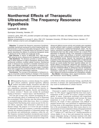
  • 4. streaming relates to the movement of objects from one place to another as a function of the force of the wave. In terms of ultrasound therapy, phonophoresis is commonly used to move medication transdermally. Second, cavitation relates to the os- cillation of microscopic gas bubbles that may, in turn, affect the cell or cellular process. However, the frequency resonance hypothesis relates to the absorption of ultrasound by proteins and protein complexes that may directly result in alterations Figure 1. The resonating mechanical force produced by the ultra- to signaling mechanisms within the cell, either by inducing a sound wave may induce transient conformational shifts. Molecules conformational shift or by disrupting a multimolecular com- are normally found in one of two 3-dimensional shapes, an ‘‘active’’ plex. or ‘‘inactive’’ conformation. In the original experiment investigating whether ultrasound could alter protein activity, the researchers reported no effect with respect to the monomer or dimer form of the enzyme creatine kinase.61 However, Chetverikova et al61 reported that ultrasound decreased the activity of the dimeric and tetrameric forms of creatine kinase and suggested that the decrease in activity was due to the disruption of the multimolecular forms of creatine kinase (represented in Figure 2). The authors in- ferred that ultrasound did not directly affect enzyme activity and that the primary acousto-biological interaction appeared Figure 2. Resonant or shearing forces produced by ultrasound to be occurring at a higher level of organization complexity. may result in the disjunction of a multimolecular complex with a However, more recent investigations7–16,54,55 have shown that subsequent loss of function or decrease in activity. ultrasound increases thrombolysis, demonstrating that ultra- sound can increase enzyme activity (represented in Figure 1). These data support the tried research saying, ‘‘The absence of proof is not the proof of absence.’’ The concept of the absorption of ultrasonic energy by en- zymatic proteins leading to changes in the enzymes’ activity is not novel61,62; however, the demonstration that ultrasound can increase or decrease protein activity and possible gene regulation is more recent.7–16,54–56 While considerable data ex- Figure 3. Resonant or shearing mechanical forces produced by ist, a model suggesting a molecular mechanism for how the ultrasound may dislodge an inhibitor molecule, leading to activa- tion of a signal-transduction pathway. absorption of ultrasound by proteins affects the cell function is novel and is presented here for the first time. Frequency resonance is one possible explanation of why The frequency resonance hypothesis suggests that the en- exposure to ultrasound increased enzymatic activity, resulted ergy provided to the enzyme by the ultrasound wave may in- in thrombolysis, and did not alter the activity of the enzymes duce transient conformational shifts in certain enzymatic pro- creatine kinase, lactate dehydrogenase, hexokinase, and pyru- teins, altering the enzyme’s activity (ie, kinases or vate kinase.7–16,54,55,61 A simple analogy would be 2 tuning phosphatases) and the overall function of the cell (Figure 1). forks located at one end of a room, with different frequencies Alternatively, ultrasound’s resonating force may result in the A and B. At the opposite end of the room is a third tuning dissociation of functional multimolecular complexes (Figure fork with the same frequency as fork A. Fork A is struck, 2) or the release of a sequestered molecule by dislodging an generating a sound. The sound waves travel through the air inhibitor molecule from the multimolecular complex (Figure and are absorbed at the other end of the room by fork A to 3). In essence, the mechanism of ultrasound’s action in Figures produce the same resonating sound, while fork B remains si- 2 and 3 is the same. Ultrasound disrupts a multimolecular lent. The possibility exists that thrombolysis is affected by the complex. However, Figure 2 represents a functionally active frequencies ‘‘designated’’ as therapeutic, while creatine kinase, complex, while Figure 3 represents a functionally sequestered lactate dehydrogenase, hexokinase, and pyruvate kinase are molecule. One can view an inhibitor molecule as a ‘‘safety not.7–16,54,55,61 Importantly, these reports do not directly dem- block’’ that functionally inhibits or sequesters a protein from onstrate that a conformational shift is occurring in these en- working. When the safety block is released, the protein is then zymes but support the hypothesis. operable. On the surface, it may appear that the observed decrease in Shearing forces produced by ultrasound may also play a role creatine kinase activity and an increase in thrombolytic activity in the dissociation of multimolecular complexes. Hypotheti- are contradictory; however, a fundamental difference exists. cally, frequency resonance may imply that different frequen- The decrease in activity of the creatine kinase was, most likely, cies (1 MHz, 3 MHz, 45 kHz, and others) establish unique a result of the disruption of the multimolecular dimeric or resonant or shearing forces (or both). Moreover, various fre- tetrameric (or both) forms of creatine kinase (Figure 2), while quencies may affect combinations of proteins or multimolec- the increased thrombolytic activity may be more associated ular complexes in different ways, lending to the possibility of with activation through harmonic resonance (Figure 1). targeted effects at the cellular and molecular levels. In any case, the mechanical effects of ultrasound may result The frequency resonance hypothesis differs from acoustic in either the activation or inactivation of an enzymatic protein streaming and cavitation at the basic levels. First, acoustic or a dissociation of a protein complex, leading to alterations in 296 Volume 37 • Number 3 • September 2002
  • 5. signal transduction. The frequency resonance hypothesis may However due to pharmocokinetics (ie, administration, absorp- describe the molecular mechanism or mechanisms responsible tion, distribution, and elimination of a drug), higher concen- for alterations in cellular membrane properties,34,39,41,46,49 in- trations and multiple doses per day are normally required to creases in protein production,* and modulation of enzyme ac- achieve clinical efficacy. tivity.7–16,54–56 While general recommendations for ultrasound treatment Frequency resonance and shearing forces on multimolecular suggest 5 to 10 minutes of exposure and 1 to 3 treatments per complexes may combine to produce the nonthermal effects of day, clinical treatments are almost exclusively done once a therapeutic ultrasound. Collectively, the experiments reviewed day. The possibility exists that clinical treatment protocols here support the frequency resonance hypothesis and demon- commonly employed for ultrasound are not sufficient for ther- strate that therapeutic ultrasound may modulate signal-trans- apeutic efficacy. In the review by Robertson and Baker,63 both duction pathways and gene products associated with the in- of the methodologically acceptable studies showing clinical flammatory response and cells directly involved in the healing efficacy used pulsed ultrasound (1:4) with treatment times of response (see Table). 15 minutes, resulting in energy densities of 60 and 150 J/cm2, respectively.64,65 Conversely, the remaining 5 studies lacking efficacy employed pulsed ultrasound and exposure times of 2 Clinical Implication of Ultrasound Research at the to 10 minutes, resulting in overall energy densities of 2 to 40 Cellular and Molecular Levels J/cm2.66–70 The frequency resonance hypothesis may suggest The purpose of this paper is to raise the awareness that that different ultrasonic frequencies (1 MHz, 3 MHz, 45 kHz, therapeutic levels of ultrasound (1 MHz, 3 MHz, and 45 kHz) and other) may require different durations of exposure (time), stimulate cellular and molecular effects within cells that are different energy densities (J/cm2), or both, to reach therapeutic centrally involved in the inflammatory and healing processes efficacy. (Table).1,5,7–16,34,40–42,45–55 Cumulatively, these reports pro- The identification and scientific understanding of therapeu- vide important information that may lead to a better under- tic ultrasound’s nonthermal mechanisms may lead to a com- standing and clinical application of therapeutic ultra- prehensive and effective clinical strategy. Further research is sound.1,5,7–16,34,40–42,45–55 Currently, no clear guidelines exist needed on 2 fronts: (1) cellular and molecular research to de- that provide the clinician with protocols directing when in the termine whether the mechanical mechanisms proposed by the injury and healing response ultrasound should be adminis- frequency resonance hypothesis can be elucidated and provide tered, nor are there guidelines on the frequency, intensity, insight into a comprehensive strategy for the clinical indica- treatment times, or number of treatments required for efficacy. tions of therapeutic ultrasound at various frequencies, and (2) In a recent review of the literature, a wide spectrum of ultra- methodologically sound clinical research designed to provide sound treatment protocols was found.63 In the 10 papers found meaningful input and outcome measures related to clinical ef- to be scientifically acceptable, a broad range of treatment set- ficacy. Both avenues of research should strive to establish time tings and methods was used, including (1) nine different clin- and dose-dependent response curves. ical indications, (2) five different frequencies, (3) continuous and pulsed output, (4) W/cm2 ranging from 0.02 to 2.6, (5) ACKNOWLEDGMENTS treatment time ranging from 2 to 15 minutes, and (6) energy density ranging from 2 to 150 J/cm2.64–70 Due to the variety A portion of the time spent developing this manuscript was sup- of clinical indicators and methods employed,63–70 sufficient ported by a grant from the National Athletic Trainers’ Association Research and Education Foundation. clinical data are currently not available to generate scientifi- cally sound recommendations for treatment protocols. The ultrasound treatment protocols employed to investi- REFERENCES gate the cellular and molecular effects (see Table) were 1. Dyson M. Mechanisms involved in therapeutic ultrasound. Physiotherapy. usually a 10-minute exposure at 1 or 3 MHz and 0.1 to 1.5 1987;73:116–120. W/cm2.5,39–42,45,48,50,53,56 The experimental protocol inves- 2. Kitchen SS, Partridge CJ. A review of therapeutic ultrasound. Physio- tigating thrombolysis ranged from 10 to 200 minutes of ex- therapy. 1990;76:593–600. posure at 0.17 to 3.4 MHz and 0.25 to 8.0 W/cm2, both 3. Hadjiargyrou M, McLeod K, Ryaby JP, Rubin C. Enhancement of fracture continuous and intermittent.7–16 A third experimental pro- healing by low intensity ultrasound. Clin Orthop. 1998;355(suppl):216– tocol employing a frequency of 45 kHz and an intensity 229. range of 5 to 100 mW/cm2 resulted in increased production 4. Kristiansen TK, Ryaby JP, McCabe J, Frey JJ, Roe LR. Accelerated heal- ing of distal radial fractures with the use of specific, low-intensity ultra- of IL-1, IL-8, vascular endothelial growth factor, fibrino- sound: a multicenter, prospective, randomized, double-blind, placebo-con- blast growth factor-b, and collagen, promotion of bone heal- trolled study. J Bone Joint Surg Am. 1997;79:961–973. ing, and acceleration of thrombolysis.5,45,54,55 5. Reher P, Elbeshir NL, Harvey W, Meghji S, Harris M. The stimulation The logical argument is that in vitro effects cannot be di- of bone formation in vitro by therapeutic ultrasound. Ultrasound Med rectly applied to clinical treatment protocols. However, from Biol. 1997;23:1251–1258. a cellular biology point of view, a strong argument can be 6. Rantanen J, Thorsson O, Wollmer P, Hurme T, Kalimo H. Effects of made that if a stimulus reaches a critical threshold (eg, con- therapeutic ultrasound on the regeneration of skeletal myofibers after ex- sider depolarization thresholds) the cell will respond, regard- perimental muscle injury. Am J Sports Med. 1999;27:54–59. less of whether the cell is in vitro or in vivo (eg, insulin, 7. Blinc A, Francis CW, Trudnowski JL, Carstensen E. Characterization of histamine, aspirin, or any of the proteins listed in the Table). ultrasound-potentiated fibrolysis in vitro. Blood. 1993;81:2636–2643. 8. Francis CW, Onundarson PT, Carstensen EL, et al. Enhancement of fi- Importantly, cells in tissue culture usually respond to nano- binolysis in vitro by ultrasound. J Clin Invest. 1992;90:2063–2068. gram or microgram per milliliter quantities of a stimulus. 9. Harpaz D, Chen X, Francis CW, Marder VJ, Metzer RS. Ultrasound en- hancement of thrombolysis and reperfusion in vitro. J Am Coll Cardiol. *References 5, 37, 39–42, 45, 48, 50, 53, 57. 1993;21:1507–1511. Journal of Athletic Training 297
  • 6. 10. Higazi AA, Katz I, Mayer M, Weiss AT. The effect of ultrasonic irradi- beta in the regulation of the immune response. Clin Immunol Immuno- ation and temperature on fibrinolytic activity in vitro. Thromb Res. 1993; pathol. 1992;65:1–9. 69:251–253. 37. Hogg N, Landis RC. Adhesion molecules in cell interactions. Curr Opin 11. Kornowski R, Meltzer RS, Chernine A, Vered Z, Battler A. Does external Immunol. 1993;5:383–390. ultrasound accelerate thrombolysis? Results from a rabbit model. Circu- 38. Pardi R, Inverardi L, Bender JR. Regulatory mechanisms in leukocyte lation. 1994;89:339–344. adhesion: flexible receptors for sophisticated travelers. Immunol Today. 12. Kudo S. Thrombolysis with ultrasound effect. Tokyo Jikeikai Med J. 1992;13:224–230. 1989;104:1005–1012. 39. Chokshi SK, Rongione AJ, Freeman I, Gal D, Gunwald AM, Alliger W. 13. Lauer CG, Burge R, Tang DB, Bass BG, Gomez ER, Alving BM. Effect Ultrasonic energy produces endothelium dependent vasomoter-relaxation of ultrasound on tissue-type plasminogen activator-induced thrombolysis. in vitro [abstract]. Circulation. 1989;80(suppl II):565. Circulation. 1992;86:1257–1264. 40. Fischell TA, Abbas MA, Grant GW, Siegel RJ. Ultrasonic energy: effects 14. Luo H, Steffen W, Cercek B, Arunasalam S, Maurer G, Siegel RJ. En- on vascular function and integrity. Circulation. 1991;84:1783–1795. hancement of thrombolysis by external ultrasound. Am Heart J. 1993; 41. Maxwell L, Collecutt T, Gledhill M, Sharma S, Edgar S, Gavin JB. The 125:1564–1569. augmentation of leucocytes adhesion to endothelium by therapeutic ultra- 15. Nilsson AM, Odselius R, Roijer A, Olsson SB. Pro- and antifibrinolytic sound. Ultrasound Med Biol. 1994;20:383–390. effects of ultrasound on streptokinase-induced thrombolysis. Ultrasound 42. Steffen W, Cumberland D, Gaines P, et al. Catheter-delivered high inten- Med Biol. 1995;21:833–840. sity, low frequency ultrasound induces vasodilation in vivo. Eur Heart J. 16. Olsson SB, Johansson B, Nilsson AM, Olsson C, Roijer A. Enhancement 1994;15:369–376. of thrombolysis by ultrasound. Ultrasound Med Biol. 1994;20:375–382. 43. Paul WE, Seder RA. Lymphocyte responses and cytokines. Cell. 1994; 17. Williams R. Production and transmission of ultrasound. Physiotherapy. 76:241–251. 1987;73:113–116. 44. Sporn MB, Roberts AB, Wakefield LM, Assoian RK. Transforming 18. Draper DO, Schulthies S, Sorvisto P, Hautala AM. Temperature changes growth factor-beta: biological function and chemical structure. Science. in deep muscles of humans during ice and ultrasound therapies: an in 1986;233:532–534. vivo study. J Orthop Sports Phys Ther. 1995;21:153–157. 45. Doan N, Reher P, Meghji S, Harris M. In vitro effects of therapeutic 19. Draper DO, Castel JC, Castel D. Rate of temperature increase in human ultrasound on cell proliferation, protein synthesis and cytokine production muscle during 1-MHz and 3-MHz continuous ultrasound. J Orthop Sports by human fibroblasts, osteoblasts and monocytes. J Oral Maxillofac Surg. Phys Ther. 1995;22:142–150. 1999;57:409–419. 20. Ashton DF, Draper DO, Myrer JW. Temperature rise in human muscle 46. Dyson M, Luke DA. Induction of mast cell degranulation in skin by during ultrasound treatments using Flex-All as a coupling agent. J Athl ultrasound. IEEE Trans Ultrason Ferroelectr Freq Control. 1986;33:194– Train. 1998;33:136–140. 201. 21. Chan AK, Myrer JW, Measom G, Draper DO. Temperature changes in 47. Hogan RD, Franklin TD, Fry FJ, Avery KA, Burke KM. The effect of human patellar tendon in response to therapeutic ultrasound. J Athl Train. ultrasound on microvascular hemodynamics in skeletal muscle: effect on 1998;33:130–135. arterioles. Ultrasound Med Biol. 1982;8:45–55. 22. ter Haar G. Therapeutic ultrasound. Eur J Ultrasound. 1999;9:3–9. 48. Johns LD, Colloton PA, Neuenfeldt J. Effects of therapeutic ultrasound 23. Love LA, Kremkau FW. Intracellular temperature distribution produced on T cell proliferation and IL-2 production [abstract]. J Athl Train. 1999; by ultrasound. J Accoust Soc Am. 1980;67:1045–1050. 34(suppl):S24. 24. Nyborg WL. Physical Mechanisms for Biological Effects of Ultrasound. 49. Mortimer AJ, Dyson M. The effect of therapeutic ultrasound on calcium Washington, DC: Department of Health, Education, and Welfare; 1978. uptake in fibroblasts. Ultrasound Med Biol. 1988;14:499–506. FDA Publication 78–8062. 50. Reher P, Doan N, Bradnock B, Meghji S, Harris M. Effect of ultrasound 25. Nyborg WL. Ultrasonic microstreaming and related phenomena. Br J on the production of IL-8, basic FGF and VEGF. Cytokine. 1999;11:416– Cancer Suppl. 1982;45:156–160. 423. 26. Stewart HG, Stratmeyer ME. An Overview Of Ultrasound: Theory, Mea- 51. Vivino AA, Boraker DK, Miller D, Nyborg W. Stable cavitation at low surement, Medical Applications, and Biological Effects. Washington, DC: ultrasonic intensities induces cell death and inhibits 3H-TdR incorporation Department of Health, Education, and Welfare; 1982. DHEW Publication by Con-A-stimulated murine lymphocytes in vitro. Ultrasound Med Biol. 82–8190. 1985;11:751–759. 27. Michlovitz SL, ed. Thermal Agents in Rehabilitation. Philadelphia, PA: 52. Young SR, Dyson M. The effect of therapeutic ultrasound on angiogen- FA Davis Co; 1986:3–17,141–176. esis. Ultrasound Med Biol. 1989;16:261–269. 28. Dyson M, Pond JB, Joseph J, Warwick R. The stimulation of tissue re- 53. Young SR, Dyson M. Macrophage responsiveness to therapeutic ultra- generation by means of ultrasound. Clin Sci. 1968;35:273–285. sound. Ultrasound Med Biol. 1990;16:809–816. 29. Loch EG, Fischer AB, Kuwert E. Effect of diagnostic and therapeutic 54. Gaul GB. Ultrasound thrombolysis. Thromb Heamost. 1999;82(suppl 1): intensities of ultrasonics on normal and malignant human cells in vitro. 157–179. Am J Obstet Gynecol. 1971;110:457–460. 55. Tachibana K, Tachibana S. Prototype therapeutic ultrasound emitting 30. Maeda K, Murao F. Studies on the influence of ultrasound irritation of catheter for accelerating thrombolysis. J Ultrasound Med. 1997;16:529– the growth of cultured cell in vitro. In: White D, Brown RE, eds. Ultra- 535. sound in Medicine. Volume 3B. New York, NY: Plenum Press; 1977: 56. Unger EC, McCreery TP, Sweitzer RH. Ultrasound enhances gene ex- 2045. pression of liposomal transfection. Invest Radiol. 1997;32:723–727. 31. Pizzarello DJ, Wolsky A, Becker MH, Keegan AF. A new approach to 57. Zur O, Shneyour Y, Dan U, Chemke J, Shneyour A, Shalev E. A possible testing the effects of ultrasound on tissue growth and differentiation. On- effect of ultrasound on RNA synthesis in cultured amniocytes. Acta cology. 1975;31:226–232. Obstet Gynecol Scand. 1993;72:243–245. 32. Ross P, Edmonds PD. Ultrasound induced protein synthesis as a result of 58. Karin M, Smeal T. Control of transcription factors by signal transduction membrane damage. J Ultrasound Med. 1983;2(suppl):47. pathways: the beginning of the end. Trends Biochem Sci. 1992;17:418– 33. Webster DF, Pond JB, Dyson M, Harvey W. The role of cavitation in the 422. in vitro stimulation of protein synthesis in human fibroblasts by ultra- 59. Stabel S, Parker PJ. Protein kinase C. Pharmacol Ther. 1991;51:71–95. sound. Ultrasound Med Biol. 1978;4:343–351. 60. Shirakawa F, Mizel SB. In vitro activation and nuclear translocation of 34. Anderson DW, Barrett JT. Depression of phagocytosis by ultrasound. Ul- NF-kappa B catalyzed by cyclic AMP-dependent protein kinase and pro- trasound Med Biol. 1981;7:267–273. tein kinase C. Mol Cell Biol. 1989;9:2424–2430. 35. Pinamonti S, Gallenga PE, Masseo V. Effects of pulsed ultrasound on 61. Chetverikova EP, Pashovkin TN, Rosanova NA, Sarvazyan AP, Williams human erythrocytes in vitro. Ultrasound Med Biol. 1982;8:631–638. AR. Interaction of therapeutic ultrasound with purified enzymes in vitro. 36. Sasaki H, Pollard RB, Schmitt D, Suzuki F. Transforming growth factor- Ultrasonics. 1985;23:183–188. 298 Volume 37 • Number 3 • September 2002
  • 7. 62. Sarvazyan AP. Acoustic properties of tissues relevant to therapeutic ap- 67. Hashish I, Hai H, Harvey W, Feinmann C, Harris M. Reproduction of plications. Br J Cancer Suppl. 1982;45:52–54. postoperative pain and swelling by ultrasound treatment: a placebo effect. 63. Robertson VJ, Baker KG. A review of therapeutic ultrasound: effective- Pain. 1988;33:303–311. ness studies. Phys Ther. 2001;81:1339–1350. 68. Grant A, Sleep J, McIntosh J, Ashurst H. Ultrasound and pulsed electro- 64. Ebenbichler GR, Resch KL, Nicolakis P, et al. Ultrasound treatment for magnetic energy treatment for perineal trauma: a randomized placebo- treating the carpal tunnel syndrome: randomised ‘‘sham’’ controlled trial. controlled trial. Br J Obstet Gynaecol. 1989;96:434–439. BMJ. 1998;316:731–735. 69. ter Riet G, Kessel AG, Knipschild P. A randomized clinical trial of ultra- 65. Ebenbichler GR, Erdogmus CB, Resch KL, et al. Ultrasound therapy for calcific tendinitis of the shoulder. N Engl J Med. 1999;340:1533–1538. sound in the treatment of pressure ulcers. Phys Ther. 1996;76:1301–1311. 66. Hashish I, Harvey W, Harris M. Anti-inflammatory effects of ultrasound 70. Nykanen M. Pulsed ultrasound treatment of the painful shoulder: a ran- therapy: evidence for a major placebo effect. Br J Rheumatol. 1986;25: domized, double-blind, placebo-controlled study. Scand J Rehabil Med. 77–81. 1995;27:105–108. Journal of Athletic Training 299