SlideShare ist ein Scribd-Unternehmen logo
1 von 30
Overall, the tongue has  five  major functions: Tongue  – The tongue is formed mainly of interlacing  skeletal muscle  tissue.  When the mouth is opened, only a small portion of the entire tongue is actually seen.  Visible portion of the tongue
1.  The tongue is the  main organ of  taste . 2.  The tongue plays a  major role in  speech .  4.  The tongue  cleans  the teeth . 3.  The tongue presses against the roof of the mouth and  blocks off the oral cavity during the act of  swallowing  to prevent food from shooting back out the mouth.
5.  The most important function of the tongue is in its use as a food manipulator. The tongue pushes the food between the teeth, shapes the chewed food into a nice glob (called a  bolus ) and then pushes the mass into the oropharynx.
Salivary glands  –The three sets of salivary glands are: 1.  Parotid glands The function of the salivary glands is to produce about  one liter  of  saliva   per day. 2.  Submandibular glands 3.  Sublingual glands Parotid gland Submandibular  gland Sublingual gland
Saliva  is a thin, watery, fluid about 99.5% water by composition.  It has a somewhat slippery feel which is due to protein substance called  mucin . It also contains  inorganic salts , the digestive enzyme  salivary amylase  and a  bactericidal enzyme  called  lysozyme . The pH of saliva is about 6.4 to 7.0 (or about neutral). The functions of saliva: 1.  It is a solvent, which permits the taste buds of the tongue to operate. 2.  Saliva keeps the lining of the mouth moist and along with the tongue helps to keep the teeth clean.
3.  Mechanical digestion – Saliva helps to  moisten  and  lubricate  food as the teeth grind and the tongue manipulates it.  This is important because this action helps to protect the inner surface of the esophagus from damage by food particles. 4.  Chemical digestion – Saliva begins the chemical digestive process of carbohydrates.  This is done by converting large, complex carbohydrate molecules to smaller, simpler ones through the action of  Salivary Amylase .
The chemical digestive function of saliva is not very effective for several reasons:  1.  If chewing is not adequate, food is not thoroughly mixed with saliva so the chemical digestion from salivary amylase does not get a chance to occur.  2.  Beverages are often used to moisten food or “wash” food down which prevents adequate chewing, dilutes the saliva, thus not allowing amylase a chance to work. 3.  Many drinks are quite acidic and salivary amylase is deactivated by acid.
Teeth  – An adult set is 32 teeth. This includes: 1.  Eight (8)  incisors  for cutting. incisors
Teeth  – An adult set is 32 teeth. This includes: 1.  Eight (8)  incisors  for cutting. 2.  Four (4)  canines  or  cuspids  for ripping or tearing. canines
Teeth  – An adult set is 32 teeth. This includes: 1.  Eight (8)  incisors  for cutting. 2.  Four (4)  canines  or  cuspids  for ripping or tearing. 3.  Eight (8)  bicuspids  or  premolars  for tearing and crushing.  bicuspids
Teeth  – An adult set is 32 teeth. This includes: 1.  Eight (8)  incisors  for cutting. 2.  Four (4)  canines  or  cuspids  for ripping or tearing. 3.  Eight (8)  bicuspids  or  premolars  for tearing and crushing.  4.  Twelve (12)  tricuspids  or  molars , four of which are the “wisdom teeth,” for crushing or grinding. tricuspids
The function of the teeth is to  masticate  (chew) food which means to grind and/or pulverize food.  The process of  mastication  (chewing) begins by grasping the food and cutting or tearing off a piece with the “front” teeth. The food is then ground to a mash between the “back” teeth.  It ends with the tongue shaping the pulverized food into a bolus ready for  deglutition  (the process of “swallowing”).
Deglutition  – “Swallowing” is a voluntary action in the beginning but involuntary once begun.  In other words, once it begins, it cannot be stopped by voluntary mechanisms.  It involves the tongue, pharynx, esophagus, brain and associated nerves and is divided into three stages: Buccal Stage  – this first stage is the  voluntary  phase where masticated & moistened food is shaped into a  bolus  and directed by the tongue to the back of the mouth. Through a series of voluntary muscle actions, the nasopharynx is closed off and the food is forced into the pharynx. At this point, reflex mechanisms take over and swallowing cannot be stopped.
Pharyngeal Stage  – this second stage is an  automatic  phase where the food is passed through the pharynx to the esophagus. At this stage, several  involuntary  mechanisms controlled by reflex centers in the lower pons and medulla oblongata take over to assure that the food will pass into the esophagus rather than the trachea: 1.  The larynx rises and the epiglottis folds over the trachea opening. 2.  The tongue closes off the oral cavity by pressing against the roof of the mouth. 3.  The nasopharynx is blocked by the soft palate.
Esophageal Stage  – this third stage is an  automatic  phase where food passes through the esophagus and to the stomach. After food enters the esophagus,  peristaltic waves  aided by gravity propel the food to the stomach. When the pressure in the esophagus increases to greater than that in the stomach the  cardiac sphincter  relaxes, allowing food to pass through and into the stomach. The stomach then mixes food with  gastric juice  produced by  gastric glands . There are actually three different types of cells in  gastric glands , each secreting a different constituent of  gastric juice .
Mucous or Goblet Cells  – These secrete thin  mucus  which is mixed with the food. Chief cells  – These secrete  pepsinogen  which is the  inactive  form of the enzyme,  pepsin . Pepsin is an enzyme which splits protein molecules into smaller units called  peptides . It is able to do this because the hydrochloric acid helps to unravel proteins and activate the enzyme.  This means pepsinogen is transformed from inactive to active when it comes in contact with an acid environment of pH 3 or less.  The stomach has a pH of 1 to 2 with an HCl concentration of approximately 0.017 molar. ****Note – The functions of HCl are to activate pepsinogen to pepsin, “unravels” large molecules, and kill bacteria.
Parietal cells  – These cells secrete  hydrochloric acid  (HCl) and a substance called  intrinsic factor , which helps in the body’s absorption of vitamin B 12 .  These four things together (mucus, pepsinogen/pepsin, hydrochloric acid, and intrinsic factor) make up the majority of the composition gastric juice.  They are not the only components of gastric juice, however, just the most predominant.  Other enzymes in gastric juice are  rennin  (coagulates milk),  gastric lipase  (fat digestion), but these two enzymes are not very effective in acid environments so they are  important in children , but not in adults.
The secretion of gastric juice is a continuous process, however it does increase and decrease according to need. The regulation of this secretion can be divided into 3 phases: Cephalic phase  – During this stage the sight, smell, taste or even the thought of food (especially a favorite food) stimulates a flow of gastric secretions called  appetite juice , which is high in  HCl  and  pepsinogen . Gastric phase  – During this stage the food in the stomach stimulates the secretion of the hormone  gastrin . This hormone starts a loop that stimulates additional secretions gastric juice until the volume of food diminishes, thus reducing all secretions.
Intestinal phase  – During this stage the stomach is “empty” so only small amounts of secretions are produced.  This secretion contains mostly  mucus  and lack HCl, pepsinogen, and intrinsic factor for the most part. This phase continues until the small intestine empties 6 to 8 hours later or until one thinks about eating again. Thing that inhibit gastric juice flow: 1.  Unappetizing food  – The sight, smell, taste, or even suggestion of rotten, smelly, disgusting or disliked food immediately reduces appetite juice flow which shuts down the feeling of hunger.
2.  Excess acid  concentration in the stomach decreases gastric juice production by decreasing the release of gastrin. 3.  Fat laden   chyme  in the duodenum causes the release of a hormone called  cholecystokinin . This hormone inhibits both gastric secretions and gastric movements. 4.  Strenuous exercise  diverts blood to the heart and muscles and energy production is not used for digestion.
5.  Cold temperatures  inhibit stomach action by delaying peristaltic waves and stopping gastric juice secretion. Anything that decreases gastric juice flow slows down or delays digestion.  The draw back to delaying digestion is that microorganisms can ferment (“rot”) sugars in the food, producing large amounts of smelly gas (mostly hydrogen sulfide, H 2 S and methane, CH 4 ) that can cause severe bloating, cramps, and diarrhea.
The three accessory organs not yet discussed are the  liver ,  gall bladder , and  pancreas . Liver Gall Bladder Pancreas Note – This picture gives a perspective on the location of these accessory organs with respect to each other and the stomach.
The Liver  – The liver has only  one function  directly related to the mechanical or chemical digestion of food.  That function is to  produce   bile  and send it to the gall bladder. Liver Gall Bladder
Bile  – This yellowish green liquid serves only one digestive purpose.  This purpose is to  emulsify fats . This means that bile  physically  (mechanically NOT chemically) breaks up fat into smaller and smaller “blobs” which increases the total surface area of the fat and allows the  lipase  enzymes to chemically digest the fat more easily. ***Remember  –  Bile  emulsifies fat
Gall Bladder  –  located “underneath” the liver.  Has four functions which are all related to bile: Gall Bladder 1.  Receives bile  secretions from the liver. 2.  Stores bile  for secretion to the small intestine when dietary fat is consumed. 3.  Concentrates bile  by removing water 4.  Discharges bile  into the duodenum when fat laden chyme is present.
Pancreas  – This organ has both an exocrine (digestive) and endocrine (hormonal) function. Pancreas The  endocrine  portion of the pancreas consists of clusters of cells scattered along the organ called  islets of Langerhans  and they contain two types of secretory cells.  Alpha  cells secrete  glucagon  and  Beta  cells secrete  insulin .
Glucagon  stimulates the conversion of liver glycogen (animal starch), which is a large carbohydrate storage molecule, to   blood   glucose.  In other words, glucagon  raises blood sugar levels  when necessary.  Insulin  stimulates glycogen storage and cellular glucose use.  In other words, insulin  lowers blood sugar levels .  These hormones are secreted directly into the blood stream (this is what distinguishes the function as endocrine – all hormones are secreted directly into the blood stream).
The  excretory   portion of the pancreas consists of structures called  acini  (pancreatic acinar cells)which are “grape like” clusters of secretory cells that produce and empty their secretions into microscopically sized ducts.  These smaller ducts form a network that finally empty into a large common duct called the pancreatic duct, which empties into the duodenum.
This mixture of secretions called  pancreatic juice  contains digestive enzymes:  carboxypeptidase ,  trypsin  and  chymotrypsin  (“proteases” or protein digesting enzymes),  Sucrase ,  Lactase , and  Maltase  (“amylases” or carbohydrate digesting enzymes),  Pancreatic Lipase  (which breaks down fats),  RNase  and  DNase  (“nucleases” which break down nucleic acids DNA and RNA), and  sodium bicarbonate  (Sodium Hydrogen Carbonate or NaHCO 3 ) which  neutralizes the HCl  from the stomach. The enzymes trypsin, chymotrypsin, and carboxypeptidase are secreted as  trysinogen ,  chymotrypsinogen , and  procarboxypeptidase .
Pancreatic juice actually consists of two portions:  aqueous juice  which contains mostly  carbonate ions  and  enzyme juice  which contain the enzymes. The small intestine is lined with cells that secrete large amounts of mucus called goblet cells and are also lined with special mucus secreting glands that secrete large amounts of thick, alkaline mucus which helps to  neutralize the acidic secretions of the stomach. Some final thoughts on digestion: Enzymes that are active in the small intestine are not active in acid environments and those that are in the small intestine itself are some peptidases, sucrase, maltase, lactase and intestinal lipase.

Weitere ähnliche Inhalte

Was ist angesagt?

Human digestive system
Human digestive systemHuman digestive system
Human digestive systemKush Sehgal
 
Functioning organisms - 02 Digestive system
Functioning organisms - 02 Digestive systemFunctioning organisms - 02 Digestive system
Functioning organisms - 02 Digestive systemIan Anderson
 
Digestive System_ST.ppt
Digestive System_ST.pptDigestive System_ST.ppt
Digestive System_ST.pptShama
 
Chapt17 digestive system
Chapt17 digestive systemChapt17 digestive system
Chapt17 digestive systembholmes
 
Introduction to Digestive System
Introduction to Digestive System Introduction to Digestive System
Introduction to Digestive System Pharmacy Universe
 
The digestive system
The digestive systemThe digestive system
The digestive systemEnigmatic You
 
The Digestive System - Anatomy and Physiology - Biology and Geology 3rd CSE -...
The Digestive System - Anatomy and Physiology - Biology and Geology 3rd CSE -...The Digestive System - Anatomy and Physiology - Biology and Geology 3rd CSE -...
The Digestive System - Anatomy and Physiology - Biology and Geology 3rd CSE -...Víctor Gil García
 
Digestion System Hinojosa
Digestion System HinojosaDigestion System Hinojosa
Digestion System Hinojosaulia
 

Was ist angesagt? (20)

Human Digestive System
Human Digestive SystemHuman Digestive System
Human Digestive System
 
2 1 D Ithe Digestive System Powerpoint
2 1 D Ithe Digestive System Powerpoint2 1 D Ithe Digestive System Powerpoint
2 1 D Ithe Digestive System Powerpoint
 
Human digestive system
Human digestive systemHuman digestive system
Human digestive system
 
Functioning organisms - 02 Digestive system
Functioning organisms - 02 Digestive systemFunctioning organisms - 02 Digestive system
Functioning organisms - 02 Digestive system
 
Digestive System
Digestive SystemDigestive System
Digestive System
 
Digestive System_ST.ppt
Digestive System_ST.pptDigestive System_ST.ppt
Digestive System_ST.ppt
 
Digestive system Mouth
Digestive system MouthDigestive system Mouth
Digestive system Mouth
 
DIGESTIVE SYSTEM
DIGESTIVE SYSTEM DIGESTIVE SYSTEM
DIGESTIVE SYSTEM
 
Chapt17 digestive system
Chapt17 digestive systemChapt17 digestive system
Chapt17 digestive system
 
Small intestine and large intestine
Small intestine and large intestineSmall intestine and large intestine
Small intestine and large intestine
 
Introduction to Digestive System
Introduction to Digestive System Introduction to Digestive System
Introduction to Digestive System
 
Urinary system
Urinary systemUrinary system
Urinary system
 
The digestive system
The digestive systemThe digestive system
The digestive system
 
The Digestive System - Anatomy and Physiology - Biology and Geology 3rd CSE -...
The Digestive System - Anatomy and Physiology - Biology and Geology 3rd CSE -...The Digestive System - Anatomy and Physiology - Biology and Geology 3rd CSE -...
The Digestive System - Anatomy and Physiology - Biology and Geology 3rd CSE -...
 
Digestion System Hinojosa
Digestion System HinojosaDigestion System Hinojosa
Digestion System Hinojosa
 
Digestive system
Digestive systemDigestive system
Digestive system
 
Digestive system
Digestive systemDigestive system
Digestive system
 
ppt on Digestive system akki
ppt on Digestive system akkippt on Digestive system akki
ppt on Digestive system akki
 
Human digestive system
Human digestive systemHuman digestive system
Human digestive system
 
Digestive system
Digestive systemDigestive system
Digestive system
 

Ähnlich wie Acc organs digestion

Gi physiology
Gi physiologyGi physiology
Gi physiologysumi r
 
Digestive system
Digestive system Digestive system
Digestive system DeepaMadhu2
 
Nutrition & digestive system
Nutrition & digestive systemNutrition & digestive system
Nutrition & digestive systemitutor
 
Basic ap chapter 23 powerpoint 2017
Basic ap chapter 23 powerpoint   2017Basic ap chapter 23 powerpoint   2017
Basic ap chapter 23 powerpoint 2017Kathy Richards
 
The Digestive System.pptx
The Digestive System.pptxThe Digestive System.pptx
The Digestive System.pptxSanthi Dasari
 
Organs of the Digestive System.pptx
Organs of the Digestive System.pptxOrgans of the Digestive System.pptx
Organs of the Digestive System.pptxFredereck Dominion
 
Mastication, degluttition, digestion, assimilation
Mastication, degluttition, digestion, assimilationMastication, degluttition, digestion, assimilation
Mastication, degluttition, digestion, assimilationNeelam Bhandari
 
Digestive system (jane)
Digestive system (jane)Digestive system (jane)
Digestive system (jane)imzventura17
 
Digestive System
Digestive SystemDigestive System
Digestive Systemjhanavip13
 

Ähnlich wie Acc organs digestion (20)

Digestive system.pptx
Digestive system.pptxDigestive system.pptx
Digestive system.pptx
 
Digestive System
Digestive SystemDigestive System
Digestive System
 
Gi physiology
Gi physiologyGi physiology
Gi physiology
 
Digestive system
Digestive system Digestive system
Digestive system
 
10-Biology.pdf
10-Biology.pdf10-Biology.pdf
10-Biology.pdf
 
Digestive system
Digestive systemDigestive system
Digestive system
 
Nutrition & digestive system
Nutrition & digestive systemNutrition & digestive system
Nutrition & digestive system
 
Basic ap chapter 23 powerpoint 2017
Basic ap chapter 23 powerpoint   2017Basic ap chapter 23 powerpoint   2017
Basic ap chapter 23 powerpoint 2017
 
HUMAN DIGESTIVE SYSTEM
HUMAN DIGESTIVE SYSTEMHUMAN DIGESTIVE SYSTEM
HUMAN DIGESTIVE SYSTEM
 
113-Digestion.ppt
113-Digestion.ppt113-Digestion.ppt
113-Digestion.ppt
 
Digestive System
Digestive SystemDigestive System
Digestive System
 
The Digestive System.pptx
The Digestive System.pptxThe Digestive System.pptx
The Digestive System.pptx
 
Organs of the Digestive System.pptx
Organs of the Digestive System.pptxOrgans of the Digestive System.pptx
Organs of the Digestive System.pptx
 
Human Digestive System
Human Digestive SystemHuman Digestive System
Human Digestive System
 
Lek 13-14.pptx
Lek 13-14.pptxLek 13-14.pptx
Lek 13-14.pptx
 
Mastication, degluttition, digestion, assimilation
Mastication, degluttition, digestion, assimilationMastication, degluttition, digestion, assimilation
Mastication, degluttition, digestion, assimilation
 
Digestion!
Digestion!Digestion!
Digestion!
 
Digestive system
Digestive systemDigestive system
Digestive system
 
Digestive system (jane)
Digestive system (jane)Digestive system (jane)
Digestive system (jane)
 
Digestive System
Digestive SystemDigestive System
Digestive System
 

Mehr von Michael Wrock

Mehr von Michael Wrock (20)

Protection of the cns
Protection of the cnsProtection of the cns
Protection of the cns
 
Deep brain structures
Deep brain structuresDeep brain structures
Deep brain structures
 
Muscle contraction relaxation notes
Muscle contraction  relaxation notesMuscle contraction  relaxation notes
Muscle contraction relaxation notes
 
Action potential notes
Action potential notesAction potential notes
Action potential notes
 
Review motor end plate nmj notes
Review motor end plate  nmj notesReview motor end plate  nmj notes
Review motor end plate nmj notes
 
Articulations
ArticulationsArticulations
Articulations
 
Respiratory review part 2
Respiratory review part 2Respiratory review part 2
Respiratory review part 2
 
Respiratory review part 1
Respiratory review part 1Respiratory review part 1
Respiratory review part 1
 
Laryn trachea
Laryn  tracheaLaryn  trachea
Laryn trachea
 
Intro nose pharynx
Intro nose  pharynxIntro nose  pharynx
Intro nose pharynx
 
Bronchial tree alveoli lungs review
Bronchial tree alveoli lungs  reviewBronchial tree alveoli lungs  review
Bronchial tree alveoli lungs review
 
Breathing
BreathingBreathing
Breathing
 
Volumes capacities 2
Volumes  capacities 2Volumes  capacities 2
Volumes capacities 2
 
Axial skeleton parts 1 2
Axial skeleton parts 1  2Axial skeleton parts 1  2
Axial skeleton parts 1 2
 
Appendicular skeleton
Appendicular skeletonAppendicular skeleton
Appendicular skeleton
 
Axial skeleton parts 3 5
Axial skeleton parts 3   5Axial skeleton parts 3   5
Axial skeleton parts 3 5
 
Reflexes
ReflexesReflexes
Reflexes
 
Tissue
TissueTissue
Tissue
 
Neuroglia
NeurogliaNeuroglia
Neuroglia
 
Reflexes
ReflexesReflexes
Reflexes
 

Acc organs digestion

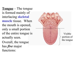
  • 1. Overall, the tongue has five major functions: Tongue – The tongue is formed mainly of interlacing skeletal muscle tissue. When the mouth is opened, only a small portion of the entire tongue is actually seen. Visible portion of the tongue
  • 2. 1. The tongue is the main organ of taste . 2. The tongue plays a major role in speech . 4. The tongue cleans the teeth . 3. The tongue presses against the roof of the mouth and blocks off the oral cavity during the act of swallowing to prevent food from shooting back out the mouth.
  • 3. 5. The most important function of the tongue is in its use as a food manipulator. The tongue pushes the food between the teeth, shapes the chewed food into a nice glob (called a bolus ) and then pushes the mass into the oropharynx.
  • 4. Salivary glands –The three sets of salivary glands are: 1. Parotid glands The function of the salivary glands is to produce about one liter of saliva per day. 2. Submandibular glands 3. Sublingual glands Parotid gland Submandibular gland Sublingual gland
  • 5. Saliva is a thin, watery, fluid about 99.5% water by composition. It has a somewhat slippery feel which is due to protein substance called mucin . It also contains inorganic salts , the digestive enzyme salivary amylase and a bactericidal enzyme called lysozyme . The pH of saliva is about 6.4 to 7.0 (or about neutral). The functions of saliva: 1. It is a solvent, which permits the taste buds of the tongue to operate. 2. Saliva keeps the lining of the mouth moist and along with the tongue helps to keep the teeth clean.
  • 6. 3. Mechanical digestion – Saliva helps to moisten and lubricate food as the teeth grind and the tongue manipulates it. This is important because this action helps to protect the inner surface of the esophagus from damage by food particles. 4. Chemical digestion – Saliva begins the chemical digestive process of carbohydrates. This is done by converting large, complex carbohydrate molecules to smaller, simpler ones through the action of Salivary Amylase .
  • 7. The chemical digestive function of saliva is not very effective for several reasons: 1. If chewing is not adequate, food is not thoroughly mixed with saliva so the chemical digestion from salivary amylase does not get a chance to occur. 2. Beverages are often used to moisten food or “wash” food down which prevents adequate chewing, dilutes the saliva, thus not allowing amylase a chance to work. 3. Many drinks are quite acidic and salivary amylase is deactivated by acid.
  • 8. Teeth – An adult set is 32 teeth. This includes: 1. Eight (8) incisors for cutting. incisors
  • 9. Teeth – An adult set is 32 teeth. This includes: 1. Eight (8) incisors for cutting. 2. Four (4) canines or cuspids for ripping or tearing. canines
  • 10. Teeth – An adult set is 32 teeth. This includes: 1. Eight (8) incisors for cutting. 2. Four (4) canines or cuspids for ripping or tearing. 3. Eight (8) bicuspids or premolars for tearing and crushing. bicuspids
  • 11. Teeth – An adult set is 32 teeth. This includes: 1. Eight (8) incisors for cutting. 2. Four (4) canines or cuspids for ripping or tearing. 3. Eight (8) bicuspids or premolars for tearing and crushing. 4. Twelve (12) tricuspids or molars , four of which are the “wisdom teeth,” for crushing or grinding. tricuspids
  • 12. The function of the teeth is to masticate (chew) food which means to grind and/or pulverize food. The process of mastication (chewing) begins by grasping the food and cutting or tearing off a piece with the “front” teeth. The food is then ground to a mash between the “back” teeth. It ends with the tongue shaping the pulverized food into a bolus ready for deglutition (the process of “swallowing”).
  • 13. Deglutition – “Swallowing” is a voluntary action in the beginning but involuntary once begun. In other words, once it begins, it cannot be stopped by voluntary mechanisms. It involves the tongue, pharynx, esophagus, brain and associated nerves and is divided into three stages: Buccal Stage – this first stage is the voluntary phase where masticated & moistened food is shaped into a bolus and directed by the tongue to the back of the mouth. Through a series of voluntary muscle actions, the nasopharynx is closed off and the food is forced into the pharynx. At this point, reflex mechanisms take over and swallowing cannot be stopped.
  • 14. Pharyngeal Stage – this second stage is an automatic phase where the food is passed through the pharynx to the esophagus. At this stage, several involuntary mechanisms controlled by reflex centers in the lower pons and medulla oblongata take over to assure that the food will pass into the esophagus rather than the trachea: 1. The larynx rises and the epiglottis folds over the trachea opening. 2. The tongue closes off the oral cavity by pressing against the roof of the mouth. 3. The nasopharynx is blocked by the soft palate.
  • 15. Esophageal Stage – this third stage is an automatic phase where food passes through the esophagus and to the stomach. After food enters the esophagus, peristaltic waves aided by gravity propel the food to the stomach. When the pressure in the esophagus increases to greater than that in the stomach the cardiac sphincter relaxes, allowing food to pass through and into the stomach. The stomach then mixes food with gastric juice produced by gastric glands . There are actually three different types of cells in gastric glands , each secreting a different constituent of gastric juice .
  • 16. Mucous or Goblet Cells – These secrete thin mucus which is mixed with the food. Chief cells – These secrete pepsinogen which is the inactive form of the enzyme, pepsin . Pepsin is an enzyme which splits protein molecules into smaller units called peptides . It is able to do this because the hydrochloric acid helps to unravel proteins and activate the enzyme. This means pepsinogen is transformed from inactive to active when it comes in contact with an acid environment of pH 3 or less. The stomach has a pH of 1 to 2 with an HCl concentration of approximately 0.017 molar. ****Note – The functions of HCl are to activate pepsinogen to pepsin, “unravels” large molecules, and kill bacteria.
  • 17. Parietal cells – These cells secrete hydrochloric acid (HCl) and a substance called intrinsic factor , which helps in the body’s absorption of vitamin B 12 . These four things together (mucus, pepsinogen/pepsin, hydrochloric acid, and intrinsic factor) make up the majority of the composition gastric juice. They are not the only components of gastric juice, however, just the most predominant. Other enzymes in gastric juice are rennin (coagulates milk), gastric lipase (fat digestion), but these two enzymes are not very effective in acid environments so they are important in children , but not in adults.
  • 18. The secretion of gastric juice is a continuous process, however it does increase and decrease according to need. The regulation of this secretion can be divided into 3 phases: Cephalic phase – During this stage the sight, smell, taste or even the thought of food (especially a favorite food) stimulates a flow of gastric secretions called appetite juice , which is high in HCl and pepsinogen . Gastric phase – During this stage the food in the stomach stimulates the secretion of the hormone gastrin . This hormone starts a loop that stimulates additional secretions gastric juice until the volume of food diminishes, thus reducing all secretions.
  • 19. Intestinal phase – During this stage the stomach is “empty” so only small amounts of secretions are produced. This secretion contains mostly mucus and lack HCl, pepsinogen, and intrinsic factor for the most part. This phase continues until the small intestine empties 6 to 8 hours later or until one thinks about eating again. Thing that inhibit gastric juice flow: 1. Unappetizing food – The sight, smell, taste, or even suggestion of rotten, smelly, disgusting or disliked food immediately reduces appetite juice flow which shuts down the feeling of hunger.
  • 20. 2. Excess acid concentration in the stomach decreases gastric juice production by decreasing the release of gastrin. 3. Fat laden chyme in the duodenum causes the release of a hormone called cholecystokinin . This hormone inhibits both gastric secretions and gastric movements. 4. Strenuous exercise diverts blood to the heart and muscles and energy production is not used for digestion.
  • 21. 5. Cold temperatures inhibit stomach action by delaying peristaltic waves and stopping gastric juice secretion. Anything that decreases gastric juice flow slows down or delays digestion. The draw back to delaying digestion is that microorganisms can ferment (“rot”) sugars in the food, producing large amounts of smelly gas (mostly hydrogen sulfide, H 2 S and methane, CH 4 ) that can cause severe bloating, cramps, and diarrhea.
  • 22. The three accessory organs not yet discussed are the liver , gall bladder , and pancreas . Liver Gall Bladder Pancreas Note – This picture gives a perspective on the location of these accessory organs with respect to each other and the stomach.
  • 23. The Liver – The liver has only one function directly related to the mechanical or chemical digestion of food. That function is to produce bile and send it to the gall bladder. Liver Gall Bladder
  • 24. Bile – This yellowish green liquid serves only one digestive purpose. This purpose is to emulsify fats . This means that bile physically (mechanically NOT chemically) breaks up fat into smaller and smaller “blobs” which increases the total surface area of the fat and allows the lipase enzymes to chemically digest the fat more easily. ***Remember – Bile emulsifies fat
  • 25. Gall Bladder – located “underneath” the liver. Has four functions which are all related to bile: Gall Bladder 1. Receives bile secretions from the liver. 2. Stores bile for secretion to the small intestine when dietary fat is consumed. 3. Concentrates bile by removing water 4. Discharges bile into the duodenum when fat laden chyme is present.
  • 26. Pancreas – This organ has both an exocrine (digestive) and endocrine (hormonal) function. Pancreas The endocrine portion of the pancreas consists of clusters of cells scattered along the organ called islets of Langerhans and they contain two types of secretory cells. Alpha cells secrete glucagon and Beta cells secrete insulin .
  • 27. Glucagon stimulates the conversion of liver glycogen (animal starch), which is a large carbohydrate storage molecule, to blood glucose. In other words, glucagon raises blood sugar levels when necessary. Insulin stimulates glycogen storage and cellular glucose use. In other words, insulin lowers blood sugar levels . These hormones are secreted directly into the blood stream (this is what distinguishes the function as endocrine – all hormones are secreted directly into the blood stream).
  • 28. The excretory portion of the pancreas consists of structures called acini (pancreatic acinar cells)which are “grape like” clusters of secretory cells that produce and empty their secretions into microscopically sized ducts. These smaller ducts form a network that finally empty into a large common duct called the pancreatic duct, which empties into the duodenum.
  • 29. This mixture of secretions called pancreatic juice contains digestive enzymes: carboxypeptidase , trypsin and chymotrypsin (“proteases” or protein digesting enzymes), Sucrase , Lactase , and Maltase (“amylases” or carbohydrate digesting enzymes), Pancreatic Lipase (which breaks down fats), RNase and DNase (“nucleases” which break down nucleic acids DNA and RNA), and sodium bicarbonate (Sodium Hydrogen Carbonate or NaHCO 3 ) which neutralizes the HCl from the stomach. The enzymes trypsin, chymotrypsin, and carboxypeptidase are secreted as trysinogen , chymotrypsinogen , and procarboxypeptidase .
  • 30. Pancreatic juice actually consists of two portions: aqueous juice which contains mostly carbonate ions and enzyme juice which contain the enzymes. The small intestine is lined with cells that secrete large amounts of mucus called goblet cells and are also lined with special mucus secreting glands that secrete large amounts of thick, alkaline mucus which helps to neutralize the acidic secretions of the stomach. Some final thoughts on digestion: Enzymes that are active in the small intestine are not active in acid environments and those that are in the small intestine itself are some peptidases, sucrase, maltase, lactase and intestinal lipase.