
Empfohlen
Weitere ähnliche Inhalte
Was ist angesagt?
Was ist angesagt? (20)
Andere mochten auch
Andere mochten auch (11)
Ähnlich wie Lecture2 heam monitoring_em_25_2
Ähnlich wie Lecture2 heam monitoring_em_25_2 (20)
Lecture2 heam monitoring_em_25_2
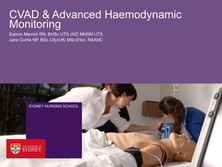
- 1. CVAD & Advanced Haemodynamic Monitoring Eamon Merrick RN, BHSc UTS (NZ) MHSM UTS Jane Currie NP, BSc City(UK) MScS'ton, RAANC SYDNEY NURSING SCHOOL
- 2. Outcomes › This lecture will: • introduce Central Venous Access Devices (CVAD), arterial lines (a or art-line), and invasive haemodynamic monitoring • overview the indications, management, and potential complications of CVAD/ arterial lines • discuss the principles of invasive haemodynamic monitoring • review your role in the safe management of patients with CVAD/ arterial lines 2
- 3. Definition › Central Venous Access Device (CVAD) • A catheter introduced via a large vein into the superior vena cava or right atrium for the administration of parental fluids, medications, or for the measurement of Central Venous Pressure (CVP) - 15-20cm long single or multipurpose catheter - Multiple lumens with multiple openings along the length of the catheter- proximal, medial, and distal. 3
- 4. Basilic and Femoral Insertions › Peripherally Inserted Central Venous Catheter (PICC) › Tend to be used for long-term access › Inserted into the cephalic, basilic, or brachial (cubital?) - Minimal risk of pneumothorax or vessel perforation during insertion - Durable and flexible - Less risk of infection - Vigorous exercise (swimming) should be avoided - Infusion pump should be used due to lumen size 4
- 5. Where are you likely to encounter CVAD › There is a good chance that you will care for a patient with a CVAD if you work in a critical care setting i.e. emergency or intensive/ critical care- also likely in oncology settings 5
- 6. Why? › When might it be useful to have a CVAD? - Remembering they are introduced via a large vein into the superior vena cava or right atrium. 6
- 7. Indications › Administration of fluid › Monitoring of hydration status › Monitoring of cardiac status › Difficulties in finding alternative intravenous access › Monitoring the effects of cardio-active agents i.e. inotropes, vasodilators › Administration of potent medications › Administration of Trans-Parental Nutrition (TPN)- why? › Haemodialysis 7
- 8. Insertion Points › Internal Jugular - Preferred site- improve chances of placement success and less risk of complications - Easiest access with the shortest and straightest route - Less risk of injury to the vagus nerve and carotid artery Although… increase risk of occlusion and irritation due to head movement and problems with maintaining a intact dressing. 8
- 9. Location of the Internal Jugular Internal jugular 9
- 10. External Jugular and Subclavian › External jugular Easy insertion Varies from person- person in size Angles into the subclavian (bendy insertion) Subclavian Preferred for long-term Less vessel irritation and risk of occlusion due to rapid flow Secures to chest wall Potential for superior vena cava obstruction Complications include pneumothorax an thrombosis 10
- 11. External Jugular, Subclavian, Brachiocephalic (innominate), Superior vena cava External Jugular Subclavian Brachiocephalic Superior vena cava 11
- 12. Basilic and Femoral Insertions › Basilic Usual site for the insertion of PICCs due to the shortest and straightest of the vein Femoral Good approach for children- tip rests in inferior vena cava Increase risk of thrombosis and infection 12
- 14. Insertion and Management/ Risk Factors › Anxiety › Obesity › Coagulopathy › Anti-platelet medications › Hypotension › Proximal surgery › Previous CVAD at site › Infection at site › Lymph node dissection or removal › History of DVT in limb › Pulmonary of pulmonary catheter- left bundle branch block 14
- 15. Catheter Selection › Planned approach based on the individual needs of the patient - Minimise the number of lumens (why?) - Should anti-microbial catheters be used? - Multiple infusions and TPN, why is this a consideration? - How long will the CVAD be insitu? - Will this be a out-patient? 15
- 16. Review- Central Venous Access Devices › Central lines - 15-20cm long single or multipurpose catheter - Multiple lumens with multiple openings along the length of the catheter- proximal, medial, and distal. - Used for: - Alternative of peripheral access - Administration of IV Fluids - Administration of IV medications - Administration of Trans-Parental Nutrition (TPN) - Monitoring of Central Venous Pressure (CVP) 16
- 17. Central Venous Access Devises- Complications › During Insertion - Pneumothorax- can occur with subclavian and jugular approaches where the plural cavity is punctured - Haemorrhage- damaged vessel on insertion (patients with coagulopathies/ thrombocytopenia are at more risk) - Air embolism - Cardiac tamponade- Poor placement of the catheter tip can result in a puncture or swelling of the myocardium - Dysrhythmias- due to a irritation of the right atrium 17
- 18. Central Venous Access Devises- Complications › Post-Insertion - Air embolism - Occlusion/ Thrombosis- inadequate flushing can result in a build up of fibrin or medication particulate- think incompatible medications - Infection- contamination at time of insertion, insertion site or lumen, and/or the giving set. - Pain- potentially indicative of poor placement and infection - Dislodgement- normally accidental (not always)- what would do in this situation? 18
- 19. Central Venous Access Devises- Removing › Removal - Aseptic technique - Consider the individual needs of the patient - Patient in a supine position with head tilted down- why? - Patient to remain in a supine position for 30- 60 minutes - Document removal and note presence (or otherwise) of intact tip - Blood cultures and culture of tip if infective process suspected 19
- 20. Preventing Occlusion How can nursing professionals prevent the occlusion of a CVAD? › Matthew, D., Mitchell, B. J. A., Williams, K., & Umscheid, C. A. (2009). Heparin flushing and other interventions to maintain patency of central venous catheters: a systematic review. Journal of Advanced Nursing, 65(10), 2007-2021. 20
- 21. Preventing Infection How can nursing professionals prevent infection/ sepsis in patients with a CVAD? › New South Wales Health Department of Health. (2011). Central venous access device insertion and post insertion care. Sydney. › Australian New Zealand Intensive Care Society. (2012). Central Line Insertion and Maintenance Guideline: ANZICS Safety and Quality Committee. Available from: http://www.anzics.com.au/downloads/doc_download/649-anzics-central-line-insertion-a-maintenance-guideline-april-2012 21
- 22. Air Embolism Identify: Sudden onset of cyanosis Tachypnoea Coughing Tachycardia Hypotension 22
- 23. Air Embolism Act: Clamp the line Turn the patients head to the opposing side of the line Lie the patient flat on her/his left hand side Get help! Ask the patient to perform the ‘Valsalva’ manoeuvre (if conscious) Goal: Immediate medical management- possibly requiring aspiration, mechanical ventilation (100% O2), and pharmacologic circulatory support. 23
- 24. Advanced Haemodynamic Monitoring Central Venous Pressure, Arterial Lines, and Wave Forms SYDNEY NURSING SCHOOL
- 25. Central Venous Pressure (CVP) Continuous monitoring of of the pressure of blood in the right atrium or superior vena cava Best interpreted as a trend- the cardiac function of the patient must be considered Useful for estimating right ventricular pre-load- not systemic pressure/ perfusion Remember that left-sided output determines systemic pressure and perfusion 25
- 26. Central Venous Pressure (CVP) › Raised CVP may indicate - Right ventricular failure - Cardiac tamponade - Tricuspid valve incompetence - Infusions in progress - Catheter tip that is occluded or displaced 26
- 27. Central Venous Pressure (CVP) › Depressed CVP may indicate - Ascites - Vasodilation of peripheral veins - Vasodilating medications - Decrease circulatory blood volume (hypervolemia, dehydration) 27
- 28. Central Venous Pressure Waveform 28
- 29. Cardiac Cycle Systole Diastole LV a c x v y 29
- 30. Arterial Lines Allows: The invasive real-time monitoring of blood pressure useful when administering inotropic or cariotropic medications The drawing of blood gases The calculation of Mean Arterial Pressure (MAP) The assessment of compromised aortic valves Garretson, S. (2005). Haemodynamic monitoring: arterial catheters. Nursing Standard, 19(31), 55-64. Watson, C. A., & Wilkinsin, M. B. (2011). Monitoring central venous pressure, arterial pressure and pulmonary wedge pressure. Anesthesia and Intensive Care Medicine, 13(3), 116-120. 30
- 31. Modified Allens Test Checks the patency of the radial and ulnar arteries Preformed prior to and post insertion of a arterial line 1. Elevate the hand and make a fist for 30 seconds 2. Apply pressure to both the ulnar and radial arteries 3. The hand is opened and should appear blanched 4. Release pressure on the ulnar artery and check for refill 31
- 32. Arterial Lines: Complications Haemorrhage Damage to artery Embolism Distress and anxiety Pain Obstruction (often caused by poor wrist placement) Necrosis 32
- 33. Arterial Line Arterial Line Setup External Link 33
- 34. Arterial Waveform 34
- 35. Mean Arterial Pressure Mean Arterial Pressure (MAP) is the pressure felt by the internal organs- it is not an average… as the duration of diastole exceeds that of systole A MAP of at least 60mmHg is required to perfuse the coronary arteries, brain and kidneys. A ‘normal’ range is about 70mmHg to to 110mmHg ((2 ´ diastolic) + (systolic)) 3 35
- 36. Cardiac Cycle a c x v y 36
Hinweis der Redaktion
- CVADs are placed in large vessels to permit frequent, continuous, or intermittent therapy- avoiding multiple venipunctures. For example the sub-clavian or jugular vien.CVADs are useful for the administration of drugs that are vesicants, blood products, and parenteral nutrition.The major benefit of CVADs is that they can allow for central haemodynamic monitoring and reduce the risk of extravasation- however they lead to an increased risk of systemic infection.PICCs- inserted into a vien in the arm rather than the neck- can be used from one to six months. PICCs tend to lead to lower infection rates- however phlebitis cans occur (usually 7-10 dys after insertion).
- Also known as long-lines- identify each of the veins and identify the ‘cubital fossa’
- Include: cancer (because chemo tends to be irritating of vesicant), infections (for the long-term administration of antibiotics), nutritional replacement (because it is possible to infuse higher concentrations of dextrose than in PVC’s), Renal failure (for heamodyalsis [especially acute] or continuous renal replacement therapy), Shock (able to infuse high volumes of fluids and electrolytes).
- At the base of the neck the internal jugular joins with the subclavian vein- and the joins the brachiocephalic vein which descends to the superior vena cavaCan any tell me what the name is of this other vein (external jugular)
- In this case long term means no more than 14 days- a recent study of 850 patients has demonstrated that pt’s tend to become pyrexic at 13.4 days- which means what? Infection!!!!!!!!!
- Draw in connectors - have students suggest
- At this point have two students come up and demonstrate the location and route of the PICC and CVC based on whomever is holding it at the time- see if they insert to the Left side or the right sideHave the students then discuss what some of the considerations may be for their nursing care if ask to assist with the insertion of CVD
- Discuss why each of these points are important
- Other types and names you will come across:PICC Lines – what are these useful for?Hickman's- used for long-term access (chemo)- lines should be hep locked and clamped to prevent air embolism- blood back flowPortocaths- Implanted device- useful for blood products long term TPN- access through a needle into the skin- talk about creepy this is
- Sometimes on insertion patients will complain of a swishy noise- normally with a jugular placement – whyAlways confirm placement by xray
- Ask the students to identify how they will reduce the risk of infection
- What are some of the individual needs of patients- think about the ability of the patient to tolerate the supine positionIs this patient on anti-coagulatants is he/she wafarinised? I so what are the chemical indicators to check prior to removal- Internationalised Normalised Ratio (INR)- INR in absence of anticoagulation therapy is 0.8-1.2. The target range for INR in anticoagulant use (e.g. warfarin) is 2 to 3. In some cases, if more intense anticoagulation is thought to be required, the target range may be 2.5-3.5.[1]What would do if the tip is not present?How might you be able to tell if there is a infective process?
- The most likely complication
- Can occur if the catheter is unclamped and cap is removed!The immediately life threatening complication!!!!
- Can occur if the catheter is unclamped and cap is removed!Place the patient on her/his left hand side- keeping the air in the apex of the right ventricle and (hopefully) preventing the air from entering the pulmonary circulationValsalve increases intrathoraic pressure and can slow the heart rate- try it nowMechanical ventilation with 100% oxygen, positive end-expiratory pressure, pharmacologic circulatory support, and external cardiac massage may be required
- A normal CVP reading in a spontaneously breathing patient is 5-10 cmH2O. However, as stated earlier, its value is influenced not only by intravascular volume and venous return, but also by venous tone and intrathoracic pressure, along with right heart function and myocardial compliance.
- A normal CVP reading in a spontaneously breathing patient is 5-10 cmH2O. However, as stated earlier, its value is influenced not only by intravascular volume and venous return, but also by venous tone and intrathoracic pressure, along with right heart function and myocardial compliance.
- A= atrial contractionC= bulging of the tricuspid valveX= Atrium relaxes and tricuspid is pulled downV= Passive filling of right atrium and vena cava when tricuspid closesY= tricuspid valve opens and blood flows into right ventircleNo a wave= atrial fibilationHeart block = cannon waves, nodal waves (look like big a waves)
- Wiggers diagram- Dr Carl Wiggers from the university of MichiganIdentify each waveformTop grey Aortic pressure curve is the what we see on the arterial pressure waveform- top line grey----Dicrotic notch corresponds with the closure of the aortic valveBlue The LV waveform- look a the variation in diastole it is about 10mmhg- during contraction it goes up to 120mmhg in a normal adult- remember that the L ventricle Is how blood gets to our body- blue lineMiddle grey- Arterial pressure is the pressure within the atria- note that operates under much less pressure- grey middleRed line- Venticular volume shows the filling and empty of the venticles.ECG- importantly the events on the ECG occur just before the events on the other curves- because to have contractions you first have to have conductionWhat is systole= ejection from the ventriclesWhat is diastole= ventricular filling- look at the redventricular volume line and note how it is gradually increasingInsert first yellow box- this is contractionNote the P wave on the ECG- this gives the atria a kick- and accounts for 15-20% of venticular volume- which we can see on the red ventricular volume lineMove to the R wave on the ECG this cases venticular contraction- the pressure in the venticules exceeds that of the atria- which we can see on the ventricular pressure line- pressure is building very fast in the ventricles but the blood has nowhere to go because both the mitral and aortic valves are closed- have a look at the notch on the arterial pressure line- that is the pressure being exerted on the valve (no regugitation)- pressure builds in the ventricle and overcomes the aortic pressure – normally at 80mmhg or the diastolic pressure- it is at this point that the aortic valve opensInsert 2nd yellow box- End of systole - relaxation- note on the ventricular pressure line that the pressure keeps down until the end of relaxationAt the beginning of relaxation aortic pressure again exceeds that of ventricular pressure (aortic pressure and ventricular pressure lines) at this point the the aotic valve closes- it is at this moment that the dicotic notch appears – think of this a a rebound the blood is trying to go somewhere but as the aortic valve closes it bounces up and off causing a brief bump in the arterial pressure wave form- look at the aterial pressure line and you will note that the aortic notch occurs at the exact moment of aortic valve closure (artia pressure increases because the blood has nowhere to go) – the diacrotic notchAs the the LV empties the atria are filling – we then enter diastole where the ventricles begin to fill again
- Aterial lines are often inserted in the the (what side) LEFT radial artery
- Allens test is performed to check distal perfusion- should there be a complication from the placement of the arterial line the distal blood flow will be maintained by the alternate artery- although there is limited evidence to suggest the utility of the Allen's test prior to arterial line placement because o f the small number of people who have only one patent supply it is useful for nursing professionals who wish to check the distal circulation of a patient who has a arterial line- checking for occlusionsIf the palm remains pale a radial puncture cannot be performed!Identify that the ulnar is the proximal artery and radial is distal and the long vein in the back is the basilic vein- CVAD
- The risks are much the same as CVADs however there is one important thing to note necrosis! Never under any circumstances should you administer a medication via a art line!One other major difference is that a hemorrhage is likely to bleed out- how do you know that this is a arterial bleed?What do you do-Take universal precautions (wear sterile gloves, goggles and gown)Ensure there are no hazardous objects in the woundUse one finger, with interposed gauze, to press directly on the bleeding vessel just proximal to the bleeding pointCall for help!Maintain this for 10 minutesOnce controlled – apply the up-side-pyriamidThe occluding finger should be substituted with a dental roll or tightly folded “nugget” of gauze.A tourniquet may be temporarily applied proximally to facilitate this.Once the positioning is correct and no further bleeding is occurring, slightly larger or less folded pieces of gauze can be placed one on top of the other, creating an inverted pyramid of gauze.The layers of gauze are secured with a loose bandage. Only very light pressure need be applied to the top layer of gauze to maintain hemostasis, as the pressure is “focused” onto the bleeding point. This technique is based on the equation: Pressure=Force/AreaThe tightness of the bandage can be judged from the amount of pressure needed to maintain hemostasis when applying the top layer of gauze.
- Top grey Aortic pressure curve is the what we see on the arterial pressure waveform- top line grey----Dicrotic notch corresponds with the closure of the aortic valvegraph of aortic pressure throughout the cardiac cycle displays a small dip (the "incisure" or "dicrotic notch") which coincides with the aortic valve closure. The dip in the graph is immediately followed by a brief rise (the "dicrotic wave") then gradual decline. Just as the ventricles enter into diastole, the brief reversal of flow from the aorta back into the left ventricle causes the aortic valves to shut. This results in the slight increase in aortic pressure caused by the elastic recoil of the semilunar valves and aorta.DRAW IN THE Ventricular PRESSURE WACW FORM
- The risks are much the same as CVADs however there is one important thing to note necrosis! Never under any circumstances should you administer a medication via a art line!Back to previous slide and illustrate where MAP sits
- Ask students to identify diastole systole contraction relaxation and the dicrotic notch