SlideShare ist ein Scribd-Unternehmen logo
1 von 103
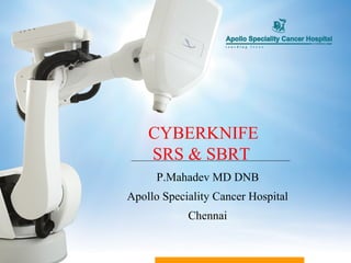
CYBERKNIFE
    SRS & SBRT
      P.Mahadev MD DNB
Apollo Speciality Cancer Hospital
            Chennai
MANAGEMENT AND DELIVERY OF IMAGE
 GUIDED HIGH DOSE RADIATION THERAPY WITH
 TUMOR ABLATIVE INTENT WITHIN A COURSE OF
 TREATMENT THAT DOES NOT EXCEED 5 DAYS
 Higher confidence in tumor targeting

 Reliable mechanisms for generating focused, sharply
  delineated dose distributions with a rapid dose fall off
 Reliable accurate patient positioning accounting for target
  motion related to time dependent organ movement
 IMAGE GUIDANCE AND EFFICIENT TRACKING
  MECHANISM
 Longer times than conventional RT, hence patient comfort
  is an issue
SRS
SBRT
IMRT FOR PROSTATE
SRT
3

            4
                5 6


        2


    1
Pitch
        Yaw   Roll
Robot is capable of delivering radiation from
different 100 nodes, with each node is capable of
giving a maximum 12 different beams.

Usage of these nodes depends on the treatment room
constraints
The table consists of 12 fixed cones and housings of
 Fixed and Iris Collimator


                                          Laser Sensor


Collimator sizes(mm): 5, 7.5, 10, 12.5, 15, 20, 25, 30,
 35, 40, 50, 60
       There are two essential features of the
    CyberKnife system that sets it apart from other
    stereotactic radiosurgery methods.
      radiation source is mounted on a precisely
  controlled industrial robot.
      The image guidance system(continuous tracking
  system)
 Eliminates the need of gating techniques and
  restrictive head frames
The Cyberknife treatment delivery is based on the
  following tracking systems
 6D_ Skull tracking system
 Fiducials tracking system
 Synchrony tracking system
 X_sight Spine tracking system
 X_sight Lung tracking system
 6D_ Skull tracking system:
 used for intra-cranial lesions up to C2
 Bony anatomy of the skull is used as reference for
 tracking
 Fiducial tracking system:
     used for soft tissues, where gold   fiducials
 can be implanted.
     Minimum of 3 nos. to be implanted
 close proximity to the lesion to be treated
 well-separated (by about 1 cm)
 non-overlapping on projections from the in-room x-
 ray imagers
Three markers are sufficient for unique spatial
 localization, but in practice 4-5 are often placed in
 case of loss or suboptimal placement of markers
•   790 fiducials
•   85% successfully placed
•   2 Patients developed pneumothorax
•   6 fiducials migrated- 3 in lung, 2 in
    liver& 1 in prostate
Respiratory-induced
 motion of tumors causes
 significant targeting
 uncertainty
   Lung, liver, pancreas,
    Prostate,kidney
Traditional radiation
 therapy margins are not
 optimized for high-dose
 radiosurgery
Imaging and Tumor Targeting
   Traditional IGRT daily set-up imaging maybe
   inadequate for
   sub-millimeter accuracy
Immobilization
Breath Holding
Imaging and Tumor Targeting
   Traditional IGRT daily set-up imaging maybe
   inadequate for
   sub-millimeter accuracy
Immobilization
Breath Holding
Gating
option for dynamic tracking without the use of
 implanted fiducials.
Tumor localization is accomplished using auto-
 mated real-time image segmentation of the in-room
 x-ray images based on the contrast of the tumor itself.
best used for lesions with sufficient contrast in
 density from the surrounding anatomy to be clearly
 visualized on both of the in-room x-ray imagers, i.e.,
 those located in the lung periphery at least 1.5 cm in
 size, and that do not overlap other dense anatomical
 structures, such as the spine, diaphragm, and heart in
 the projection views
Two features to form the basis for accuracy


   Fiducials, implanted prior to   Optical markers on a special
             treatment                     patient vest
 Prior to treatment start: creation of dynamic correlation model


    Imaging system takes positions of fiducials at                    Markers are monitored in
               discrete points of time                              real time by a camera system
 Prior to treatment start: creation of dynamic correlation model


             Imaging system takes positions of fiducials at                  Markers are monitored in real time by a camera
                        discrete points of time                                                 system


                                                              displacement
      displacement


                                               time                                                                   time
 This process repeats throughout the treatment, updating and correcting beam delivery based upon
   the patient’s current breathing pattern


                                                     displacement
     displacement


                                       time                                                     time
 X-sight Spine tracking system:used to track spine
  lesions which are close to spine from C1 to L5&sacrum
 Uses the bony anatomy of spine to track the tumors
  in close relation to spine eliminating the need for
  fiducials
 X-sight spine is now possible in prone position as well
 The appropriate tracking method has to be chosen
 during planning itself

 No treatment is possible without planning and proper
 tracking method
 Treatment planning is done on the CT images of slice
 thickness 1mm acquired at 125 kV and 400 mAs with a
 pixel size of 512 x 512

 MRI, PET and 3D-Angio images can be used to fuse
 with the primary CT images for target and OAR
 delineation
Planning System (MultiPlan) uses inverse
planning algorithm with following options
1. Conformal Planning
2. Sequential optimization
The system provides the user the option of
using either ray tracing method or Monte Carlo
 The mechanical accuracy of the system is 0.12
  mm , according to Accuray
 The system maintains sub-millimeter tracking
  accuracy, if the patient positions are within the
  following limits
 Left / Right (Lat)     10 mm
 Ant/ Post (Ver)        10 mm
 Sup/ Inf ( Long) 10 mm
 Roll (Left / Right) 10
 Pitch (Head Up / Down) 10
 Yaw ( C.W / C.C.W)     30
 The Robot will correct its position if the off set values
  are with in the specified limits

 The robot will trigger an Emergency Stop outside of
  these tolerances
Gamma knife, X-knife are probably as good.
May have an advantage for larger lesions requiring
 multiple fractions- meningioma, acoustic
 schwanomma etc
More patient friendly(frame)
Continuous image guidance
T1&T2 NSCLC – inoperable or medical
 contraindication or patient refuses surgery, ideal
 lesion <3cm & peripheral location
oligometastasis
in a uniform population of medically inoperable
 patients with peripherally located early lung cancer,
 the RTOG 0236 study dem- onstrated 98% local
 control (within the primary tumor) and 87% local-
 regional control (within the ipsilateral lobe, hilum,
 and mediastinum) at 3 years with an intensive
 regimen of 60 Gy in 3 fractions
RADIOBIOLOGICAL RATIONALE: LOW
 APLHA/BETA RATIO
GOOD RESULTS OBTAINED WITH HDR
 brachytherapy
LESS INVASIVE THAN BRACHYTHERAPY
Ju AW et al :Radiat oncol jan2013
41 pts intermediate risk
Median fu 21 mo
99% biochemical PFS
No gr3/4 bladder or bowel morbidity
No significant change in sexual QOL
BRACHYTHERAPY: 10/10.5 Gy x3 over 24 hours, each
 fraction 8 hours apart
BED : 130/142 Gy
SBRT : 7.25 Gy x5 over 5 days
BED: 123 Gy
T1   T2    PSA<10   PSA>10   GS<7   GS>7   TOTAL


HDR      30   22    35       17       40     12     52


CK       34   32    40       16       35     31     66


IMRT/I   94   186   94       186      126    154    280
GRT
MEDIAN FU      2 YR    MEDIAN
                        BIOCHEMI   PSA
                        CAL        NADIR
                         PFS
HDR         22 MONTHS   94%        0.8


CK          16 MONTHS   96%        1.0


IMRT/IGRT   48 MONTHS   89%        0.9
Fiducials placed at surgery
One planning CT with oral and IV contrast
1000cGy to +ve margins 3-4 weeks post OP
5040cGy 5-6 field IMRT6-8 weeks postOP
Concurrent Xeloda
Adjuvant Gemcitabine
 Intramedullary spinal cord AVM’s only
 Not amenable to microsurgical excision/embolisatio
 symptomatic
 Neurologic examination
 MRI
 Conventional 2D spinal angio
Spine tracking
1.25 mm contrast enhanced axial CT
Target volume traced on CT in cojunction with:MRI
     2D/3D spinal angio
 24 patients
 15 males 9 females
 Time from diagnosis to SRS:7.8 yrs
 Mean age at SRS 34 Yrs
 Presentation :12 hemorrhages
              12 had progressive pain or myelopathy
  secondary to steal or venous congestion
 13 cervical
 8 thoracic
 3 conus medullaris
 Target volume :2.8cc(0.26-15 cc)
 Marginal nidus dose :2050cGy(1600-2100)
 Prescription isodose line:79%(68-90%)
 Dmax:2580cGy
 Fractions:1 to 4
Angiographic outcome:significant AVM reduction in
 all patients >1yr post SRS
6 of 19 patients obliterate
No angio done in 5 patients
Clinical outcome:no further hemorrhages
3 PATIENTS
28 YRS OLD LADY EMBOLISATION DONE TWICE
 PRESENTED WITH SEVERE PAIN IN THE
 POPLITEAL FOSSA AND CALF REGION
56 YEARS OLD LADY WITH SUDDEN ONSET OF
 MYELOPATHY
BOTH THE PATIENTS RESPONDED WELL
25 yr old young man, repeated embolisations
 done,had no improvement
Current prescription dose to nidus is 2000 cGy
 in 2 sessions to larger lesions & 16-18 Gy for
 small (<0.7 cc) AVM
radiosurgery is a reasonable option in most
 type II spinal cord AVMs
Gerszten et al., Radiosurgery for spinal metastases: clinical
     experience in 500 cases from a single institution Volume 32,
                      Number 2, pp 193–199, 2007

 500 cases of spinal metastases treated by CyberKnife ® Radiosurgery at
   the University of Pittsburgh

 73 cervical, 212 thoracic, 112 lumbar, and 103 sacral lesions

 Long-term pain improvement occurred in 290 of 336 cases (86%)

 Long-term tumor control in 90% of lesions treated with radiosurgery as a
primary treatment modality

 Long-term tumor control in 88% of lesions that failed other therapies
Stereotactic radiosurgery is not a substitute to
 surgery but an alternative when indicated
SBRT is becoming a component in the
 multidisciplinary treatment of Cancer
In selected cases, SBRT may prove to be a curative
 modality of treatment in early cancers
Apollo hydbd feb8 2013 (cancer ci 2013) p. mahadev md

Weitere ähnliche Inhalte

Was ist angesagt?

To use or not to use the LQ model at “high” radiation doses
To use or not to use the LQ model at “high” radiation doses To use or not to use the LQ model at “high” radiation doses
To use or not to use the LQ model at “high” radiation doses Santam Chakraborty
 
Central Lung Tumour: 'Flying in NO Flying Zone'
Central Lung Tumour: 'Flying in NO Flying Zone'Central Lung Tumour: 'Flying in NO Flying Zone'
Central Lung Tumour: 'Flying in NO Flying Zone'duttaradio
 
Radiosurgery for lung cancer short version
Radiosurgery for lung cancer short versionRadiosurgery for lung cancer short version
Radiosurgery for lung cancer short versionRobert J Miller MD
 
Gynae ONCOLOGY CARTOONS
Gynae ONCOLOGY CARTOONSGynae ONCOLOGY CARTOONS
Gynae ONCOLOGY CARTOONSKanhu Charan
 
Stereotactic Body Radiation Therapy
Stereotactic Body Radiation TherapyStereotactic Body Radiation Therapy
Stereotactic Body Radiation Therapyfondas vakalis
 
Cyberknife,WORLDS NO. ONE ROBOTIC RADIOSURGERY SYSTEM
Cyberknife,WORLDS NO. ONE ROBOTIC RADIOSURGERY SYSTEMCyberknife,WORLDS NO. ONE ROBOTIC RADIOSURGERY SYSTEM
Cyberknife,WORLDS NO. ONE ROBOTIC RADIOSURGERY SYSTEMSubrata Roy
 
HIPPOCAMPUS TARGET DELINEATION
HIPPOCAMPUS TARGET DELINEATIONHIPPOCAMPUS TARGET DELINEATION
HIPPOCAMPUS TARGET DELINEATIONKanhu Charan
 
SPINE SBRT CARTOON
SPINE SBRT CARTOONSPINE SBRT CARTOON
SPINE SBRT CARTOONKanhu Charan
 
Hyperthermia in radiotherapy
Hyperthermia in radiotherapyHyperthermia in radiotherapy
Hyperthermia in radiotherapyJewel Joseph
 
Role of Image Guidance in Radiotherapy
Role of Image Guidance in RadiotherapyRole of Image Guidance in Radiotherapy
Role of Image Guidance in RadiotherapySusan Rochelle
 
Radiobiological aspects of radiotherapy precision
Radiobiological aspects of radiotherapy precisionRadiobiological aspects of radiotherapy precision
Radiobiological aspects of radiotherapy precisionAmin Amin
 
parsport trial ppt
parsport trial pptparsport trial ppt
parsport trial pptGaurav Kumar
 
PENTEC GUIDELINES FOR PAEDIATRIC RADIOTHERAPY
PENTEC GUIDELINES FOR PAEDIATRIC RADIOTHERAPYPENTEC GUIDELINES FOR PAEDIATRIC RADIOTHERAPY
PENTEC GUIDELINES FOR PAEDIATRIC RADIOTHERAPYKanhu Charan
 
Plan evaluation in Radiotherapy- Dr Kiran
Plan evaluation in Radiotherapy- Dr KiranPlan evaluation in Radiotherapy- Dr Kiran
Plan evaluation in Radiotherapy- Dr KiranKiran Ramakrishna
 
Altered fractionation schedules in radiation oncology
Altered fractionation schedules in radiation oncologyAltered fractionation schedules in radiation oncology
Altered fractionation schedules in radiation oncologyAbhishek Soni
 
Alawi treatment planning-evaluation-of-volumetric-modulated-arc-therapy-vmat-...
Alawi treatment planning-evaluation-of-volumetric-modulated-arc-therapy-vmat-...Alawi treatment planning-evaluation-of-volumetric-modulated-arc-therapy-vmat-...
Alawi treatment planning-evaluation-of-volumetric-modulated-arc-therapy-vmat-...Tagreed Alawi
 
PANCREATIC SBRT SIMULATION
PANCREATIC SBRT SIMULATIONPANCREATIC SBRT SIMULATION
PANCREATIC SBRT SIMULATIONKanhu Charan
 
Basics of linear quadratic model
Basics of linear quadratic modelBasics of linear quadratic model
Basics of linear quadratic modelAjeet Gandhi
 

Was ist angesagt? (20)

4D Radiotherapy
4D Radiotherapy4D Radiotherapy
4D Radiotherapy
 
To use or not to use the LQ model at “high” radiation doses
To use or not to use the LQ model at “high” radiation doses To use or not to use the LQ model at “high” radiation doses
To use or not to use the LQ model at “high” radiation doses
 
Central Lung Tumour: 'Flying in NO Flying Zone'
Central Lung Tumour: 'Flying in NO Flying Zone'Central Lung Tumour: 'Flying in NO Flying Zone'
Central Lung Tumour: 'Flying in NO Flying Zone'
 
Radiosurgery for lung cancer short version
Radiosurgery for lung cancer short versionRadiosurgery for lung cancer short version
Radiosurgery for lung cancer short version
 
Contouring rectal cancers
Contouring rectal cancersContouring rectal cancers
Contouring rectal cancers
 
Gynae ONCOLOGY CARTOONS
Gynae ONCOLOGY CARTOONSGynae ONCOLOGY CARTOONS
Gynae ONCOLOGY CARTOONS
 
Stereotactic Body Radiation Therapy
Stereotactic Body Radiation TherapyStereotactic Body Radiation Therapy
Stereotactic Body Radiation Therapy
 
Cyberknife,WORLDS NO. ONE ROBOTIC RADIOSURGERY SYSTEM
Cyberknife,WORLDS NO. ONE ROBOTIC RADIOSURGERY SYSTEMCyberknife,WORLDS NO. ONE ROBOTIC RADIOSURGERY SYSTEM
Cyberknife,WORLDS NO. ONE ROBOTIC RADIOSURGERY SYSTEM
 
HIPPOCAMPUS TARGET DELINEATION
HIPPOCAMPUS TARGET DELINEATIONHIPPOCAMPUS TARGET DELINEATION
HIPPOCAMPUS TARGET DELINEATION
 
SPINE SBRT CARTOON
SPINE SBRT CARTOONSPINE SBRT CARTOON
SPINE SBRT CARTOON
 
Hyperthermia in radiotherapy
Hyperthermia in radiotherapyHyperthermia in radiotherapy
Hyperthermia in radiotherapy
 
Role of Image Guidance in Radiotherapy
Role of Image Guidance in RadiotherapyRole of Image Guidance in Radiotherapy
Role of Image Guidance in Radiotherapy
 
Radiobiological aspects of radiotherapy precision
Radiobiological aspects of radiotherapy precisionRadiobiological aspects of radiotherapy precision
Radiobiological aspects of radiotherapy precision
 
parsport trial ppt
parsport trial pptparsport trial ppt
parsport trial ppt
 
PENTEC GUIDELINES FOR PAEDIATRIC RADIOTHERAPY
PENTEC GUIDELINES FOR PAEDIATRIC RADIOTHERAPYPENTEC GUIDELINES FOR PAEDIATRIC RADIOTHERAPY
PENTEC GUIDELINES FOR PAEDIATRIC RADIOTHERAPY
 
Plan evaluation in Radiotherapy- Dr Kiran
Plan evaluation in Radiotherapy- Dr KiranPlan evaluation in Radiotherapy- Dr Kiran
Plan evaluation in Radiotherapy- Dr Kiran
 
Altered fractionation schedules in radiation oncology
Altered fractionation schedules in radiation oncologyAltered fractionation schedules in radiation oncology
Altered fractionation schedules in radiation oncology
 
Alawi treatment planning-evaluation-of-volumetric-modulated-arc-therapy-vmat-...
Alawi treatment planning-evaluation-of-volumetric-modulated-arc-therapy-vmat-...Alawi treatment planning-evaluation-of-volumetric-modulated-arc-therapy-vmat-...
Alawi treatment planning-evaluation-of-volumetric-modulated-arc-therapy-vmat-...
 
PANCREATIC SBRT SIMULATION
PANCREATIC SBRT SIMULATIONPANCREATIC SBRT SIMULATION
PANCREATIC SBRT SIMULATION
 
Basics of linear quadratic model
Basics of linear quadratic modelBasics of linear quadratic model
Basics of linear quadratic model
 

Ähnlich wie Apollo hydbd feb8 2013 (cancer ci 2013) p. mahadev md

Efficiency of Fiducial Tracking of Carcinoma Prostate With Cyberknife System ...
Efficiency of Fiducial Tracking of Carcinoma Prostate With Cyberknife System ...Efficiency of Fiducial Tracking of Carcinoma Prostate With Cyberknife System ...
Efficiency of Fiducial Tracking of Carcinoma Prostate With Cyberknife System ...Subrata Roy
 
Cyberknife An expertise state of art Technology to execute Stereotactic Robot...
Cyberknife An expertise state of art Technology to execute Stereotactic Robot...Cyberknife An expertise state of art Technology to execute Stereotactic Robot...
Cyberknife An expertise state of art Technology to execute Stereotactic Robot...Subrata Roy
 
Image guided radiation therapy
Image guided radiation therapyImage guided radiation therapy
Image guided radiation therapySwarnita Sahu
 
Cyberknife Treatment Delivery Radiation Therapist Perspective
Cyberknife Treatment Delivery  Radiation Therapist PerspectiveCyberknife Treatment Delivery  Radiation Therapist Perspective
Cyberknife Treatment Delivery Radiation Therapist PerspectiveSubrata Roy
 
IMRT and 3D CRT in cervical Cancers
IMRT and 3D CRT in cervical CancersIMRT and 3D CRT in cervical Cancers
IMRT and 3D CRT in cervical CancersSantam Chakraborty
 
ROSE CASE BRAIN MET SRS
ROSE CASE BRAIN MET SRSROSE CASE BRAIN MET SRS
ROSE CASE BRAIN MET SRSKanhu Charan
 
Cyberknife At Saint Raphael’s Campus_revB
Cyberknife At Saint Raphael’s Campus_revBCyberknife At Saint Raphael’s Campus_revB
Cyberknife At Saint Raphael’s Campus_revBJustin Vinci
 
Stevens et al, Continuous monitoring of prostate position using stereoscopic ...
Stevens et al, Continuous monitoring of prostate position using stereoscopic ...Stevens et al, Continuous monitoring of prostate position using stereoscopic ...
Stevens et al, Continuous monitoring of prostate position using stereoscopic ...David Parsons
 
Calypso Medical's Prostate Cancer Treatment: Imaging Technology News
Calypso Medical's Prostate Cancer Treatment: Imaging Technology NewsCalypso Medical's Prostate Cancer Treatment: Imaging Technology News
Calypso Medical's Prostate Cancer Treatment: Imaging Technology NewsCalypso Medical
 
Stereotactic Radiosurgery/ Radiotherapy
Stereotactic Radiosurgery/ RadiotherapyStereotactic Radiosurgery/ Radiotherapy
Stereotactic Radiosurgery/ Radiotherapyumesh V
 
Assessment of Intrafraction Motion during real-time tracking in the treatment...
Assessment of Intrafraction Motion during real-time tracking in the treatment...Assessment of Intrafraction Motion during real-time tracking in the treatment...
Assessment of Intrafraction Motion during real-time tracking in the treatment...Subrata Roy
 
Radiotherapy in Cervical Cancers1.ppt
Radiotherapy in Cervical Cancers1.pptRadiotherapy in Cervical Cancers1.ppt
Radiotherapy in Cervical Cancers1.pptNeetumishti Chadha
 
Role of radiotherapy in brain tumours
Role of radiotherapy in brain tumoursRole of radiotherapy in brain tumours
Role of radiotherapy in brain tumoursAbhilash Gavarraju
 

Ähnlich wie Apollo hydbd feb8 2013 (cancer ci 2013) p. mahadev md (20)

Cyberknife
Cyberknife Cyberknife
Cyberknife
 
Efficiency of Fiducial Tracking of Carcinoma Prostate With Cyberknife System ...
Efficiency of Fiducial Tracking of Carcinoma Prostate With Cyberknife System ...Efficiency of Fiducial Tracking of Carcinoma Prostate With Cyberknife System ...
Efficiency of Fiducial Tracking of Carcinoma Prostate With Cyberknife System ...
 
Cyberknife An expertise state of art Technology to execute Stereotactic Robot...
Cyberknife An expertise state of art Technology to execute Stereotactic Robot...Cyberknife An expertise state of art Technology to execute Stereotactic Robot...
Cyberknife An expertise state of art Technology to execute Stereotactic Robot...
 
Image guided radiation therapy
Image guided radiation therapyImage guided radiation therapy
Image guided radiation therapy
 
2009.12.29, Draft of New Chapter
2009.12.29, Draft of New Chapter2009.12.29, Draft of New Chapter
2009.12.29, Draft of New Chapter
 
Cyberknife Treatment Delivery Radiation Therapist Perspective
Cyberknife Treatment Delivery  Radiation Therapist PerspectiveCyberknife Treatment Delivery  Radiation Therapist Perspective
Cyberknife Treatment Delivery Radiation Therapist Perspective
 
IMRT and 3D CRT in cervical Cancers
IMRT and 3D CRT in cervical CancersIMRT and 3D CRT in cervical Cancers
IMRT and 3D CRT in cervical Cancers
 
ROSE CASE BRAIN MET SRS
ROSE CASE BRAIN MET SRSROSE CASE BRAIN MET SRS
ROSE CASE BRAIN MET SRS
 
Brain metastasis
Brain metastasisBrain metastasis
Brain metastasis
 
Cyberknife At Saint Raphael’s Campus_revB
Cyberknife At Saint Raphael’s Campus_revBCyberknife At Saint Raphael’s Campus_revB
Cyberknife At Saint Raphael’s Campus_revB
 
Beam Direction
Beam DirectionBeam Direction
Beam Direction
 
Lung sbrt ppt
Lung  sbrt pptLung  sbrt ppt
Lung sbrt ppt
 
Stevens et al, Continuous monitoring of prostate position using stereoscopic ...
Stevens et al, Continuous monitoring of prostate position using stereoscopic ...Stevens et al, Continuous monitoring of prostate position using stereoscopic ...
Stevens et al, Continuous monitoring of prostate position using stereoscopic ...
 
Calypso Medical's Prostate Cancer Treatment: Imaging Technology News
Calypso Medical's Prostate Cancer Treatment: Imaging Technology NewsCalypso Medical's Prostate Cancer Treatment: Imaging Technology News
Calypso Medical's Prostate Cancer Treatment: Imaging Technology News
 
Stereotactic Radiosurgery/ Radiotherapy
Stereotactic Radiosurgery/ RadiotherapyStereotactic Radiosurgery/ Radiotherapy
Stereotactic Radiosurgery/ Radiotherapy
 
Assessment of Intrafraction Motion during real-time tracking in the treatment...
Assessment of Intrafraction Motion during real-time tracking in the treatment...Assessment of Intrafraction Motion during real-time tracking in the treatment...
Assessment of Intrafraction Motion during real-time tracking in the treatment...
 
Radiotherapy in Cervical Cancers1.ppt
Radiotherapy in Cervical Cancers1.pptRadiotherapy in Cervical Cancers1.ppt
Radiotherapy in Cervical Cancers1.ppt
 
Image Guided Radiotherapy
Image Guided RadiotherapyImage Guided Radiotherapy
Image Guided Radiotherapy
 
10 may sbrt
10 may sbrt10 may sbrt
10 may sbrt
 
Role of radiotherapy in brain tumours
Role of radiotherapy in brain tumoursRole of radiotherapy in brain tumours
Role of radiotherapy in brain tumours
 

Mehr von Dr. Vijay Anand P. Reddy

Hahn proton talk (cancer ci 2013) stephen m. hahn
Hahn proton talk (cancer ci 2013) stephen m. hahnHahn proton talk (cancer ci 2013) stephen m. hahn
Hahn proton talk (cancer ci 2013) stephen m. hahnDr. Vijay Anand P. Reddy
 
Kupelian 2nd talk prostate igrt hyderabad 2013 (kupelian)
Kupelian 2nd talk prostate igrt hyderabad 2013 (kupelian)Kupelian 2nd talk prostate igrt hyderabad 2013 (kupelian)
Kupelian 2nd talk prostate igrt hyderabad 2013 (kupelian)Dr. Vijay Anand P. Reddy
 
Kupelian 1st talk planning dose hyderabad 2013 (cancer ci 2013) patrick kupel...
Kupelian 1st talk planning dose hyderabad 2013 (cancer ci 2013) patrick kupel...Kupelian 1st talk planning dose hyderabad 2013 (cancer ci 2013) patrick kupel...
Kupelian 1st talk planning dose hyderabad 2013 (cancer ci 2013) patrick kupel...Dr. Vijay Anand P. Reddy
 
Sbrt liver tumors_kag(cancer ci 2013) karyn a. goodman
Sbrt liver tumors_kag(cancer ci 2013) karyn a. goodmanSbrt liver tumors_kag(cancer ci 2013) karyn a. goodman
Sbrt liver tumors_kag(cancer ci 2013) karyn a. goodmanDr. Vijay Anand P. Reddy
 
03 suh lung sbrt hyderabad feb 2013 (cancer ci 2013) john h. suh
03 suh lung sbrt hyderabad feb 2013 (cancer ci 2013) john h. suh03 suh lung sbrt hyderabad feb 2013 (cancer ci 2013) john h. suh
03 suh lung sbrt hyderabad feb 2013 (cancer ci 2013) john h. suhDr. Vijay Anand P. Reddy
 
02 suh srs hyderabad 2013 (cancer ci 2013) john h. suh
02 suh srs hyderabad 2013 (cancer ci 2013) john h. suh02 suh srs hyderabad 2013 (cancer ci 2013) john h. suh
02 suh srs hyderabad 2013 (cancer ci 2013) john h. suhDr. Vijay Anand P. Reddy
 
01 suh brain anatomy, planning and delivery hyderabad 2013 (cancer ci 2013) j...
01 suh brain anatomy, planning and delivery hyderabad 2013 (cancer ci 2013) j...01 suh brain anatomy, planning and delivery hyderabad 2013 (cancer ci 2013) j...
01 suh brain anatomy, planning and delivery hyderabad 2013 (cancer ci 2013) j...Dr. Vijay Anand P. Reddy
 
Friday intro 8.35 am intro & objectives (cancer ci 2013) dr prabhakar
Friday intro 8.35 am  intro & objectives (cancer ci 2013) dr prabhakarFriday intro 8.35 am  intro & objectives (cancer ci 2013) dr prabhakar
Friday intro 8.35 am intro & objectives (cancer ci 2013) dr prabhakarDr. Vijay Anand P. Reddy
 
02 igrt for india jan 2013 (cancer ci 2013) avraham eisbruch
02 igrt for india jan 2013 (cancer ci 2013) avraham eisbruch02 igrt for india jan 2013 (cancer ci 2013) avraham eisbruch
02 igrt for india jan 2013 (cancer ci 2013) avraham eisbruchDr. Vijay Anand P. Reddy
 
01 planning for hn india feb 2013 (cancer ci 2013) avraham eisbruch
01 planning for hn india feb 2013 (cancer ci 2013) avraham eisbruch01 planning for hn india feb 2013 (cancer ci 2013) avraham eisbruch
01 planning for hn india feb 2013 (cancer ci 2013) avraham eisbruchDr. Vijay Anand P. Reddy
 
Igrt for cervical cancer feb 8 2013 920 a cancer ci 2013
Igrt for cervical cancer feb 8 2013 920 a cancer ci 2013Igrt for cervical cancer feb 8 2013 920 a cancer ci 2013
Igrt for cervical cancer feb 8 2013 920 a cancer ci 2013Dr. Vijay Anand P. Reddy
 

Mehr von Dr. Vijay Anand P. Reddy (12)

04 hyd panel nccn cervix feb 9 2013
04 hyd panel nccn cervix feb 9 201304 hyd panel nccn cervix feb 9 2013
04 hyd panel nccn cervix feb 9 2013
 
Hahn proton talk (cancer ci 2013) stephen m. hahn
Hahn proton talk (cancer ci 2013) stephen m. hahnHahn proton talk (cancer ci 2013) stephen m. hahn
Hahn proton talk (cancer ci 2013) stephen m. hahn
 
Kupelian 2nd talk prostate igrt hyderabad 2013 (kupelian)
Kupelian 2nd talk prostate igrt hyderabad 2013 (kupelian)Kupelian 2nd talk prostate igrt hyderabad 2013 (kupelian)
Kupelian 2nd talk prostate igrt hyderabad 2013 (kupelian)
 
Kupelian 1st talk planning dose hyderabad 2013 (cancer ci 2013) patrick kupel...
Kupelian 1st talk planning dose hyderabad 2013 (cancer ci 2013) patrick kupel...Kupelian 1st talk planning dose hyderabad 2013 (cancer ci 2013) patrick kupel...
Kupelian 1st talk planning dose hyderabad 2013 (cancer ci 2013) patrick kupel...
 
Sbrt liver tumors_kag(cancer ci 2013) karyn a. goodman
Sbrt liver tumors_kag(cancer ci 2013) karyn a. goodmanSbrt liver tumors_kag(cancer ci 2013) karyn a. goodman
Sbrt liver tumors_kag(cancer ci 2013) karyn a. goodman
 
03 suh lung sbrt hyderabad feb 2013 (cancer ci 2013) john h. suh
03 suh lung sbrt hyderabad feb 2013 (cancer ci 2013) john h. suh03 suh lung sbrt hyderabad feb 2013 (cancer ci 2013) john h. suh
03 suh lung sbrt hyderabad feb 2013 (cancer ci 2013) john h. suh
 
02 suh srs hyderabad 2013 (cancer ci 2013) john h. suh
02 suh srs hyderabad 2013 (cancer ci 2013) john h. suh02 suh srs hyderabad 2013 (cancer ci 2013) john h. suh
02 suh srs hyderabad 2013 (cancer ci 2013) john h. suh
 
01 suh brain anatomy, planning and delivery hyderabad 2013 (cancer ci 2013) j...
01 suh brain anatomy, planning and delivery hyderabad 2013 (cancer ci 2013) j...01 suh brain anatomy, planning and delivery hyderabad 2013 (cancer ci 2013) j...
01 suh brain anatomy, planning and delivery hyderabad 2013 (cancer ci 2013) j...
 
Friday intro 8.35 am intro & objectives (cancer ci 2013) dr prabhakar
Friday intro 8.35 am  intro & objectives (cancer ci 2013) dr prabhakarFriday intro 8.35 am  intro & objectives (cancer ci 2013) dr prabhakar
Friday intro 8.35 am intro & objectives (cancer ci 2013) dr prabhakar
 
02 igrt for india jan 2013 (cancer ci 2013) avraham eisbruch
02 igrt for india jan 2013 (cancer ci 2013) avraham eisbruch02 igrt for india jan 2013 (cancer ci 2013) avraham eisbruch
02 igrt for india jan 2013 (cancer ci 2013) avraham eisbruch
 
01 planning for hn india feb 2013 (cancer ci 2013) avraham eisbruch
01 planning for hn india feb 2013 (cancer ci 2013) avraham eisbruch01 planning for hn india feb 2013 (cancer ci 2013) avraham eisbruch
01 planning for hn india feb 2013 (cancer ci 2013) avraham eisbruch
 
Igrt for cervical cancer feb 8 2013 920 a cancer ci 2013
Igrt for cervical cancer feb 8 2013 920 a cancer ci 2013Igrt for cervical cancer feb 8 2013 920 a cancer ci 2013
Igrt for cervical cancer feb 8 2013 920 a cancer ci 2013
 

Apollo hydbd feb8 2013 (cancer ci 2013) p. mahadev md

  • 1. CYBERKNIFE SRS & SBRT P.Mahadev MD DNB Apollo Speciality Cancer Hospital Chennai
  • 2. MANAGEMENT AND DELIVERY OF IMAGE GUIDED HIGH DOSE RADIATION THERAPY WITH TUMOR ABLATIVE INTENT WITHIN A COURSE OF TREATMENT THAT DOES NOT EXCEED 5 DAYS
  • 3.  Higher confidence in tumor targeting  Reliable mechanisms for generating focused, sharply delineated dose distributions with a rapid dose fall off  Reliable accurate patient positioning accounting for target motion related to time dependent organ movement  IMAGE GUIDANCE AND EFFICIENT TRACKING MECHANISM  Longer times than conventional RT, hence patient comfort is an issue
  • 5.
  • 6. 3 4 5 6 2 1
  • 7. Pitch Yaw Roll
  • 8. Robot is capable of delivering radiation from different 100 nodes, with each node is capable of giving a maximum 12 different beams. Usage of these nodes depends on the treatment room constraints
  • 9.
  • 10. The table consists of 12 fixed cones and housings of Fixed and Iris Collimator Laser Sensor Collimator sizes(mm): 5, 7.5, 10, 12.5, 15, 20, 25, 30, 35, 40, 50, 60
  • 11.
  • 12.
  • 13. There are two essential features of the CyberKnife system that sets it apart from other stereotactic radiosurgery methods.
  • 14. radiation source is mounted on a precisely controlled industrial robot.  The image guidance system(continuous tracking system)  Eliminates the need of gating techniques and restrictive head frames
  • 15. The Cyberknife treatment delivery is based on the following tracking systems  6D_ Skull tracking system  Fiducials tracking system  Synchrony tracking system  X_sight Spine tracking system  X_sight Lung tracking system
  • 16.  6D_ Skull tracking system: used for intra-cranial lesions up to C2 Bony anatomy of the skull is used as reference for tracking
  • 17.
  • 18.  Fiducial tracking system: used for soft tissues, where gold fiducials can be implanted. Minimum of 3 nos. to be implanted
  • 19.  close proximity to the lesion to be treated  well-separated (by about 1 cm)  non-overlapping on projections from the in-room x- ray imagers Three markers are sufficient for unique spatial localization, but in practice 4-5 are often placed in case of loss or suboptimal placement of markers
  • 20.
  • 21. 790 fiducials • 85% successfully placed • 2 Patients developed pneumothorax • 6 fiducials migrated- 3 in lung, 2 in liver& 1 in prostate
  • 22.
  • 23.
  • 24.
  • 25.
  • 26.
  • 27.
  • 28.
  • 29.
  • 30.
  • 31.
  • 32. Respiratory-induced motion of tumors causes significant targeting uncertainty  Lung, liver, pancreas, Prostate,kidney Traditional radiation therapy margins are not optimized for high-dose radiosurgery
  • 33.
  • 34. Imaging and Tumor Targeting  Traditional IGRT daily set-up imaging maybe inadequate for sub-millimeter accuracy Immobilization Breath Holding
  • 35. Imaging and Tumor Targeting  Traditional IGRT daily set-up imaging maybe inadequate for sub-millimeter accuracy Immobilization Breath Holding Gating
  • 36. option for dynamic tracking without the use of implanted fiducials. Tumor localization is accomplished using auto- mated real-time image segmentation of the in-room x-ray images based on the contrast of the tumor itself.
  • 37. best used for lesions with sufficient contrast in density from the surrounding anatomy to be clearly visualized on both of the in-room x-ray imagers, i.e., those located in the lung periphery at least 1.5 cm in size, and that do not overlap other dense anatomical structures, such as the spine, diaphragm, and heart in the projection views
  • 38. Two features to form the basis for accuracy Fiducials, implanted prior to Optical markers on a special treatment patient vest
  • 39.  Prior to treatment start: creation of dynamic correlation model Imaging system takes positions of fiducials at Markers are monitored in discrete points of time real time by a camera system
  • 40.  Prior to treatment start: creation of dynamic correlation model Imaging system takes positions of fiducials at Markers are monitored in real time by a camera discrete points of time system displacement displacement time time
  • 41.  This process repeats throughout the treatment, updating and correcting beam delivery based upon the patient’s current breathing pattern displacement displacement time time
  • 42.
  • 43.
  • 44.
  • 45.
  • 46.
  • 47.  X-sight Spine tracking system:used to track spine lesions which are close to spine from C1 to L5&sacrum  Uses the bony anatomy of spine to track the tumors in close relation to spine eliminating the need for fiducials  X-sight spine is now possible in prone position as well
  • 48.
  • 49.  The appropriate tracking method has to be chosen during planning itself  No treatment is possible without planning and proper tracking method
  • 50.  Treatment planning is done on the CT images of slice thickness 1mm acquired at 125 kV and 400 mAs with a pixel size of 512 x 512  MRI, PET and 3D-Angio images can be used to fuse with the primary CT images for target and OAR delineation
  • 51. Planning System (MultiPlan) uses inverse planning algorithm with following options 1. Conformal Planning 2. Sequential optimization The system provides the user the option of using either ray tracing method or Monte Carlo
  • 52.  The mechanical accuracy of the system is 0.12 mm , according to Accuray  The system maintains sub-millimeter tracking accuracy, if the patient positions are within the following limits Left / Right (Lat) 10 mm Ant/ Post (Ver) 10 mm Sup/ Inf ( Long) 10 mm Roll (Left / Right) 10 Pitch (Head Up / Down) 10 Yaw ( C.W / C.C.W) 30
  • 53.  The Robot will correct its position if the off set values are with in the specified limits  The robot will trigger an Emergency Stop outside of these tolerances
  • 54.
  • 55.
  • 56.
  • 57.
  • 58. Gamma knife, X-knife are probably as good. May have an advantage for larger lesions requiring multiple fractions- meningioma, acoustic schwanomma etc More patient friendly(frame) Continuous image guidance
  • 59. T1&T2 NSCLC – inoperable or medical contraindication or patient refuses surgery, ideal lesion <3cm & peripheral location oligometastasis
  • 60.
  • 61.
  • 62.
  • 63.
  • 64.
  • 65. in a uniform population of medically inoperable patients with peripherally located early lung cancer, the RTOG 0236 study dem- onstrated 98% local control (within the primary tumor) and 87% local- regional control (within the ipsilateral lobe, hilum, and mediastinum) at 3 years with an intensive regimen of 60 Gy in 3 fractions
  • 66.
  • 67. RADIOBIOLOGICAL RATIONALE: LOW APLHA/BETA RATIO GOOD RESULTS OBTAINED WITH HDR brachytherapy LESS INVASIVE THAN BRACHYTHERAPY
  • 68.
  • 69.
  • 70.
  • 71. Ju AW et al :Radiat oncol jan2013 41 pts intermediate risk Median fu 21 mo 99% biochemical PFS No gr3/4 bladder or bowel morbidity No significant change in sexual QOL
  • 72.
  • 73.
  • 74.
  • 75. BRACHYTHERAPY: 10/10.5 Gy x3 over 24 hours, each fraction 8 hours apart BED : 130/142 Gy SBRT : 7.25 Gy x5 over 5 days BED: 123 Gy
  • 76. T1 T2 PSA<10 PSA>10 GS<7 GS>7 TOTAL HDR 30 22 35 17 40 12 52 CK 34 32 40 16 35 31 66 IMRT/I 94 186 94 186 126 154 280 GRT
  • 77. MEDIAN FU 2 YR MEDIAN BIOCHEMI PSA CAL NADIR PFS HDR 22 MONTHS 94% 0.8 CK 16 MONTHS 96% 1.0 IMRT/IGRT 48 MONTHS 89% 0.9
  • 78.
  • 79.
  • 80. Fiducials placed at surgery One planning CT with oral and IV contrast 1000cGy to +ve margins 3-4 weeks post OP 5040cGy 5-6 field IMRT6-8 weeks postOP Concurrent Xeloda Adjuvant Gemcitabine
  • 81.
  • 82.
  • 83.
  • 84.
  • 85.
  • 86.
  • 87.
  • 88.  Intramedullary spinal cord AVM’s only  Not amenable to microsurgical excision/embolisatio  symptomatic
  • 89.  Neurologic examination  MRI  Conventional 2D spinal angio
  • 90. Spine tracking 1.25 mm contrast enhanced axial CT Target volume traced on CT in cojunction with:MRI 2D/3D spinal angio
  • 91.  24 patients  15 males 9 females  Time from diagnosis to SRS:7.8 yrs  Mean age at SRS 34 Yrs  Presentation :12 hemorrhages 12 had progressive pain or myelopathy secondary to steal or venous congestion
  • 92.  13 cervical  8 thoracic  3 conus medullaris
  • 93.  Target volume :2.8cc(0.26-15 cc)  Marginal nidus dose :2050cGy(1600-2100)  Prescription isodose line:79%(68-90%)  Dmax:2580cGy  Fractions:1 to 4
  • 94. Angiographic outcome:significant AVM reduction in all patients >1yr post SRS 6 of 19 patients obliterate No angio done in 5 patients Clinical outcome:no further hemorrhages
  • 95.
  • 96.
  • 97. 3 PATIENTS 28 YRS OLD LADY EMBOLISATION DONE TWICE PRESENTED WITH SEVERE PAIN IN THE POPLITEAL FOSSA AND CALF REGION 56 YEARS OLD LADY WITH SUDDEN ONSET OF MYELOPATHY BOTH THE PATIENTS RESPONDED WELL 25 yr old young man, repeated embolisations done,had no improvement
  • 98.
  • 99.
  • 100. Current prescription dose to nidus is 2000 cGy in 2 sessions to larger lesions & 16-18 Gy for small (<0.7 cc) AVM radiosurgery is a reasonable option in most type II spinal cord AVMs
  • 101. Gerszten et al., Radiosurgery for spinal metastases: clinical experience in 500 cases from a single institution Volume 32, Number 2, pp 193–199, 2007  500 cases of spinal metastases treated by CyberKnife ® Radiosurgery at the University of Pittsburgh  73 cervical, 212 thoracic, 112 lumbar, and 103 sacral lesions  Long-term pain improvement occurred in 290 of 336 cases (86%)  Long-term tumor control in 90% of lesions treated with radiosurgery as a primary treatment modality  Long-term tumor control in 88% of lesions that failed other therapies
  • 102. Stereotactic radiosurgery is not a substitute to surgery but an alternative when indicated SBRT is becoming a component in the multidisciplinary treatment of Cancer In selected cases, SBRT may prove to be a curative modality of treatment in early cancers