Introduction to the Cell: Molecules That Make Life
1. PYF1 3/21/05 7:53 PM Page 1
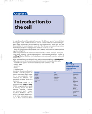
Chapter 1
Introduction to
the cell
Living cells are formed from a small number of the different types of molecules that
make up the earth. Most biomolecules contain carbon and many contain nitrogen.
Both carbon and nitrogen are very scarce in non-living entities. While cells may not
always utilize the most abundant molecules, they do use molecules whose unique
chemistry is capable of carrying out the reactions necessary for life.
There are three levels of organization to describe the molecules that make up living
organisms (Fig. 1.1).
1 The simplest level is the individual elements such as carbon, nitrogen, or oxygen.
2 The basic elements can be arranged into a series of small molecules known as
building blocks. Building blocks include compounds such as amino acids and
nucleic acids.
3 The building blocks are organized into larger compounds, known as macromole-
cules. Macromolecules comprise the different structures that are found in cells.
Four different types of macromolecules are used to construct a cell: FYI 1.1
• nucleic acids; Elements, ions, and
• proteins; trace minerals that
• lipids; make up living
• carbohydrates. systems
Each type of macromolecule is
Elements Ions Trace minerals
used for a specific purpose in
the cell. There are many exam- Oxygen Sodium Manganese
ples of macromolecules being Carbon Potassium Iron
Nitrogen Magnesium Cobalt
combined in different con-
Hydrogen Calcium Copper
figurations to form larger cell Phosphorus Chloride Zinc
structures. Sulfur Aluminum
The nucleic acids can be Iodine
subdivided into DNA and RNA. Nickel
DNA is composed of two kinds Chromium
Selenium
of building blocks, the bases Boron
(adenine, guanine, cytosine, Vanadium
and thymine) and a sugar– Molybdenum
phosphate backbone. DNA is Silicon
used by the cell as a repository Tin
Fluorine
for all of the information neces-
sary to direct synthesis of the
2. PYF1 3/21/05 7:53 PM Page 2
2 Chapter 1
Elements
Phosphate
Pyrimidines Fatty acids
Purines Amino acids Glycerol Sugars
Ribose Other components
Deoxyribose
Fig. 1.1 Three levels of Carbohydrates
Nucleic acids Proteins Lipids or
organization describe the
Polysaccharides
compounds that make up living
organisms.
macromolecules and to produce energy for this synthesis. DNA is also used to trans-
mit information from one generation to the next. RNA has a very similar composition
to DNA. The two major differences between DNA and RNA are in the sugar used in the
sugar–phosphate backbone (ribose for RNA and deoxyribose for DNA) and in one
of the bases (uracil for RNA and thymine for DNA). In Escherichia coli, DNA exists
as a double-stranded molecule. Chapter 2 describes in detail both the structure and
replication of DNA.
The RNA in the cell has at least four different functions.
1 Messenger RNA (mRNA) is used to direct the synthesis of specific proteins.
2 Transfer RNA (tRNA) is used as an adapter molecule between the mRNA and the
amino acids in the process of making the proteins.
3 Ribosomal RNA (rRNA) is a structural component of a large complex of proteins
and RNA known as the ribosome. The ribosome is responsible for binding to the
mRNA and directing the synthesis of proteins.
4 The fourth class of RNA is a catch-all class. There are small, stable RNAs whose
functions remain a mystery. Some small, stable RNAs have been shown to be in-
volved in regulating expression of specific regions of the DNA. Other small, stable
RNAs have been shown to be part of large complexes that play a specific role in the
cell. In general, RNA is used to convey information from the DNA into proteins.
Proteins are composed of amino acids. Most proteins are made from a unique
combination of 20 different amino acids (Fig. 1.2). The order in which amino acids
appear in a protein are specified by the mRNA used to direct synthesis of the protein.
All amino acids have a common core of repeating amino–carbon–carboxyl groups,
with varying side chains on the central carbon. Proteins, therefore, have a repeating
backbone with an amino terminus and carboxyl terminus. The amino acids can be
grouped together and described by physical properties such as charge (acid or basic),
size, interactions with water (hydrophobic — water “hating” or hydrophilic — water
“loving”), a specific element (sulfur containing) or structure they contain (aromatic
rings). The types of amino acids used to make up a protein specify what the protein is
capable of. Proteins perform many duties in the cell, including functioning as struc-
tural and motor components, enzymes, signaling molecules, and regulatory mole-
cules. Some proteins perform only one function while others are multifunctional.
Lipids are an unusual group of molecules that, in bacteria, are used to make the
membranes that surround a cell. One type of lipid, known as a fatty acid, is composed
3. PYF1 3/21/05 7:53 PM Page 3
Introduction to the Cell 3
Nonpolar, aliphatic R groups Aromatic R groups
COO- COO- COO- COO- COO- COO-
+ + + + + +
H3N C H H3N C H H3N C H H3N C H H3N C H H3N C H
H CH3 CH CH2 CH2 CH2
CH3 CH3 C=CH
Glycine Alanine Valine NH
Gly Ala Val
OH
COO- COO- COO-
+ + + Phenylalanine Tyrosine Tryptophan
H3N C H H3N C H H3N C H Phe Tyr Trp
CH2 CH2 H C CH3
CH CH2 CH2
Positively charged R groups
CH3 CH3 S CH3
COO- COO- COO-
CH3 + + +
Leucine Methionine Isoleucine H3N C H H3N C H H3N C H
Leu Met Ile CH2 CH2 CH2
CH2 CH2 C NH
Polar, uncharged R groups CH2 CH2 CH
C N
COO- COO- COO- CH2 NH H
+ + + +
+NH C=NH2
H3N C H H 3N C H H3N C H 3
CH2OH H C OH CH2 NH2
Lysine Arginine Histidine
CH3 SH
Lys Arg His
Serine Threonine Cysteine
Ser Thr Cys
Negatively charged R groups
COO- COO- COO-
H + +
C H3N C H H3N C H COO- COO-
+ + +
H2N CH2 CH2 CH2 H3N C H H3N C H
H2C CH2 C CH2 CH2 CH2
H2N O C COO- CH2
H2N O COO-
Proline Asparagine Glutamine Aspartate Glutamate
Pro Asn Gln Asp Glu
Fig. 1.2 The 20 common amino acids used to make proteins.
of long chains of carbon molecules attached to a smaller head group (Fig. 1.3a). The
small head group is known as the polar head group. The fatty acid chains are
extremely hydrophobic and line up with the long chains of carbons near each other.
Because of the properties of the fatty acid chains, membranes are double-sided (Fig.
1.3b). One side of a membrane is known as a leaflet. The polar head groups face the
outside surfaces of the membranes because the polar head groups are water soluble.
Other chemical groups can be added to the head groups of the lipids but, in general,
4. PYF1 3/21/05 7:53 PM Page 4
4 Chapter 1
(b)
(a)
Polar
Polar
head group
head
O group
C
Fatty acid
CH2
H2C
CH2
H2C
(c) Polar
CH2 head
H2C O group
CH2 C
H2C CH2
CH2 Saturated H2C
fatty acid CH2
H2C
CH2 HC
H2C
HC
CH2 2
H2C CH
Fig. 1.3 Lipid molecules. (a) The
2
general structure of a saturated CH2 C CH
H2
fatty acid. (b) The structure of H2C 2
membranes with two leaflets.
C CH
CH3 H2
(c) The general structure of an 3
CH
unsaturated fatty acid.
other chemical groups cannot be added to the fatty acids. If the fatty acids contain
only single bonds, they are known as saturated fatty acids. Saturated fatty acids are
flexible and can be tightly packed. If the fatty acids contain any double bonds they are
known as unsaturated fatty acids (Fig. 1.3c). Unsaturated fatty acids have a kink in
them and cannot be packed as closely. Membranes usually contain a mixture of fatty
acids to maintain the right packing density and fluidity.
Carbohydrates are composed of simple sugars (Fig. 1.4). They can be used as:
1 an immediate source of energy;
FYI 1.2 2 a stored source of energy;
The composition of 3 structural components of the cell.
an average E. coli cell In bacteria, carbohydrates that
are used as immediate sources of
Component No. of different species % of cell
energy are the simple carbohy-
Water 1 70 drates such as glucose, lactose,
Proteins 3000 or more 16.5 and galactose. Frequently, the
Nucleic acids
DNA 1 0.9
carbohydrates that a bacterium
RNA 1000 or more 6.2 utilizes as energy sources can be
Lipids 40 2.7 used to distinguish one species
Lipopolysaccharide 1 1.0 of bacteria from another. Energy
Peptidoglycan 1 0.8 is usually stored in a carbohy-
Glycogen 1 0.8
drate that is a long polymer of
Polyamines 2 0.1
Metabolites, cofactors, and ions 800 or more 1.0 glucose known as glycogen.
Structurally, carbohydrates
Note: Although a nearly infinite number of different species of proteins, nucleic acids,
are used to make a protective
polysaccharides, etc. are possible, only a tiny fraction of them are found in a given cell type.
covering for the outside of the
5. PYF1 3/21/05 7:53 PM Page 5
Introduction to the Cell 5
cell called the capsule or
capsular polysaccharide.
Monosaccharides Arabinose
Carbohydrates are also Glucose
added to the polar head Fructose
groups of the fatty acids on Galactose
Ribose
the face of the membrane
Deoxyribose
that is exposed to the
outside of the cell. The Disaccharides Lactose
Maltose
fatty acid attached carbo-
hydrates help protect the Polysaccharides Glycogen
cell from detergents and
antibiotics. A complex CH2OH CH2OH
mixture of carbohydrates is O OH O OH
HO
used to make the cell wall.
Cell walls maintain the OH OH
HO
shape of the cell. In some
OH OH
species of bacteria, the cell
wall is located outside Galactose Glucose
the membrane. In other
species, the cell wall is loc-
H2O
ated underneath the outer
membrane. CH2OH CH2OH
Each of the four types HO O O OH
of macromolecules pro- Fig. 1.4 Types and general
OH O OH structures of carbohydrates. The
vides unique functions to
polymerizing of galactose with
the cell. For some of the OH OH glucose results in the formation
cell’s requirements, a single Lactose of lactose upon liberation of one
type of macromolecule suf- H2O molecule.
fices. In other situations, a
mixture of macromolecules is required. It is interesting that beginning with a limited
number of elements and ending with only four major classes of large molecules, the
5000–6000 different compounds needed to make a cell can be constructed.
The bacterial cell: a quick overview
Escherichia coli is a simple, single-celled organism. It is most commonly found in one
of two places, either as a normal, though minor, component of the human intestinal
tract or outside the body in areas contaminated by feces. E. coli can grow in either the
absence or presence of oxygen. It is easy to grow in the laboratory and grows in the ab-
sence of other organisms. It can be grown on chemically defined media and divides as
quickly as every 20 to 30 minutes. Many of its genetic variants are non-disease caus-
ing. These attributes are the main reasons that E. coli has been so extensively and
intensively studied. The last 50 years of research have made it one of the best-under-
stood life forms.
E. coli is a Gram-negative, rod-shaped bacteria. It is 1 to 2 microns in length and 0.5
to 1 micron in diameter. The cell is surrounded by two membranes (the inner and
outer membranes) and as such has four cellular locations, the outer membrane, the
inner membrane, the space between the two membranes or the periplasm, and
the space surrounded by the inner membrane called the cytoplasm (Fig. 1.5). Each
6. PYF1 3/21/05 7:53 PM Page 6
6 Chapter 1
Fig. 1.5 The four cellular
locations of the Gram-negative Outer membrane
E. coli cell. The outer membrane
Periplasm
contains lipids,
lipopolysaccharides, and protein Inner membrane
molecules. The periplasm or 1–2 mm (cytoplasmic membrane)
periplasmic space is an aqueous Cytoplasm
environment containing many
kinds of proteins. The inner
membrane contains lipids and
proteins. The proteins used to
generate energy are located here.
The cytoplasm contains the
DNA and is the site of synthesis
for most of the macromolecules
0.5–1 mm
in the cell.
of these locations has unique contents and properties that provide specific functions
to the cell.
The outer membrane is the main barrier of the cell to certain kinds of toxic sub-
stances. The outer membrane is the cellular surface that interacts with the outside
environment. Detergents, dyes, hydrophobic antibiotics, and bile salts from the in-
testines that could be toxic to the cell cannot cross this membrane. It is composed of
the very common double layer of lipids with the polar head groups of the fatty acids
facing outward (Fig. 1.6a). One unusual aspect of the outer membrane is that the
FYI 1.3 lipids on the outer leaflet of the membrane are different from the lipids on the inner
The Gram stain leaflet. This means that the cell can distinguish the inside of the cell from the outside
of the cell by the composition of the membrane leaflets.
The Gram stain was discovered Attached to the outer membrane and facing away from the cell are specialized
in 1884 by Christian Gram. It lipids, known as lipopolysaccharides or LPS, that contain large carbohydrate side
distinguishes two major
chains (Fig. 1.6b). LPS is essential for the cell to prevent toxic compounds from pass-
groups of bacteria, Gram-
positive and Gram-negative. ing through the outer membrane. Because the LPS is very dense, with six to seven fatty
The major difference in these acid chains per molecule, it is very hard for any compound to get through. LPS con-
groups of bacteria is the nature tains predominantly saturated fatty acids, which favor tight packing of the chains
of their cell wall. In a Gram and a greater impermeability of the membrane. In mutants that are partially defective
stain, actively growing cells are
in LPS, the outer membrane is much more permeable and the cells become very frag-
heat-fixed, stained with the
basic dye, crystal violet, and
ile. The carbohydrate side chains of LPS are usually negatively charged. The side
then with iodine. They are chains allow the cells growing in the intestinal tract to avoid being engulfed by intes-
briefly decolorized by tinal cells, attacked by the immune system, or degraded by digestive enzymes. The
treatment with a solvent side chains may also allow the bacterial cells to colonize the surface of human cells in
such as acetone or alcohol. the intestinal tract.
Gram-positive cells are not
decolorized and remain
Anchored in the outer membrane and facing the outside of the cell are a number of
stained a deep blue-black. specialized structures. Many different types of hair-like projections, known as fim-
Gram-negative cells are briae (singular = fimbria), can cover the cell surface (Fig. 1.7). A single cell can express
completely decolorized and several types of fimbriae at the same time and there can be between 100 and 1000 fim-
appear red. The Gram stain briae per cell. Fimbriae are made of protein and are not generally associated with help-
remains one of the fixtures for
ing the cell move. Rather, fimbriae usually bind to a specific component of eukaryotic
classifying bacteria.
cells and allow the bacteria to adhere to the eukaryotic cell. Some of the more
7. PYF1 3/22/05 5:01 PM Page 7
Introduction to the Cell 7
(a) Outside Mg++ or Ca++
Outer LPS
leaflet
Inner
Phospholipids
leaflet
Inside
Porins Outer membrane proteins
(b)
OAc Galf
Abe
Glc
O-antigen Man
Rha - Ac
Rha
Gal . . . Glc bGlcNAc - Glc
n n
Glc - Glc NAc Glc
Gal Glc
Glc - Gal Glc - Gal
Fig. 1.6 (a) Structure of E. coli’s
Core outer membrane. (b) The
Hep - P Hep - P lipopolysaccharide of E. coli and
Salmonella. Mutants defective in
the O antigen lose their
Hep - P - P - EtN Hep - P - P - EtN
virulence. Mutants defective in
the parts of the core closest to
KDO - KDO - KDO - P KDO - KDO - KDO - P the lipid A are hypersensitive to
dyes, bile salts, antibiotics,
EtN EtN
detergents, and mutagens.
P - GlcN - GlcN - P . . . AraNH3 P - GlcN - GlcN - P Mutants defective in lipid A are
dead. OAc, O-acetyl; Abe,
abequose; Man, D-mannose;
Rha, L-rhamnose; Gal, D-
Lipid A galactose; GlcNAc, N-acetyl-D-
glucosamine; Hep,
L-glycero-D-manno-heptose;
KDO, 2-keto-3-deoxy-octonic
acid; EtN, ethanolamine;
P, phosphate; GlcN, D-
Salmonella enterica serovar typhimurium Escherichia coli glucosamine; AraNH3, 4-
aminoarabinose.
8. PYF1 3/21/05 7:53 PM Page 8
8 Chapter 1
Right-handed
helical structure
Fig. 1.7 Structure of E. coli
fimbriae. Note the right-handed
helical structure. Each subunit of
the fimbriae is a single identical
protein.
FYI 1.4
The colonization of
human cell surfaces common receptors for fimbriae are blood group antigens, collagens, and eukaryotic
by bacteria cell surface sugars.
The flagella (singular = flagellum) are anchored in the inner membrane and face
Virtually every exposed surface the outside of the cell. They differ from the fimbriae in that they are corkscrew-like
of the body is inhabited by
and 5 to 10 microns in length. Flagella are present in fewer numbers (1 to 10 per cell)
some type of bacteria. Usually
each exposed surface has a and they have a motor at their base that is imbedded in the membrane (Fig. 1.8). The
mixed population of many flagella rotate in both the clockwise and counter-clockwise directions. The result of
different species of bacteria. flagella rotation is to move the cell. Flagella movement is coupled to a sensing system
Different bacteria live on in the cytoplasm that results in the net movement of the cell towards a favorable en-
different cell surfaces: for
vironment and away from a harmful one. This directed movement of a bacterial cell is
example, the skin is widely
inhabited by Staphylococcus known as chemotaxis.
epidermis while E. coli is a Some E. coli cells have an additional surface structure that has a unique function. It
natural but minor resident of is also a hair-like structure 2 to 3 microns in length and is called the F pilus (plural =
the colon. Problems for pili) (Fig. 1.9). It is used to build a bridge between two cells, the male that contains the
humans arise when either the
F pilus and the female that does not. A single strand of DNA from the male chromo-
number of a certain type of
bacteria increases dramatically
some is transferred to the female cell. The transferred DNA is replicated and the
or the bacteria end up in a part double-stranded DNA can be incorporated into the chromosome of the female cell. F
of the body where bacteria do pilus mediated transfer of genetic information between cells is called conjugation
not normally reside. Some (see Chapter 10). Conjugation has been instrumental in our understanding of E. coli
parts of the body contain and continues to provide insights into exchange of DNA between cells. It has been
extremely large numbers of
bacteria. The colon contains
shown that conjugation can be used to transfer DNA from E. coli to other bacterial
~109 bacteria per gram. A few species and even into eukaryotic cells.
simple calculations lead to the A second surface structure that is loosely attached to the outer membrane and com-
conclusion that there are about pletely surrounds the cell is the capsule or capsular polysaccharide. The capsule
10 times more bacterial cells is a thick slime layer that is sometimes present on the cell. Cells use the capsule to
than there are human cells in
protect themselves from dehydration or osmotic shock when they are outside the
the human body!
intestines and to avoid being eaten by macrophages when they are inside the body.
9. PYF1 3/22/05 5:01 PM Page 9
Introduction to the Cell 9
Hook/Filament junction Cap
Filament ~10 mm
Hook
LPS
Distal rod
L-ring
Outer membrane
P-ring Peptidoglycan
Proximal
rod
MS-ring Inner membrane
Motor/Switch
Fig. 1.8 The base of the E. coli
Motor flagella. Note the motor
proteins C-ring structure anchored in the
membrane.
Imbedded in the outer membrane are a number of different types of proteins (Fig.
1.10). Some act as receptors for specific substances and are used to transport these sub-
stances through the membrane. Some of the proteins form gated holes or pores
through the membrane. The pores allow passive diffusion across the outer membrane
of chemicals required by
the cell for growth. Pores
that go all the way through
the membrane must be
gated or closed off when
not in use. The pores must F-pilus
be gated so that small mol-
ecules do not leak out of the
cell or toxic compounds do LPS
not leak into the cell. Other
proteins have no transport Outer membrane
A A
function and appear to play A A V
K Fig. 1.9 Assembly of the F pilus.
a structural role. It is gener- H A A Peptidoglycan The proteins A, B, C, E, F, G, H, I,
ally thought that the outer L, Q, U, V, and W are needed for
F W
membrane must contain a C U F pilus biogenesis. Additional F
certain amount of protein. encoded proteins are needed for
If one of the protein species I A B E L QX G Inner membrane mating pair stabilization (two
proteins), surface exclusion (two
is missing from the mem-
proteins), and DNA synthesis
brane, the cell responds by (four proteins). There are 13 F
increasing the amounts of C encoded proteins with no
the other proteins. known function.
10. PYF1 3/21/05 7:53 PM Page 10
10 Chapter 1
Porins Structural Specific
protein transporter
Fig. 1.10 The porins, structural proteins, and specific transport receptors spanning E. coli’s outer
membrane. Porins are composed of three identical subunits with three channels, one through each
subunit. They are responsible for passive diffusion of small molecules. Examples of porins are OmpF and
OmpC. Structural proteins are required in the outer membrane. When they are missing, the outer
membrane is destabilized. An example of a structural protein is OmpA. Specific transporters only allow
specific compounds to pass through them. Many have structural similarities to porins. Examples include
LamB that transports maltodextrin sugars, BtuB that transports vitamin B, and FadL that transports fatty
acids.
FYI 1.5
KHO antigens
Anchored to the inner face of the outer membrane and jutting out into the
Many different subspecies of E. periplasm is a rigid structure called the peptidoglycan layer or cell wall (Fig.
coli can be isolated. More than 1.11a,b,c). The peptidoglycan surrounds the cell and is required to maintain the rod
50 years ago, Kauffman and shape of the cell. The peptidoglycan is arranged in rings that go around the short axis
coworkers described a system of the cell. The penicillin antibiotics interfere with the assembly of the peptidoglycan
to classify E. coli based on their
rings into the completed structure. When a cell is growing in the presence of peni-
outer membrane surface
structures. The system uses cillin, the rings are not connected to one another so that when the cell grows, the
three major surface structures rings come apart and the cell explodes. Thus, the peptidoglycan maintains the cell
that are capable of eliciting an shape and protects the cell from the pressure differences between the inside and out-
immune response in side of the cell.
mammals: the
The periplasmic space or periplasm is an aqueous compartment that resembles
lipopolysaccharide or O
antigen, the flagella or H
the aqueous environment outside the cell. It contains proteins to help concentrate
antigen, and the capsule or K nutrients from outside the cell to inside the cell. Other periplasmic proteins sense the
antigen. To date, 173 different external environment and transduce this information across the inner membrane to
O groups, 56 H groups, and the cytoplasm. These sensing systems allow the cell to respond to the environment,
80 K groups have been found. either protecting itself from harmful conditions or taking advantage of beneficial
The most widely used
laboratory strain is E. coli K12.
conditions. Additional components of the periplasm include proteases to degrade
In contrast to the innocuous, abnormal proteins and a system to provide for the formation of disulfide bonds in
non-pathogenic E. coli K12 specific proteins. Stable disulfide bonds are only found in the periplasm; they cannot
strain, outbreaks of E. coli be found in the cytoplasm. The periplasm monitors the outside of the cell and con-
O157:H7, a strain with very centrates solutes to the inside of the cell.
distinct O and H antigens,
While the outer membrane is the barrier for many toxic compounds, the inner
continue to arise in the United
States each year, usually from membrane is the major physiological barrier between the outside of the cell and the
contaminated food. Infection cytoplasm (Fig. 1.12). It contains the proteins needed to generate energy and has an
by E. coli O157:H7 can be electrical gradient, called the proton motive force, across it. The proton motive force
lethal in very young, very old, is used to generate ATP. In addition, the inner membrane contains proteins that trans-
or immunocompromised
port substances into or out of the cytoplasm, proteins that transmit signals into
individuals.
the cytoplasm, and systems to transport proteins out of the cytoplasm. The inner
11. PYF1 3/21/05 7:53 PM Page 11
Introduction to the Cell 11
(c) N-acetylglucosamine
(a) N-acetylmuramic acid
CH2OH
O CH2OH
Porin Lipoprotein OH O OH
O
HO
NH
Outer membrane H NH
CO O
C=O
Peptidoglycan CH3 CH3 CH
CH3
C=O
Inner membrane NH
L-alanine
CH3 CH
C=O
COOH NH
(b) M M M M M M M CH HOOC CH3
CH2 CH D-alanine
G G G G G G G
D-glutamic acid
CH2 NH
M M M M M M M
C=O C=O
G G G G G G G
N CH
M M M M M M M H
CH2
G G G G G G G
CH2
m-diaminopimilic acid
M = N-acetylmuramic acid CH2
G = N-acetylglucosamine CH
= Peptide H2N COOH
Fig. 1.11 (a) The location of peptidoglycan in E. coli. (b) The structure of peptidoglycan strands. (c) The chemical structure of peptidoglycan.
membrane is the location of many proteins involved in cell division. Some of these di-
vision proteins are only found in limited places in the inner membrane, such as in a
ring around the center of the short axis of the cell. The inner membrane is the site
of many and varied biochemical reactions, signaling reactions, and transport
activities.
The cytoplasm is the hub of cellular activities. It is the location of the single E. coli
chromosome, a circular double-stranded molecule of 4,639,221 base pairs. All DNA
replication and repair of damaged DNA takes place here. Transcription of the DNA
into messenger RNA (mRNA) and translation of the mRNA into protein occur in the
cytoplasm. Decisions about which gene products to make are made in the cytoplasm.
Signals from the environment are received and acted upon in the cytoplasm. In the
cytoplasmic compartment, food sources are broken down or stored and energy is gen-
erated. Because the cytoplasm contains the DNA, it is the site of synthesis of the
majority of the molecules that the cell makes.
12. PYF1 3/21/05 7:53 PM Page 12
12 Chapter 1
Lipids are orientated
in this fashion to
form inner membrane
Anchor proteins
for flagella ñ
dedicated
transport systems
ATP driven transport
Transport systems ATP independent transport
High affinity transporters
F
D
General protein export
Secretion machinery G Polysaccharide export
A Secretion of proteins to
E outside of cell
Y
Fig. 1.12 The inner membrane
of E. coli is composed of ATP
approximately 50% protein and ATP generation H+
50% phospholipid. The major ADP
+ Pi
types of phospholipid are Q
phosphotidylethanolamine,
phosphotidylglycerol, and 4H+ 8H+
cardiolipin. The inner Electron transport H2O Q2
membrane contains the proteins O2 S
for transport systems, the 2H+
protein export machinery, ATP SH2
generation machinery, and Q
electron transport, to name a
few.
13. PYF1 3/21/05 7:53 PM Page 13
Introduction to the Cell 13
How do cells grow? 1 - 2 mm
In order to reproduce, cells
0.5 - 1 mm
must grow and divide.
Growth is not random, but
rather follows a very regular Growth
pattern (Fig. 1.13). The di-
ameter of most rod-shaped
bacteria does not change
2 - 4 mm
during growth. It is the
length of the cylinder that
increases until the cell is ap- 0.5 - 1 mm
proximately double in size.
At this point, E. coli builds a Septum
physical barrier, called the
septum, between the two
daughter cells. Subsequent-
ly, the daughter cells are Fig. 1.13 Cell growth. During
growth, all components of the
physically separated from
cell must be doubled. The
one another. resulting daughter cells are
During growth, all com- physically separated from one
ponents of the cell must be another by a septum.
FYI 1.6
Measuring cell growth the same, there are not enough cells in the on the agar represents the growth from a
suspension to measure. If it is less it is because single cell. The colonies are counted and the
Measuring cells as they grow and divide the cells have scattered the light. How much number of bacteria in each sample is
provides a reliable and quantitative indication less is proportional to the number of cells. For determined. Frequently, the OD600
of the health of the population. Cell growth is example, if the OD600 is 0.25 in one culture measurements and the VCC are both carried
directly related to the nutrients the cells are and 0.5 in a second culture, the second culture out. This allows determination of the
consuming. Measurements of cell growth on has twice as many cells. OD600 is used to correlation of an OD value with an exact count
different energy sources or in mutant cells can make measurements on populations of cells of the number of viable cells. The combination
be directly compared. For energy sources, this and to compare the relative numbers of cells in of OD and VCC measurements eliminates the
has allowed the determination of which these populations. drawbacks of only taking OD readings.
combinations of nutrients are most or least The main drawback of this method is that it Both the OD600 and the VCC take
beneficial to the cells. For different mutant does not distinguish between live and dead measurements on populations of cells. If a
cells, it indicates how important the gene is to cells or between cells and large particles of change occurred at the level of a single cell,
the cell and if there are conditions where it is debris. It also relies on the cells being these measurements would miss it. A third
more or less important. approximately the same size. If a mutant cell method, called flow cytometry, was devised
Cell growth can be measured in three forms long filaments or other odd-shaped to measure the light scatter of single cells. A
different ways. First, the optical density cells, the OD will not give a reading that is stream of media with bacteria in it is passed
(OD) of cells growing in liquid media can be proportional to the number of cells. The across a microscope slide field. Aimed at the
measured in a spectrophotometer. This strengths of this method are that it is rapid and field is a beam of visible light and opposite the
technique is based on the fact that small easy so that many samples can be measured in beam is a detection system that is attached to
particles (cells, in this case) scatter light a short amount of time. OD measurements do a computer. As individual cells pass through
approximately proportionally to their number. not kill cells so after an OD measurement is the beam, the amount of light they scatter is
A suspension of cells is put into a holder or made, the cells can be used for further recorded. Several thousand cells can be
cuvette in the spectrophotometer and a beam experimentation. measured per second. This is a very powerful
of 600 nm wavelength light is passed through The second way to measure bacterial technique but the main drawback is the cost
the suspension. The amount of light that growth is by a viable cell count (VCC). A of the flow cytometer. For certain types of
comes out of the suspension is measured. The sample is taken, diluted and spread on solid experiments where measurements on
amount of light coming out is either the same media, and incubated overnight. The next individual cells are required, it is the only way
as the amount that went in or it is less. If it is day, each colony, or group of cells, that form the data can be obtained.
14. PYF1 3/21/05 7:53 PM Page 14
14 Chapter 1
FYI 1.7 doubled. For components that are present in very large numbers such as ribosomes (2
The growth curve ¥ 104 molecules/cell), membrane lipids (2 ¥ 107 molecules/cell), or outer membrane
pore proteins (2 ¥ 105 molecules/cell), doubling is approximate. Because of the mole-
When E. coli, or any bacteria, is cules’ abundance, the numbers do not have to be exact. For cell components that are
placed in a nutrient-rich broth, present in small numbers, doubling must be exact. In an E. coli cell that is dividing
they begin to grow. Invariably, every 30 minutes, there is a single circular chromosome. The chromosome must be
they follow a simple pattern of
replicated only once so that each daughter cell can inherit one molecule. The dupli-
growth. First, the cells must
prepare for growth and make cation of the chromosome is a carefully controlled and regulated event. Different cell
sure they have all the necessary components are present in specific numbers with some being rare, some at interme-
proteins required for growth, diate levels, and some in very high numbers. Many different mechanisms are em-
this is called the lag phase. ployed to make sure the cell has the correct number of any given component and that
Next, cells begin growing
the daughter cells inherit the right amount. The rarer a component is, the more effort
logarithmically and double at a
constant rate, this is log or the cell invests to ensure that it is divided evenly.
exponential growth. Once cells The growth of a cell is ultimately dictated by the growth of the membranes and the
have used all the available peptidoglycan layer that make up the cell’s boundaries. For both the inner and the
nutrients, they begin to slow outer membrane, new lipids are added randomly into the existing membranes, there
down and prepare for a period
is no one specific zone of growth (Fig. 1.14). Practically, this means that the cell can-
of inactivity known as the
stationary phase. If the nutrient
not use the growth of the membrane to mark specific places along the membrane or to
deprivation lasts long enough, move molecules along it. How the cell localizes specific structures along the mem-
the cells will begin to die. brane and how it measures its length are not known. The fact that daughter cells are
Historically, most experiments born at approximately the same size and very rarely are mistakes made indicates that
have been carried out on the measuring system is accurate.
exponentially growing cells at
37°C. In the past few years, the
Growth of the peptidoglycan cell wall occurs randomly around the peptidoglycan
genes that respond to different along the long axis of the cell. The peptidoglycan that forms the caps of the cells and
growth conditions have been the septum is not exactly the same as that on the long axis of the cell and is synthe-
investigated. New families of sized by different enzymes. For cells dividing every 48 minutes, 50,000 muro-
genes have been discovered peptides or strands of
that are required for stationary
peptidoglycan per minute
phase, cold shock, and heat
shock, to name a few. are added to the growing
cell wall. The new strands
are inserted next to pre-
existing muropeptides at
~200 different sites in the
cell wall (Fig. 1.15). The
Newly inserted lipids enzymes that synthesize
the peptidoglycan reside
Pre-existing lipids
in the inner membrane
and move in a unidirec-
tional fashion around the
cell. They can circumnavi-
gate the cell in about 8
minutes. Excluding the
poles, a cell has ~1100
rings of muropeptides that
maintain its shape.
As the membranes grow,
Fig. 1.14 Growth of
the volume of the cyto-
membranes. Lipid insertion into
growing membranes does not
plasm also increases. The
occur at a specific place on the cytoplasmic contents in-
membrane, rather it is random. cluding the DNA and the
15. PYF1 3/21/05 7:53 PM Page 15
Introduction to the Cell 15
several thousand different
Old strands
protein species must
double in number. Because
proteins are regulated inde-
pendently of each other,
there is no singular mecha-
nism for their doubling.
The vast majority of the
proteins in the cytoplasm
freely diffuse throughout
the boundaries of the cyto-
plasm. When the division
septum is laid down, ap-
proximately half of the con-
tents of the cytoplasm are
trapped on one side of the
septum and half on the
other side. There are a few
known cytoplasmic pro-
teins that are not freely dif- Fig. 1.15 Growth of
fusable but they are the peptidoglycan. Each strand is
exception rather than the New strand actually a ribbon that is in a
rule. Aside from the DNA, helical conformation.
the components of the
cytoplasm are numerous enough to be divided approximately in half.
What is genetics?
The main goal of biology is to understand how a living cell works. Genetics is specifi-
cally concerned with the characteristics of the cell (size, shape, ability to grow on a
certain sugar, specific internal structures, etc.) and how they are passed from one gen-
eration to the next. Early geneticists described physical characteristics or pheno-
types of the cell and followed their inheritance without knowing that they were
encoded by the DNA. After the discovery of DNA and the proof that it is the molecule
used to transmit information from generation to generation, geneticists began to cor-
relate the phenotype with the site in the DNA (or gene) that is responsible for it. The
information in the DNA is referred to as the genotype and the characteristic it speci-
fies is described as the phenotype.
One of the main characteristics defining genetics is that genetics studies the cell
when the cell is intact and growing. Genetics does not attempt to separate each mole-
cule and study the behavior of an individual molecule in a test tube. Rather, genetics
focuses on a specific molecule and isolates that molecule in the cell by making a
mutation in the DNA or gene that specifies the molecule. The behavior of the mutant
cell is then compared to a wild-type, or normal cell. From the behavior patterns of
the two cells, the function of the molecule is inferred. The greater the number of
specific tests that can be devised for the wild-type and mutant cells, the more reliable
is the assignment of the function for the gene being studied. Genetics always tries to
find the simplest, logical argument to describe what has been observed.
16. PYF1 3/21/05 7:53 PM Page 16
16 Chapter 1
Summary
E. coli is one of the best-understood life forms. As a alterations are possible. Each of these topics can
simple, single-celled organism with a completely dramatically influence how to go about isolating
sequenced genome, much is known about E. coli. Our mutants and what interpretations can be drawn from
understanding of E. coli’s lifestyle and the molecular the mutants. Next, it is important to know what tools
structures needed to support that lifestyle has are available to study the mutants. How can they be
contributed to E. coli’s widespread use as a model moved from cell to cell? How can the mutants be
organism for techniques involving molecular genetic cloned and subjected to the tools of molecular
analysis. When undertaking a genetic analysis, the biology? In the chapters that follow, these subjects
first goal is to make mutants that affect the specific will be examined in more detail. Each of the chapters
pathway or molecule of interest. To make mutants, contributes one more piece to the puzzle of how we
it is necessary to understand DNA metabolism, how study biological systems using a genetic point of view.
mutants arise or can be induced, and what kinds of
Study questions
1 What are the building blocks for each of the four macromolecules?
2 What are differences between DNA and RNA? Similarities?
3 What are the functions of messenger RNA, transfer RNA, and ribosomal
RNA?
4 What two features of a fatty acid molecule dictate how it is oriented in a
membrane?
5 What are the differences between carbohydrates used as energy sources and
carbohydrates used as structural components of the cell?
6 What is the function of the outer membrane? The inner membrane?
7 How many different cell surface structures can be present on the outer
surface of a bacterial cell? What is the function of each?
8 In what cellular compartment are disulfide bonds formed?
9 What is the function of the septum and where is the septum located?
References
Gerhardt, P., Murray, M.G.E., Wood, W.A., and Krieg, N.R. eds. 1994. Methods for General and Molecular
Bacteriology. Washington, DC: ASM Press.
Inouye, M. ed. 1986. Bacterial Outer Membranes as Model Systems. New York: Wiley.
Joklik, W., Willett, H., Amos, D., and Wilfert, C. eds. 1992. Zinsser Microbiology. Norwalk, CN: Appleton and
Lange. Chapter 3: Bacterial morphology and ultrastructure.
Neidhardt, F.C., Curtiss III, R., Ingraham, J.L., Lin, E.C.C., Low, K.B., Hagasanik, B., Rexnikoff, W.S., Riley,
M., Schaechter, M., and Umbarger, H.E. eds. 1996. Escherichia coli and Salmonella typhimurium: Cellular
and Molecular Biology, 2nd edn., Washington, DC: ASM Press. Part One, Chapters 3 through 13. Each of
these chapters explores a part of the cell in detail. Each chapter is fully referenced.