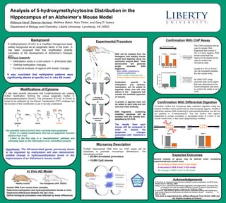
Dynamin I plays dual roles in the activity-dependent shift in exocytic mode i...
finalposterapril13
1. Analysis of 5-hydroxymethylcytosine Distribution in the
Hippocampus of an Alzheimer’s Mouse Model
Rebecca Haraf, Deanna Harrison, Matthew Baker, Noor Taher, and Gary D. Isaacs
Department of Biology and Chemistry, Liberty University, Lynchburg, VA 24502
Modifications of Cytosine
Two possible roles of 5-hmC have currently been proposed:
• 5-hmC is a stable modification that has an epigenetic function
distinct from 5-mC
• 5-hmC is the first step in a “demethylation” pathway and
ultimately leads to the reformation of an unmodfied cytosine
TET1
TET2
TET3
O2
DMNTs
cytosin
e
5-mC 5-hmC
It has been recently discovered that 5-methylcytosine can undergo
further modification, forming the unique epigenetic marker 5-
hydroxymethylcytosine (5-hmC). The conversion of 5-mC to 5-hmC is
known to be catalyzed by Ten-Eleven Translocation (TET) oxidases, but
the function of this modification is yet to be fully understood.
Hypothesis: The AD-associated genes previously found
to be regulated by methylation will also demonstrate
notable change in hydroxymethylation levels in the
hippocampus of an Alzheimer’s mouse model.
In Vivo AD Model
Two transgenes (APP, PSEN1)Healthy Control
•Isolate DNA from mouse brain samples
•Determine methylation and hydroxymethylation levels in mice
•Determine differences between the two mice
•Define biological processes most affected by these differences
5-methylcytosine (5-mC) is a modified nitrogenous base
widely recognized as an epigenetic factor in the brain. It
has been proposed that this modification directly
correlates to the development of Alzheimer’s Disease
(AD).
Background
It was concluded that methylation patterns were
significantly altered at specific loci in vitro AD model.
Previous research:
• Methylation study in a cell culture +/- β-Amyloid (Aβ)
• Defined methylation changes
• Functional analysis of regions with drastic changes
• 20,404 annotated promoters
• 15,980 CpG islands
Microarray Description
DNA will be isolated from the
transgenic and healthy mouse
model and digested using the
restriction enzyme MseI. Heat
denaturation will generate
single stranded DNA
fragments.
Blue: ssDNA containing no
cytosine modifications
Green: ssDNA containing 5-mC
Orange: ssDNA containing 5-hmC
Experimental Procedure
Antibodies specific to
methylation and hydroxy-
methylation will be added to
separate tubes and will only
adhere to the fragments that
contain the modified cytosines
of interest.
A protein A agarose resin will
be added to each tube and will
bind the initial antibody.
DNA fragments containing
modified cytosines will be
isolated from the sample and
amplified by RT-PCR.
Confirmatio
n with ChIP
and PvuRts
1I
Control
Transgenic
Purified hippocampal DNA from our ChIP assay will be
hybridized to promoter microarrays (NimbleGen). The
microarrays contain:
Microarray Distribution
of 5-hmC
Microarray Distribution
of 5-hmC
The results from each
mouse will be compared in
order to assess the
epigenetic changes
associated with Alzheimer’s
Disease.
Expected Outcomes
Several classes of genes may be defined when comparing
experimental and control mice:
• An increase in DNA 5-hmC in AD model
• A decrease in DNA 5-hmC in AD model
• No change in DNA 5-hmC in AD model
Acknowledgements
B. Khulan et al., Comparative isoschizomer profiling of cytosine methylation: the HELP assay.
Genome Res 16, 1046 (Aug, 2006).
M. Oda et al., High-resolution genome-wide cytosine methylation profiling with simultaneous
copy number analysis and optimization for limited cell numbers. Nucleic Acids Res 37,
3829 (Jul, 2009).
This work is supported by the Jeffress Memorial Trust (Grant J-998) and
the Virginia Academy of Science.
Oakley, H., et al., Intraneuronal beta-amyloid aggregates, neurodegeneration, and neuron loss
in transgenic mice with five familial Alzheimer's disease m utations: potential factors
in amyloid plaque formation. J Neurosci 26, 10129 (Oct. 2006).
The ChIP procedure will be
used to isolate DNA
fragments that contain 5-
mC or 5-hmC regions. PCR
primers that flank the
regions to be tested will be
used to confirm the
microarray data.
Levels of 5-mC or 5-hmC
can be compared between
the two mouse lines.
An initial ChIP using
neuronal cells and a region
that was determined to be
hypermethylated was done
to confirm the ability to do a
ChIP.
Projects a loss of
hydroxymethylatio
n in transgenic
Demonstrates pull-
down of specific
DNA sequence
due to methylation
Region to be tested
NA
NA NA5-hmC 5-hmC
Control Transgenic
IncreasingDNA
FoldEnrichment
Confirmation With Differential Digestion
To further confirm the microarray data, restriction digestion using the
enzyme PvuRts1I will be performed on the microarray regions. PvuRts1I
selectively cleaves DNA containing 5-hydroxymethylcytosine, but will not
cut cytosine or 5-methylcytosine. This will confirm that regions
suspected to contain increased or decreased levels of 5-hmC in the
mouse model have, in fact, been epigenetically modified.
C C G G
G G C C
Gene Y
No Enzyme
Gene X
FoldEnrichment
No Enzyme
Gene Y
FoldEnrichment
C C G G
G G C C
Gene X
5-hm
C
5-hm
C
Hinweis der Redaktion
define methylation and hydr. in both mice
fix figure font sizes
methylation levels in a cell culture treated with β-Amyloid (Aβ) to induce “Alzheimer’s-like” characteristics. Differential digestion and gene-specific PCR was used to locate the regions displaying the greatest increase in methylation (hypermethylation) and the greatest decrease in methylation (hypomethylation) in the AD state.