
Empfohlen
Empfohlen
Weitere ähnliche Inhalte
Was ist angesagt?
Was ist angesagt? (20)
Andere mochten auch
Andere mochten auch (20)
Ähnlich wie Oct angiography
Ähnlich wie Oct angiography (20)
Kürzlich hochgeladen
Kürzlich hochgeladen (20)
Oct angiography
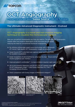
- 1. Now you can perform angiography any time you need to. The optional OCT Angiography module offers non-invasive observation of the microvascular structures reducing the need for conventional fluorescein angiography. Swept Source OCT’s ultra-fast scan captures large quantities of data for excellent visualization Easier recognition of abnormalities by using layer by layer “tissue peeling” intuitive graphical user interface Improved patient’s comfortably*1 - no dyes or dilation required, rapid capture Direct comparison with fundus images in IMAGEnet 6 Integral*3 *1 Compared to conventional fluorescein angiography *2 Observation & photography of anterior segment can be performed only when the optional anterior segment attachment kit is used *3 Optional software *4 DRI OCT Triton plus : OCT /Anterior OCT (Option)/ OCT Angiography (Option) /Color / Red-Free / FA / FAF DRI OCT Triton : OCT /Anterior OCT (Option)/ OCT Angiography (Option) / Color / Red-Free OCT Angiography is a novel and non-invasive imaging technique to visualize the microvascular network BRVOCNV GA By utilizing cutting-edge swept source technology with a wavelength of 1 micron, high-quality OCT Angiography images are acquired All-in-one powerful diagnostic tool (SS OCT /SS OCT Anterior image*2 / SS OCT Angiography image*3 / Color fundus /Red-free /FA / FAF)*4 increasing clinical efficiency See, Discover, Explore Superficial layer OCT Angiography - now combined with Swept Source DRI OCT Triton The Ultimate Advanced Diagnostic Instrument - Evolved Courtesy of SriniVas R. Sadda, M.D., Doheny Eye Institute, UCLA Courtesy of SriniVas R. Sadda, M.D., Doheny Eye Institute, UCLA Choroid Choroid
- 2. *OCT Angiography is optional software *Viewing OCT Angiography image is possible only in combination with IMAGEnet 6 Integral *Not available for sale in the US Diabetic Retinopathy Glaucoma Screen image with IMAGEnet 6 Integral (example) Image Examples 75-1 Hasunuma-cho, Itabashi-ku, Tokyo 174-8580, Japan. Phone:+81-(0)3-3558-2522/2502 Fax:+81-(0)3-3965-6898 www.topcon.co.jp Courtesy of Dr. A. Ishibazawa and Prof. A. Yoshida (Asahikawa Medical Univ., Japan) Superficial layer Deep plexus layer Superficial layer Deep plexus layer (Scan: 3.0 x 3.0mm) (Scan: 3.0 x 3.0mm) See, Discover, Explore Superficial layer Deep plexus layer Normal Eye (Scan: 3.0 x 3.0mm) Printed in Japan 2015 07-50 NP E133-1 GE 2015