Female reproductive system
•Download as PPTX, PDF•
30 likes•4,685 views

A brief and understandable presentation on Female reproductive system

Recommended
More Related Content
What's hot
What's hot (20)
Similar to Female reproductive system
Similar to Female reproductive system (20)
More from Chintan Chavda
Recently uploaded
Recently uploaded (20)
Female reproductive system
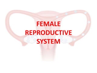
- 2. Female Reproductive System Ovaries Fallopian Tubes Uterus Vagina
- 4. Ovary • Two in number. • The ovary is a small, oval-shaped endocrine gland located on either side of the uterus. • The ovary produces ovum (egg) and hormones like estrogen and progesterone. • Each month, one of the ovaries releases a mature egg, known as an oocyte. • Ovarian follicle is the basic unit of female reproductive biology and is made up of roughly spherical collection of cells found in the ovary. They contain a single oocyte or egg.
- 5. Ovary • A baby girl is born with about 10,00,000 ovarian follicles. • By the time a girl reaches puberty and her menstrual cycle begins, only about 4,00,000 ovarian follicles are left to develop into mature eggs. • During childhood, approximately half of the ovarian follicles are absorbed by the body.
- 7. Fallopian Tube • Also known as Oviducts or Uterine Tubes. • The fallopian tubes stretch from the uterus to the ovaries and measure about 8 to 10 cm (4 to 6 inches) in length. • The ends of the fallopian tubes lying next to the ovaries feather into ends called fimbria (Latin for "fringes" or "fingers"). • Millions of tiny hair-like cilia line the fimbria and interior of the fallopian tubes. The cilia beat in waves hundreds of times a second catching the egg at ovulation and moving it through the tube to the uterine cavity.
- 8. Fallopian Tube • Cells in the tube's inner lining or endothelium nourish the egg and lubricate it's path during its stay inside the fallopian tube. • The egg and sperm meet and the egg is fertilized inside the fallopian tube. • If an egg doesn't become fertilized within 24 to 36 hours after ovulation, it will deteriorate and be removed by the body's immune system like any other dead cell in the body.
- 9. Uterus • The Uterus is a hollow, pear-shaped organ that is the home to a developing fetus. • The reproductive function of the uterus is to accept a fertilized ovum which passes through the utero-tubal junction from the fallopian tube. • It then becomes implanted into the endometrium, and derives nourishment from blood vessels which develop exclusively for this purpose. • The fertilized ovum becomes an embryo, attaches to a wall of the uterus, creates a placenta, and develops into a fetus until childbirth.
- 10. Uterus • The Uterus is divided into two parts: - Cervix (Neck of the Uterus) - Corpus (Body of the Uterus)
- 11. Cervix • The Cervix (or neck of the uterus) is the lower, narrow portion of the uterus where it joins with the top end of the vagina. • Cervix serves 2 major functions: - First, it maintains its firmness (physical integrity) during pregnancy as the uterus dramatically enlarges. - Second, in preparation for labor and delivery, the cervix softens and becomes more distensible, a process called Cervical Ripening.
- 12. Corpus • Corpus of Uterus is composed of 3 layers: • The outer layer of the uterine wall is called Perimetrium. • The Myometrium is the middle layer of the uterine wall consisting of smooth muscle cells and supporting stromal and vascular tissue. • The inner layer of the uterine wall is called Endometrium or Uterine lining. Shedding of the functional endometrial lining is responsible for menstrual bleeding.
- 13. Vagina • The vagina is a canal that joins the cervix (the lower part of uterus) to the outside of the body. • During childbirth, the vagina provides the channel to deliver the infant from the uterus to its independent life outside the body of the mother. So, it is also known as the birth canal. • The vagina provides a path for menstrual blood and tissue to leave the body.
- 14. Thank You