1. Egg Injection Techniques & Tricks
E2 injection techniques as developed by Karina Cramer and interpreted by Sue
Hendricks.
Windowing Techniques
q Set eggs in the morning. Use eggs ~52 hours after they were set
(so, +4 hours 2 days later; stage 12-14). I get the best success
with eggs that are set within a couple days of arrival (eggs arrive
on Tuesday, so Thursday and Friday give me the best success).
q Bring up eggs on egg cardboard crate. Turn so that egg is on
side
q Allow eggs to rest for ~10 minutes
q Swab liberally with 70% ETOH
q Wipe all instruments with 70% ETOH
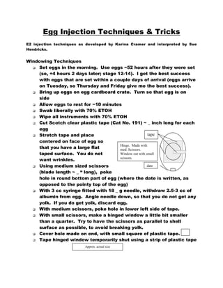
q Cut Scotch clear plastic tape (Cat No. 191) ~ _ inch long for each
egg
tape
q Stretch tape and place
q
q
q
q
q
q
centered on face of egg so
Hinge. Made with
that you have a large flat
med. Scissors.
Window cut with small
taped surface. You do not
scissors.
want wrinkles.
date
Using medium sized scissors
(blade length ~ _ “ long), poke
hole in round bottom part of egg (where the date is written, as
opposed to the pointy top of the egg)
With 3 cc syringe fitted with 18 _ g needle, withdraw 2.5-3 cc of
albumin from egg. Angle needle down, so that you do not get any
yolk. If you do get yolk, discard egg.
With medium scissors, poke hole in lower left side of tape.
With small scissors, make a hinged window a little bit smaller
than a quarter. Try to have the scissors as parallel to shell
surface as possible, to avoid breaking yolk.
Cover hole made on end, with small square of plastic tape.
Tape hinged window temporarily shut using a strip of plastic tape
Approx. actual size
2. Injection Techniques
q Turn on picospritzer, set to 10 ms pulses
q Turn on Fluorescence lamp if necessary
q Turn on air, set to 10 psi
q Fill pulled pipettes with dyes using Microfil Syring Needle
a. Pull pipettes on David Kopf Vertical Pipette Puller Model
700C (downstairs) using the heater set @ 65, and solenoid
set at 50.
q Place windowed egg on wax ring under dissection scope
q Open window, and use tape that kept it closed to keep it open
q Using your probe or fine forceps, pop any bubbles on surface of
egg. Discard any egg in which the yolk has broken.
q You should be able to see the embryo (although it may take some
practice) but to see it better, we inject India ink under the
embryo
q India ink injection:
a. 1:30 dilution of store-bought stationary India ink in sterile
PBS. We generally make 30 mls at a time, in a 50 ml tube.
It’s the particulate carbon matter in the ink that’s
important, so you cannot filter the ink to make it sterile. It
doesn’t need to be sterile, so you’re all good.
b. Fill syringe with ink, attach 30g _ inch needle.
c. Using forceps or your fingers, grasp the needle and bend it
to make a 90° angle
d. Expel any bubbles in syringe
e. Insert bent needle into the yolk just under the yolk
membrane a little bit away from the embryo. Avoid
inserting needle into any membranes associated with the
embryo
f. Expel ink gently under embryo. You should be able to see
the embryo nicely now
q Stage embryo (count somites)
q Using a probe, or the tip of your fine forceps gently remove the
top membrane from over the embryo. Do this by scraping the tip
across the surface moving from say 4 o’clock to 11 o’clock. (do
not start directly over the embryo! Remove more membrane than
that, so you don’t poke the embryo by mistake)
3. q
q
q
q
q
a. The membrane is VERY difficult to see; in fact you can only
see it when you remove it. So, this takes practice.
b. You’ll be able to tell if you didn’t remove the membrane
because it’ll be difficult to do the next step, and your
pipettes will continually get plugged.
Open the roof plate over the area you plan to inject using your
probe
Lower pipette filled with dye of choice using the
micromanipulator. [I use DiI & DiD (D282&D7757 both from
Probes)]
Break very tip of pipette using forceps
Inject at will
If your pipette becomes clogged, try swabbing tip with a tiny
piece of kimwipe soaked in 70% ETOH. If that doesn’t work, then
break the tip again. Obviously, you don’t want to have to break
the tip very often, or too much dye will explode out of your
pipette during injections.
For Rhombomeric Injections:
q Embryos should have at least 13 somites, and can be used up to
about 21 somites (head will have turned at this point).
q Inject embryos
Top membrane, removed
only if their
from over embryo
otocysts are
clearly visible.
This is the most
salient landmark
for R5/R6
injections.
Grossly speaking,
R5 is found as the
R4
R5
rostral half of the
R6
otocyst, while R6
otocyst
is even with the
q
caudal half of the
otocyst.
Also, early embryos in particular may show a clearly delineated
R4
4. q
q
q
q
For lateral injections, aim &
“Imaginary” line: the
dorsal part of the
lower pipette at the dorsal
rhombomeric region
is more opaque than
surface of the Rhombomere.
the rest of the neural
tube
This one’s easy
For medial injections, aim &
lower pipette at the most
extreme medial edge of the
dorsal boundary of the
otocyst
rhombomere. As you insert
your pipette, it will actually
enter the embryo on the slope
of the neural tube. You want
this injection to be separate
and distinct from the lateral
otocyst
injection, but if you try to lower
the pipette into the neural tube,
your injection will turn out to be WAY too ventral. This one is
very difficult.
Because the embryo turns, using slightly older embryos may be
beneficial for these medial injections…it’s easier to get at, but as
a consequence, your lateral injection is now harder. Pick your
poison.
It’s a good idea to take a picture, and draw the ‘cross-section’ of
your injection for fine tuning later
Caring for your data-generating eggs
q After injection, close the window with the strip of plastic tape.
q Write the experimental number and your initials on the egg, near
the date.
q Cut 2 pieces of electrical tape just longer than the width of the
plastic tape piece already on the egg
q Cover window completely with electrical tape, taking care to
slightly stretch the tape as you’re placing it to get a tight seal.
Smooth out any wrinkles, and make sure the seal around the
window is good and tight
q Place eggs in table-top incubator which should be set to 100° F
and have plenty of water in all the reservoirs. Do not rotate
eggs. In fact, ensure that they remain level.
5. q
q
q
q
Make sure to check water level daily, and ensure that the
cardboard egg crates do not get soaked.
I get very good survival rates (85-100%), so I never peek before
the date of sacrifice. It’s up to you though.
Dissect in PBS, postfix in 4% Para overnight. Embed in 3%
Agarose, section on vibratome.
As you’re learning the technique, you should dissect your newly
injected E2 embryos, postfix in 4% para for 2 hours-overnight,
cryoprotect in 30% sucrose and section on cryostat at 10mm. Do
this to ensure accurate injection placement (see the crosssection diagram above)
6. Materials
Product
Pipettes
Glass filament, thin-wall 1.2mm x
.9mm 4”
Scotch plastic tape
Scotch Super 33+ vinyl electrical
tape
Microfil Syringe Needle
Cat. Number
616000
Vendor
A-M Systems
191
05400706132
MF28G67-5
3M,
3M, get through
University Stores
World Precision
Instruments
DiI (DiIC18(3))
D-282
Molecular Probes
DiD (DiI C18(5))
D-7757
Molecular Probes
India Ink, Fountain Pen Ink
46030(723)
Higgins
Tools:
q 55 Dumont Fine Forceps
q Wax ring (~2” diameter)
q Medium scissors
q Small scissors (Roboz 5840)
q Sharp probe (Roboz RS6100)
q 70% ETOH
q 18g _ inch needles
q 30g _ inch needles
q 3 cc syringes
q Kimwipes
Address
131 Business Park Loop
PO Box 850
Carlsborg, WA 98324
Phone Number
1800.426.1306
175 Sarasota Center
Blvd
Sarasota, FL 34240
PO Box 22010
Eugene, OR 97402
PO Box 22010
Eugene, OR 97402
Bookstore
941.371.1003
f941.377.5428
q
q
541-465-8300
541-465-8300
tabletop incubator, held at 100°F with
full water reservoir
black plastic bag (for egg waste)