Atopic dermatitis: mechanism of disease
•Als PPTX, PDF herunterladen•
67 gefällt mir•43,881 views

This document summarizes the mechanisms of atopic dermatitis (AD). It discusses the epidemiology of AD and notes that it commonly affects children under 5 years old. The pathophysiology involves genetic, environmental, immunological, and epidermal factors. Key aspects of the pathophysiology discussed include the role of skin barrier dysfunction and genes involved in barrier function like filaggrin. It also examines the role of the immune system in AD, focusing on the predominance of TH2 cytokines and immune cells like dendritic cells, T lymphocytes, mast cells, and eosinophils that perpetuate the inflammatory response in AD.

Empfohlen
Weitere ähnliche Inhalte
Was ist angesagt?
Was ist angesagt? (20)
Ähnlich wie Atopic dermatitis: mechanism of disease
Ähnlich wie Atopic dermatitis: mechanism of disease (20)
Mehr von Chulalongkorn Allergy and Clinical Immunology Research Group
Mehr von Chulalongkorn Allergy and Clinical Immunology Research Group (20)
Kürzlich hochgeladen
Kürzlich hochgeladen (20)
Atopic dermatitis: mechanism of disease
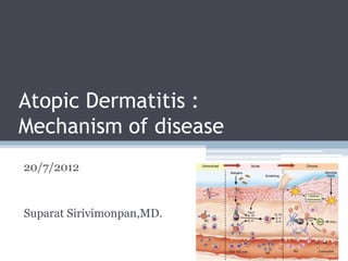
- 1. Atopic Dermatitis : Mechanism of disease 20/7/2012 Suparat Sirivimonpan,MD.
- 2. Introduction • AD is a chronic relapsing inflammatory skin disease • More than 50% develop asthma • 75% develop AR • complex interrelationship of ▫ genetic, environmental, immunologic, and epidermal factors Mark Boguniewicz, Donald Leung.Middleton’s Allergy 7’th edition 893-1999
- 3. Epidemiology • Affects 15-30% of children, 2-10% of adult • 45% begin within the first 6 mo • 60% begin during the first yr • 85% begin before 5 yrs • Up to 70%: spontaneous remission before adolescence NJEM 2008;358:1483-94 NJEM 2008;358:1483-94
- 4. Acute AD • Intensely pruritic, erythematous papule associated with excoriations, vesiculation, and serous exudate • Pathology : spongiosis (intercellular epidermal edema), superficial epidermal hypertrophy and acantholysis • marked infiltration of CD4 activated memory T cells, APCs, (LCs, inflammatory dendritic epidermal cells (IDECs), macrophages), and degranulated mast cell Histology: Spongiotic area within the epidermis
- 5. Chronic AD • thickened plaques with increased lichenification • Pathology : marked epidermal hyperplasia, acanthosis • macrophage-dominated mononuclear cell infiltrate in dermis, and perivascular accumulation of lymphocytes in smaller numbers than seen in acute AD Hyperplastic of epidermis with hyperkeratosis Adv Immunol.2009;102;135-226
- 6. Pathophysiology of AD • Genetics • Barrier function of the skin • Immunopathologic mechanism • Autoimmunity NJEM 2008;358:1483-94
- 7. Genetics of AD • atopic dermatitis–specific genes • related loci on chromosomes 3q21,1q21,16q,17q25, 20p,3p26 • loci associated with psoriasis , two disease are rarely linked genes expressed in skin play important role • do not overlap with allelic variants that are frequent in allergic asthma Adv Immunol.2009;102;135-226 NJEM 2008;358:1483-94
- 8. Genetic of AD • two major groups of genes • (1) genes involved in skin barrier function : FLG, SCCE, SPINK5 • (2) genes involved in the immune response Adv Immunol.2009;102;135-226
- 9. Genes involved in skin barrier function • strong genetic linkage to Chromosome 1q21 : human EDC (epidermal differentiation complex ) • Mutations in the FLG (encode filaggrin) gene located on chromosome 1q21.3 ▫ identified in ichthyosis vulgaris, AD Adv Immunol.2009;102;135-226
- 10. N Engl J Med 2011;365:1315-27.
- 11. • 425 Singaporean Chinese patients • All patients : Dx by two paediatric dermatologists according to the U.K. Working Party’s diagnostic criteria for AD • examined for palmar hyperlinearity and keratosis pilaris • AD severity was graded as mild, moderate or severe according to SCORAD • mean SD age was 10 5.18 years (range 1–21) • 68% of patients were male • Control : Genomic DNA from 440 Singaporean Chinese individuals was obtained from the Singapore Bio Bank • Unknown IV ⁄AD status • mean SD age = 44 14 years (range 1–80), 44.1 males Br J Dermatol2011;165:106–114
- 13. • Irish AD cohort is slightly enriched for cases of severe AD (47.8 defined by the Nottingham Eczema Severity Score) compared with the Singaporean Chinese cohort (39.5 contributing factor Br J Dermatol2011;165:106–114
- 14. • palmar hyperlinearity : PPV 34.1 , NPV 85.5 • keratosis pilaris : PPV 31.6%, NPV 79.3 • indicates that patients with AD with the absence of palmar hyperlinearity and ⁄or keratosis pilaris are unlikely to carry FLG-null mutations Br J Dermatol2011;165:106–114
- 15. FLG • Null mutations in the FLG gene are predisposing factor for early-onset AD, which persists into adulthood • FLG expression is also reduced in AD patients with no FLG mutations due to local expression of Th2 cytokines IL-4 ,IL-13 : downregulate FLG expression in keratinocytes Adv Immunol.2009;102;135-226
- 16. Genes involved in skin barrier function SCCE (stratum corneum chymotryptic enzyme) • 19q13 • play a central role in desquamation by cleaving proteins of the SC SPINK5 gene • 5q32 • encodes LEKTI (lympho-epithelial kazal-type related inhibitor) : regulates proteolysis in terminal keratinocyte differentiation • Mutations in SPINK5 : Netherton’s syndrome ▫ many features of AD including dermatitis, eosinophilia, and high IgE level Adv Immunol.2009;102;135-226
- 17. Genes involved in skin barrier function • E420K single nucleotide polymorphism (SNP) variant in the SPINK5 gene ▫ significant association with disease severity ▫ presence of food allergy in children with AD • Other protease inhibitors with similar roles to SPINK5 are encoded by a cluster of genes on chromosome 20q12, : linked to AD Adv Immunol.2009;102;135-226
- 18. Genes involved in the immune response Chromosome 5q31-33 • cytokine genes cluster • Th2 : IL-4, IL-5, and IL-13 • CD14 antigen • IL-12b subunit (IL-12 p40) (Th1 cytokines) Chromosome 16q12 • SNPs within the a chain of the IL-4 receptor gene Adv Immunol.2009;102;135-226 J Allergy Clin Immunol 2006;118:24-34
- 19. Genes involved in the immune response Chromosome 11q22.2–22.3 • IL-18 Chromosome 1q31–32 • IL-10 gene , anti-inflammatory responses Chromosome 11q13 • FcƐRI gene • associate with AD and asthma Chromosome 12q24 • IL-31, associated with itching Adv Immunol.2009;102;135-226
- 20. Genes involved in the immune response • Promoter region of lymphocyte- attracting chemokine (17q)1 ▫ RANTES (17q11) (regulated on activation, normal T-cell expressed and secreted) ▫ Eotaxin 1 (17q21.1-q21.2) • Gain-of-function polymorphisms in the α subunit of IL-4 receptor (16q12) 2 • Polymorphisms of the gene encoding IL-18 (11q22) 2 1 J Allergy Clin Immunol 2006;118:24-34 2 NJEM 2008;358:1483-94
- 21. J Allergy Clin Immunol 2006;118:24-34
- 22. Barrier function of the skin • Physical barrier • Innate immune system NJEM 2008;358:1483-94
- 23. Barrier function of the skin • Cornified envelop (CE) :several proteins ▫ filaggrin, loricrin, trichohyalin, small proline-rich proteins, involucrin and keratin intermediate filaments ▫ cross-linked extensively by transglutaminases ▫ epidermal differentiation complex (EDC) ▫ a cluster of genes on human chromosome 1q21 • SC lipid composition : ▫ ceramides (45–50% by weight) ▫ Cholesterol (25%) ▫ free fatty acids (10–15%) ▫ less than 5% each of several other lipids : most important = cholesterol sulfate J Clin Invest.2006;116:1150-58 Adv Immunol.2009;102;135-226
- 24. • Profilaggrin : giant inactive precursor, highly phosphorylated polypeptide • main constituent of keratohyalin F granules : granular cell layer • profilaggrin is dephosphorylated and proteolytically cleaved by serine proteases, into multiple filaggrin polypeptides • filaggrin binds to keratin in a structure aligned parallel to the outer surface of the epidermis Adv Immunol.2009;102;135-226
- 26. Filaggrin • filaggrin peptides are further degraded into hydrophilic amino acids, including urocanic acid, pyrrolidone carboxylic acid, and alanine 1 • Trans-urocanic acid : protection against ultraviolet radiation,modulates immune function 2 • Pyrrolidone carboxylic acid :derivative of glutamine 2 • Arginine residues in filaggrin are converted to citrulline proteolysis short peptides a pool of hygroscopic amino acids and derivatives : natural moisturizing factor (NMF) 2 ▫ involves caspase 14 and other proteases 1 Adv Immunol.2009;102;135-226 2 N Engl J Med 2011;365:1315-27.
- 27. Profilaggrin • histidine-rich and glutamine-rich proteins • modulate pH of the stratum corneum • promote the retention of moisture • possibly exert antimicrobial activity against staphylococcus N Engl J Med 2011;365:1315-27.
- 28. N Engl J Med 2011;365:1315-27.
- 29. Physical barrier proteases, and protease inhibitors • Balance • Proteases, especially SC tryptic and chymotryptic enzymes : desmosome breakdown and corneocyte desquamation • skin proteases is controlled by protease inhibitors ▫ SPINK5, a gene which encodes a putative serine protease inhibitor, lymphoepithelial Kazal-type-related inhibitor (LEKTI) Adv Immunol.2009;102;135-226
- 30. Physical barrier • Changes in pH • Changes in the cornified envelope proteins involucrin and loricrin or lipid composition • Decrease in ceramide • Underlying inflammation can alter the expression of genes such as FLG increase in skin penetration of allergen and increased TEWL + pruritus inflammation and sensitization NJEM 2008;358:1483-94
- 31. Innate immune system • protect skin against infection ▫ Pathogen associated molecular pattern (PAMP) molecules VS Pattern recognition receptors (PRRs) ▫ Antimicrobial peptides (AMPs) Adv Immunol.2009;102;135-226
- 32. PAMPs and PRRs • Toll-like receptors (TLRs) (TLR1-11) • 39 C-type lectins • nucleotide-binding oligomerization domain–like receptors • peptidoglycan-recognition proteins (PGRPs) (4) • expressed on macrophages, DCs, PMNs, mucosal epithelial, and endothelial cells NJEM 2008;358:1483-94 Adv Immunol.2009;102;135-226
- 33. Antimicrobial peptides (AMPs) • expressed by keratinocytes and cells that form sebaceous and sweat glands, mast cells, circulating cells (PMNs,NK) • Antimicrobial action against bacteria, viruses, and fungi • 2 major classes ▫ cathelicidins ▫ defensins Adv Immunol.2009;102;135-226
- 34. Innate immune system • TH2 cytokine (prominent in AD) : IL-4, IL-13 down-regulates antimicrobial peptides in skin difficult to manage microbial infections • Lesional and normal looking skin is extensively colonized by bacteria such as Staphylococcus aureus or fungi such as malassezia • predisposed to eczema herpeticum and eczema vaccinatum (decrease cathelicidin potent antiviral activity) NJEM 2008;358:1483-94
- 35. Immunopathologic mechanisms • Early-onset atopic dermatitis : absence of IgE-mediated allergic sensitization • IgE mediated sensitization often occurs several weeks or months after the lesions appear ▫ some children (mostly girls) —no IgE-mediated allergic sensitization • The initial mechanisms that induce skin inflammation in patients with atopic dermatitis are unknown NJEM 2008;358:1483-94
- 36. Immunopathologic mechanisms • neuropeptide-induced, irritation-induced, or pruritus-induced scratching, which releases proinflammatory cytokines from keratinocytes NJEM 2008;358:1483-94
- 38. Key cell in AD • T lymphocytes • Dendritic cells (DCs) • Keratinocytes • Mast cells • Eosinophils J Allergy Clin Immunol -
- 39. T lymphocyte • LCs and DCs present antigen to recirculating naive T cells • proliferation and differentiation into memory/effector cells that express skin homing receptors such as ▫ cutaneous lymphocyte antigen (CLA) ▫ CCR4, and CCR10 • Antigen-specific effector CD4 T cells leave LN into the circulation and re-enter the skin, via their skin specific receptors proliferate and secrete cytokine Adv Immunol.2009;102;135-226
- 40. T lymphocyte • Skin homing cutaneous lymphocyte antigen (CLA +) T cell ▫ CLA : inducible carbohydrate modification of P-selectin glycoprotein ligand-1, memory T cells (absent on naïve) ▫ facilitates binding of T cells to E-selectin ▫ distinct capacity to home to the skin through expression of the skin-homing receptor CLA 90% of infiltrating T cells ▫ IL-4 and IL-13 like TH2 ▫ produce IL-31 Pruritus J Allergy Clin Immunol 2006;117:418-25
- 42. • Cytokines : ▫ TNF-a and IL-1 ▫ keratinocytes, mast cells, and DCs ▫ expression of vascular endothelial cell adhesion molecules esp. VCAM-1, E-selectin, CD54 • Chemokines : ▫ Keratinocyte, LC ▫ RANTES, MCP-4, eotaxin : Eo, TH2 type lymphocyte ▫ IL-16 : CD4+ T cell ▫ CTACK/CCL27 : CTA+ T cell ▫ CCL1, CCL18 • extravasation of inflammatory cells J Allergy Clin Immunol Mark Boguniewicz, Donald Leung.Middleton’s Allergy 7’th edition 083-1099 -
- 43. Predominant TH2 profile • TLSP : keratinocyte , LC • TH1 cells : high IFN-ɣ–producing increased apoptosis both TH1 and TH2 (TH1 > TH2 cells) • Increased expression of Fas, Fas-ligand, tumor necrosis factor receptor-II, and caspase activation was detected on TH1 cells • IFN-ɣ keratinocyte apoptosis J Allergy Clin Immunol
- 44. T lymphocyte • more chronic : expression of IFN-ɣ, IL-12, GM-CSF • The mechanism : switch from Th2 to Th1 type ▫ is not well understood ▫ IDEC (Inflammatory dendritic epidermal cells) : IL12,18 ▫ could be related to microbial products TLR2 is important for Th1 response to cutaneously introduced antigen Adv Immunol.2009;102;135-226
- 45. T lymphocyte : T reg T regulatory (CD4+CD25+FOXP3) • Immunosuppressive function • Inhibit development of both TH1 and TH2 responses • Increased in peripheral blood with normal immunosuppressive activity • decreased in lesion J Allergy Clin Immunol 2006;117:418-25 • Staphylococcal superantigens subvert Treg cell function ▫ > 90% of patients have S. aureus colonization ▫ enhanced effector T cell activation skin inflammation Adv Immunol.2009;102;135-226
- 46. IL-31 • cutaneous and peripheral blood CLA+ T cells : source • correlation between IL-31 serum levels with the severity of AD • staphylococcal enterotoxin rapidly induces IL-31 expression in lymphocytes,monocytes and macrophages (but not on dendritic) infection lead to exacerbation of pruritus • IL-31 induces expression of the inflammatory T cell attracting chemokines CCL17/TARC and CCL22/MDC (keratinocytes) • IL-31 enhanced secretion of IL-1, IL-6 and IL-18 and up- regulated CD86 expression Adv Immunol.2009;102;135-226 Allergy 2010; 65: 712–721
- 47. T lymphocyte : Th17 cell • markedly in acute than chronic AD lesions • number of Th17 cells is increased in the peripheral blood of AD • IL-17 serum levels are elevated • IL-17 stimulates keratinocytes to produce GM-CSF, TNF-a, IL- 8, CXCL10, and vascular endothelial growth factor (VEGF), and HBD-2 • produce IL-22 • marked synergistic effect between IL-17 and IL-22 was observed on IL-8 and AMP production Adv Immunol.2009;102;135-226
- 48. Dendritic Cells • 2 myeloid dendritic cells (high-density display of FcεRI) ▫ Langerhans’ cells : normal skin ▫ Inflammatory dendritic epidermal cells(IDEC) : only in inflamed skin • Epidermal dendritic cells bear IgE and express its high-affinity receptor(FcεRI) • In skin lesions, dendritic cells of the plasmacytoid lineage (potent antiviral activity IFN-α production) are almost absent viral infection Adv Immunol.2009;102;135-226 NJEM 2008;358:1483-94
- 49. Keratinocyte • cutaneous immune responses • production of proinflammatory cytokines, chemokines, AMPs activation and recruitment of DCs, T cells, other leukocyte amplify and maintain skin inflammation • Itch induced scratching :release of proinflammatory cytokines and chemokines • Atopic keratinocyte-derived GM-CSF : proliferation and differentiation of peripheral blood monocytes into mature DCs in presence of IL-4 Adv Immunol.2009;102;135-226
- 50. Keratinocyte • IL-1 and IL-18 ▫ inactive precursors in keratinocytes ▫ converted to active forms by CASP1 enzyme after stimulation by danger signals ▫ amount of active IL-18 in serum increases with exacerbation of their disease • TSLP ▫ Microbial products, physical injury, or inflammatory cytokines, including proinflammatory (TNF-α and J Allergy Clin Immunol IL-1α) and Th2 (IL-4 or IL-13) cytokines induce - TSLP ▫ polarizes human DCs to skew the T cell response to Th2 Adv Immunol.2009;102;135-226
- 51. Keratinocyte • activated skin-infiltrating T cell can upregulate Fas expression on keratinocytes keratinocyte apoptosis spongiosis (key pathogenic event in AD) • overexpress numerous chemokines activation and recruitment ▫ CCL20/MIP-3α, CCL27, CCL17/TARC, CCL22/MDC - attract DCs and T cells ▫ CCL20/MIP-3α - recruitment of CCR6-expressing immature DCs and memory/effector T Adv Immunol.2009;102;135-226
- 52. Keratinocyte • Keratinocyte-derived AMPs (antimicrobial peptide) : innate ▫ β-defensins (HBD-2 and HBD-3), and cathelicidin, hCAP18/ LL- 37 • Expression of HBD-2, HBD-3, and hCAP18/LL-37 is significantly decreased in acute and chronic AD skin lesions compared to psoriasis skin lesions • IL-13 and IL-4 inhibit production of HBD-3 by keratinocytes • Th2 cytokine may suppress innate immune response against bacterial and viral pathogens • Decreased AMPs in AD skin may contribute to its susceptibility to infections Adv Immunol.2009;102;135-226
- 53. Mast cell • early stage AD : normal numbers but undergo degranulation in the affected skin • late stage AD : increased number, but without degranulation • Mast cell-derived histamine, mast cell proteinases enzymes tryptase and chymase (MCC), and other inflammatory mediators pruritus and inflammation • Histamine upregulates production • inflammatory cytokines by keratinocytes • Increased proteinase activity skin barrier defect Adv Immunol.2009;102;135-226
- 54. Eosinophil • common findings in AD ▫ Peripheral blood eosinophilia ▫ elevated serum levels of eosinophil granule proteins, basic proteins eosinophil cationic protein (ECP), eosinophil-derived neurotoxin (EDN) major basic protein (MBP) • correlate with disease activity • increased expression : eosinophil chemotactic factor CCL11/eotaxin and of IL-5 and IL-5Rα in acute and chronic skin lesions , blood in AD patients J Allergy Clin Immunol - Adv Immunol.2009;102;135-226
- 55. Eosinophil • Eosinophils are recruited and activated by IL-5 and IL-13 inflammation and tissue damage • Inhibition of eosinophil apoptosis in AD : IL-5 ,GM-CSF • Eosinophils : switching TH1 response by IL- 12 • more pronounced in chronic AD J Allergy Clin Immunol - Adv Immunol.2009;102;135-226
- 56. NK cell • release of perforin and granzyme • Release proinflammatory cytokines such as IFN-g, TNF-a, GM-CSF, IL-5, and IL-8 recruitment of other innate immune cells • Circulating CD56+CD16+ NK cells are reduced but increases in lesional skin with severity of disease • NK : functionally defective in AD ▫ MHC-nonrestricted cytotoxicity against standard NK- sensitive target cells ▫ reduced release of IFN-ɣ • Decreased NK activity skin infection Adv Immunol.2009;102;135-226
- 57. PMNs • lack of detectable PMNs in skin lesion • PMN chemotactic defect ▫ found to correlate with markers of AD disease severity, serum IgE levels, and skin bacterial infection • PMN functions are impaired especially during infectious period ▫ impaired phagocytosis, reduced capacity to produce reactive oxygen species, impaired release of β-glucuronidase, defective leukotriene B4 production and release ▫ absent deposition of extracellular PMN granule proteins (lactoferrin and PMN elastase) • contribute to the susceptibility of AD skin to infection Adv Immunol.2009;102;135-226
- 59. Autoimmunity in Atopic Dermatitis • In addition to IgE antibodies against food and aeroallergens • Serum from patients with severe atopic dermatitis contain IgE antibodies against proteins from keratinocytes and endothelial cells ▫ manganese superoxide dismutase ▫ calcium-binding proteins • serum levels of these IgE autoantibodies correlate with disease severity • Scratching probably releases intracellular proteins from keratinocytes • These proteins could be molecular mimics of microbial structures induce IgE autoantibodies NJEM 2008;358:1483-94
- 60. Autoimmunity in Atopic Dermatitis • 25% of adults AD : IgE antibodies against self-proteins • these patients, early-onset atopic dermatitis, intense pruritus, recurrent bacterial skin infections, and high serum IgE levels are hallmarks of disease • IgE antibodies against self-proteins can be detected in patients with atopic dermatitis as early as 1 year of age • IgE antibodies against autoantigens in skin can perpetuate the allergic inflammation NJEM 2008;358:1483-94
- 61. Gene–Gene and Gene–Environment Interactions in the Natural History of Atopic Dermatitis NJEM 2008;358:1483-94
- 62. Thank you for your attention…