The Proteases: Mechanisms and Applications
•Download as PPT, PDF•
9 likes•11,349 views

1. Serine proteases use a catalytic triad of serine, histidine, and aspartate residues to hydrolyze peptide bonds through a nucleophilic attack by the serine residue. 2. Site-directed mutagenesis experiments have demonstrated the importance of these catalytic residues and the oxyanion hole for stabilizing the reaction intermediate. Mutating these residues reduces catalytic activity by several orders of magnitude. 3. Recent evidence suggests additional mechanisms such as low barrier hydrogen bonds and substrate assisted catalysis may contribute to the efficiency of serine protease catalysis.

Recommended
More Related Content
What's hot
What's hot (20)
Viewers also liked
Viewers also liked (20)
Similar to The Proteases: Mechanisms and Applications
Similar to The Proteases: Mechanisms and Applications (20)
Recently uploaded
Recently uploaded (20)
The Proteases: Mechanisms and Applications
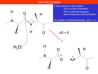
- 1. THE PROTEASES 3 big problems for the kinetics: H2O is a bad nucleophile NH is a bad leaving group loss of resonance during reaction No problem for thermodynamics ( ∆G << 0 ) N N H O H H OH H2O: N O O H OH H3 N H - + ∆G < 0
- 2. N N H O H H OH H2O Hydrolyzed under very harsh conditions, in acid (HCl 6 M, 110°C, 24-72 h) or base (KOH 1 M 100°C 24 h) The peptide bond is « stable » under physiological conditions t1/2 102 years despite its thermodynamic instability THE PROTEASES
- 3. 5 biological strategies to solve the problems, 5 classes of proteases 1. Serine proteases Trypsin in mammalian digestion Coagulation factors (Thrombine) 2. Cysteine proteases Papaine Cathepsines (in the lysosomes) 3. Aspartyl proteases Pepsine (in our stomach) AIDS virus protease 4. Metal ion proteases (Zn2+ ) Carboxypeptidases 5. Threonine proteases Proteasome N N H O H H OH H2O THE PROTEASES 3 big problems for the kinetics: H2O is a bad nucleophile NH is a bad leaving group loss of mesomery during reaction
- 4. Where the proteases act? Exo-proteases (exo-peptidases, cut amino acids from the N- or from the C-terminal of proteins/peptides) Endo-proteases (cut in the interior of proteins/peptides) Specificity Non-specific (Proteinase K, used for stability studies of proteins) Specific proteases (Trypsine X-X-Arg↓X-X et X-X-Lys↓X-X recognize 1 side chain Very specific (Thrombine LeuValProArg↓GlySer ) recognize 6 side chains THE PROTEASES
- 5. IMPORTANT HINT! The protease best substrates are UNFOLDED proteins. Compact protein domain are not hydrolyzed, instead the connecting domains are hydrolyzed Essential application in protein biochemistry and imunology for domain preparation by controlled proteolysis Classical experiment: Porter 1955, preparation of immunoglobulin fragments by treatment with papain or pepsin THE PROTEASES Disulfide bonds
- 6. THE PROTEASES SDS gel electrophoresis
- 7. Stanley B. Prusiner • strange pathogen (resistance to UV, heat etc…) •transmission to mouse (incubation time 150-300 days) • 1975-77: transmission to hamster (70 days) P r P C P r P s e n P r P 2 7 - 3 0 P r P r e s P r P S C 1 2 3 2 infectedinfected proteinproteinasease KK The presence of the Prion protein is demonstrated by the resistence to proteolysis
- 8. Blood clotting Blood clotting results from a cascade of reactions. In a cascade, a signal initiates a series of steps, each of them catalyzed by an enzyme. At each step the signal is amplified. In blood clotting the activated form of one clotting factor catalyzes activation of the next. Very small amounts of the initial factors trigger the cascade, => rapid response to trauma (e.g., damage to a blood vessel).
- 10. Conversion of fibrinogen to fibrin causes clotting. The final step of clotting is conversion of fibrinogen to fibrin by thrombin, a protease. Fibrinogen has 6 protein chains (2x Aα, Bβ and γ), folded into globular units connected by rods. Thrombin cleaves 4 peptides from the Aα and Bβ chains in the central globule, resulting in fibrin monomer (αβγ)2.
- 11. Carboxyl ends of the β- and γ chains interact with the newly exposed N-terminal regions => polymerization (protofibrils).
- 13. Fibrils are stabilized by cross-linking: formation of amide bonds between lysine and glutamine by transglutaminase, which is activated from protransglutaminase by thrombin. The network of fibrils forms the clot.
- 14. Activation of thrombin. Thrombin activates fibrinogen, but how is thrombin activated ? Thrombin is activated by proteolytic activation of prothrombin with factors Xa (also a protease) and Va. Activation removes a gla and 2 kringle domains. Modular structure of prothrombin
- 15. Use of chromogenic substrates for studying the proteases Thrombine (enzyme in blood coagulation) Natural substrate: le fibrinogen (a large protein, about 2000 residues) Benzoyl-Phe-Val-Arg ↓NH NO2 NH2 NO2 The product (p-nitro-aniline) est yellow (λ 380 nm)
- 17. 1. The serine proteases Proteases having an essential serine in the active site Protéases Trypsine Chymotrypsine Elastase Subtilisine (Bacilus subtilis) Same mechanism for esterases Lipases Esterases (acétyl)choliesterase N O O H H OH H2O Amides and esters have similar structure and reactivity N N H O H H OH R H2O O N
- 18. Identification of active serine in serine proteases An Unusually Reactive Serine in Chymotrypsin Chymotrypsin is inactivated by treatment with diisopropylphosphofluoridate (DIPF), which reacts only with serine 195 among 28 possible serine residues. No reaction with the unfolded enzyme, nor with free serine
- 19. Identification of active serine in serine proteases Reaction time PercentInhibitionofactivity(%) 100 50 0 No substrate Add substrate S + DIFP + DIFP & substrate XX Addition of Substrate protects DIFP Inhibition
- 20. substrate inactivator (TPCK) With [14 C]TPCK get 1 equiv. [14 C] bound; pepsin hydrolysis gives a [14 C] peptide with His-57 modified CH2 CH NH SO2 C CH3 CH2 CH NH SO2 C CH3 OCH3 O O CH2Cl 2.11 2.12 Evidence for Histidine Participation
- 21. The serine proteases: the specificity pocket
- 22. 4 Catalytic elements in serine proteases O-
- 23. Specificity pocket Aa 189, 216, 226 Oxyanion hole Aa 193-195 Substrate binding Aa 214-216 Catalytic triad Ser195, His57, Asp102 4 Catalytic elements in serine proteases Chymotrypsin
- 24. Chymotrypsin STRUCTURE: David BLOW 1968
- 25. Serine is the NUCLEOPHILE Histidine is a BASE: it binds the serine’s proton and decreases its pKa from 15 to about 7 The aspartate keeps the histidine in the correct orientation (an old theory: proton relay, but the proton does not move) :
- 26. An example of CONVERGENT evolution Trypsin Subtilisin but identical active site!
- 28. Thrombin and Chymotrypsin are HOMOLOGS (Almost identical structures, similar sequences
- 29. Evolution is most often DIVERGENT A few examples of CONVERGENT evolution « Ancestral » gene, duplication and separate evolution by mutation Trypsine, chymotrypsine, élastase Structure très similaire Famille de protéines Triade: Ser195, His57, Asp102 Subtilisine Structure très différente Triade: Ser221, His64, Asp32 Different genes, protein evolution To a similar active site configuration
- 30. Many serine proteases age activated by proteolysis (protection of the cells which synthetize the proteases)
- 31. Active site residues Hydrophobic pocket Serine Protease Mechanism - Chymotrypsin
- 37. This is a reaction INTERMEDIATE and not a transition state Reaction coordinate
- 38. The C-terminal part of the substrate dissociated and leaves the Acyl-enzyme
- 42. Kinetic demonstration of the serine protease mechanism: burst kinetics
- 43. Demonstration of the serine protease mechanism: site-directed mutagenesis Nature. 1988, 332(6164):564-8. Substrate: N-succinyl-L-Ala-L-Ala-L-Pro-L-Phe-p-nitroanilide Bacillus amyloliquefaciens subtilisin, these functional elements impart a total rate enhancement of at least 109 to 1010 times the non-enzymatic hydrolysis of amide bonds
- 44. Reaction mechanism of a serine protease (in this case, subtilisin) Note the three residues of the “catalytic triad”: Ser221, His64, & Asp32.
- 45. •
- 46. Subtilisine Km kcat/Km kcat Asp32 H64 S221 (µM) (M-1 s-1 ) s-1 Asp His Ser 220 250000 55 Ala His Ser 480 5 0.0024 Asp Ala Ser 390 0.1 0.000039 Asp His Ala 420 0.1 0.000042 Ala Ala Ala 420 0.1 0.000042 kSerHisAsp/knon-enzymatique = 3 750 000 000 kAlaAlaAla/knon-enzymatique = 3 000 Demonstration of the serine protease mechanism: site-directed mutagenesis
- 47. 1. When very low residual activities are expected, a very low level of contamination with other proteases is a serieus problem. How has this been avoided? Serine24 (on the protein surface) has been replaced by a Cysteine which makes possible protein purification by covalent affinity chromatography. 2. A second problem could be the mis-incorporation during traduction. An error rate of 1/1000 can be a problem !
- 48. 3. Ascertaining the role of specific amino acids in catalysis by site-directed mutagenesis can easily by interpreted if the chemical step is rate-limiting (A). If the substrate binding is rate- limiting (B), it is well possible to miss important details of the mechanism. E + S ES E + P G Reaction coordinate ∆ G ∆ G E + S ES E + P G Reaction coordinate ∆ G ∆ G A B Replacing an active-site residue will slown down reaction in A but not in B The measured rate is slower with the mutant No apparent effect!
- 49. Take home lesson: even with no catalytic residues, the enzyme still accelerates the reaction better than 1000-fold the rate of the uncatalyzed reaction. Way to bind that transition state!
- 50. Demonstration of the serine protease mechanism: site-directed mutagenesis Site-directed mutagenesis and the role of the oxyanion hole in subtilisin. Bryan P, Pantoliano MW, Quill SG, Hsiao HY, Poulos T. Proc Natl Acad Sci U S A. 1986 Jun;83(11):3743-5. Reaction intermediate is stabilized by main-chain NH in chymotrypsin: its role cannot be probed by site- directed mutagenesis Reaction intermediate is stabilized Asn side-chain in subtilisin: its role CAN be probed by site-directed mutagenesis!!!
- 51. Demonstration of the serine protease mechanism: site-directed mutagenesis In the transition state complex, the carbonyl group of the peptide bond to be hydrolyzed is believed to adopt a tetrahedral configuration rather than the ground-state planar configuration. Crystallographic studies suggest that stabilization of this activated complex is accomplished in part through the donation of a hydrogen bond from the amide side group of Asn-155 to the carbonyl oxygen of the peptide substrate. To specifically test this hypothesis, leucine was introduced at position 155. Leucine is isosteric with asparagine but is incapable of donating a hydrogen bond to the tetrahedral intermediate. The Leu-155 variant was found to have an unaltered Km but a greatly reduced catalytic rate constant, kcat, (factor of 200-300 smaller) when assayed with a peptide substrate. These kinetic results are consistent with the Asn-155 mediating stabilization of the activated complex and lend further experimental support for the transition-state stabilization hypothesis of enzyme catalysis.
- 52. A recent addition to the serine protease mechanism: the Low barrier hydrogen bonds 2.8 Å 2.55 Å 2.29 Å
- 53. A recent addition to the serine protease mechanism: the Low barrier hydrogen bonds Formation of a short (less than 2.5 angstroms), very strong, low- barrier hydrogen bond in the transition state, or in an enzyme- intermediate complex, can be an important contribution to enzymic catalysis. Formation of such a bond can supply 10 to 20 kilocalories per mole and thus facilitate difficult reactions such as enolization of carboxylate groups. Because low-barrier hydrogen bonds form only when the pKa's (negative logarithm of the acid constant) of the oxygens or nitrogens sharing the hydrogen are similar, a weak hydrogen bond in the enzyme-substrate complex in which the pKa’s do not match can become a strong, low-barrier one if the pKa’s become matched in the transition state or enzyme- intermediate complex. Low-Barrier Hydrogen Bonds and Enzymic Catalysis W. W. Cleland and Maurice M. Kreevoy
- 54. A second recent addition to the serine protease mechanism: Substrate assisted catalysis
- 55. A recent addition to the serine protease mechanism Substrate assisted catalysis
- 56. A second recent addition to the serine protease mechanism Substrate assisted catalysis
- 57. A recent addition to the serine protease mechanism Substrate assisted catalysis
- 58. Can proteases be used for protein SYNTHESIS? CHEMICAL ligation
- 59. Kaiser and co-workers demonstrated the practicality of this work by preparing a subtilisin variant, thiolsubtilisin, where the active site Ser was chemically converted to Cys (S221C)) Using activated esters to acylate the active site Cys in the presence of amine nucleophiles, it was possible to efficiently synthesize amide bonds. The ratio of aminolysis to hydrolysis is 600-fold greater for thiolsubtilisin relative to subtilisin; the variant selenolsubtilisin was later prepared by Hilvert and co-workers and shown to be 14,000-fold more effective for aminolysis than subtilisin.
- 60. Aminolysis/Hydrolysis Serine OH 1.0 Cysteine SH 600 Selenocysteine SeH 14000 Meth Enz 289, 298-313 Subtiligase: a tool for semisynthesis of proteins. Chang TK, Jackson DY, Burnier JP, Wells JA. Department of Protein Engineering, Genentech, Inc., South San Franc 94080. Aminolysis Hydrolysis
- 61. Serine hydrolases: proteases and other enzymes Asparaginase Serine proteases Esterase Penicillin acylase β-lactamase
- 62. The acetyl-cholinesterase – a serine esterase
- 63. Acetylcholinesterase: an archetype for cation–p bonding in biology? Acetylcholinesterase is often considered as the foremost example of cation–p bonding in biological molecular recognition. In its interaction with acetylcholine, it serves as an excellent model for the recognition of quaternary amines by proteins. Early kinetic, spectroscopic and chemical modification studies [17] suggested that the active site of acetylcholinesterase is divided into two subsites: the 'esteratic' site (the site of bond breaking/making) and the 'anionic' (choline binding) site. The 'anionic' site is a misnomer, as this site is in fact uncharged and lipophilic. The molecular detail of acetylcholinesterase was revealed following the determination of the crystal structure of the enzyme from Torpedo californicans [18]. A structure for the enzyme–substrate complex is not available, but the details of substrate binding can be extrapolated from the structure of the enzyme alone [18] and those of the enzyme complexed with tacrine, edrophonium and decamethonium [19].
- 64. 2. Cysteine proteases Papaïne from plants is one example Cathepsines (protease from lysosomes) S H N N H:
- 65. Protease from AIDS virus: an aspartyl protaase 3. Aspartyl proteases
- 67. 3. Aspartyl protéases Pepstatin is a potent inhibitor of aspartyl proteases. It is a hexa- peptide containing the unusual amino acid statine (Sta, (3S,4S)-4- amino-3-hydroxy-6-methylheptanoic acid), having the sequence Isovaleryl-Val-Val-Sta-Ala-Sta (Iva-Val-Val-Sta-Ala-Sta). It was originally isolated from cultures of various species of Actinomyces due to its ability to inhibit pepsin at picomolar concentrations. It was later found to inhibit nearly all acid proteases with high potency and, as such, has become a valuable research tool, as well as a common constituent of protease inhibitor cocktails. This is a TRANSITION STATE ANALOG Isovaleryl-Val- Val- Sta- Ala- Sta statine
- 70. During proteasome-catalysed transpeptidation, the energy from peptide-bond hydrolysis fuels subsequent peptide-bond ligation. When presented with the three- and six-residue components of the nine-residue peptide, the proteasome was unable to splice them together. However, when supplied with the six and seven-residue fragments that comprise the 13-residue precursor peptide, the proteasome efficiently produced the nona-peptide. These observations indicated that the proteasome can catalyse peptide-bond formation only when the process is linked to peptide-bond hydrolysis. Nucleophilic attack of peptide bonds by the hydroxyl group of an active-site threonine in the proteasome results in an acyl-enzyme intermediate, in which the peptide and the threonine are joined by an ester bond. The acyl-enzyme intermediate plays a part in the proteasome-catalysed transpeptidation event. In the first step the hydroxyl group of an active-site threonine catalyses the cleavage of a precursor peptide, generating an N-terminal and a C-terminal fragment. In the second step an active-site threonine attacks the peptide bond in the N-terminal fragment forming an acyl-enzyme intermediate with the N-terminal peptide. At this point the N-terminus of the C-terminal peptide fragment attacks the acyl-enzyme intermediate and, recycling the energy from the cleavage reaction, ligates onto the (now cleaved) N-terminal peptide. This transpeptidation model explains how peptide-bond hydrolysis and formation occur together without the net input of energy. It shows also that the splice site need not be highly conserved because, once a peptide bond has been activated at the protease active site, ligation of almost any incoming peptide with a free N- The architecture of the central chamber of the proteasome defines the catalytic specificity and also might regulate the incidence of splicing. The substrate-binding sites that flank the scissile bond favour certain amino acids and, therefore, enable certain peptides to linger in the active-site cavity, thus providing an opportunity for an N-terminal nucleophile to attack the acyl-enzyme intermediate. The determinants for protease-catalysed splicing are certainly finely controlled because the active site also must enable normal proteolytic events to occur. The question that arises in the case of proteasome-catalysed protein splicing is whether the splicing process is favoured for a functional purpose of the resulting peptides. Proteasomes may not only mediate the complete degradation of proteins, but also the processing of precursors into mature, active proteins. 5. A «new» mechanism: the threonine protease in the proteasome
- 72. Protein Splicing: Analogy to RNA Splicing Attention: this is different from typical « enzyme » in that it is single turn-over!
- 73. 1. Protein splicing is catalyzed entirely by amino acid residues contained in the intein. 2. Protein splicing is an intramolecular process (usually). 3. Protein splicing requires no coenzymes or sources of metabolic energy and therefore involves bond rearrangements rather than bond cleavage followed by resynthesis. Annu Rev Biochem. 2000;69:447-96. Protein splicing and related forms of protein autoprocessing. Paulus H. Properties of protein splicing
- 74. What do they look like? Small inteins are about 150 amino acids. (the smallest is 134 amino acids, largest is 1650)
- 75. Step 1: formation of a linear ester intermediate by NO or NS acyl rearrangement involving the nucleophilic amino acid residue at the N-terminal splice junction; Step 2: formation of a branched ester intermediate by the attack of the nucleophilic residue at the C-terminal splice junction on the linear ester intermediate; Step 3: cyclization of the asparagine residue adjacent to the C-terminal splice junction, coupled to cleavage of the branched ester intermediate to yield an excised intein with a C-terminal aminosuccinimide residue and the two exteins joined by an ester bond; Step 4: spontaneous hydrolysis of the aminosuccinimide residue and rearrangement of the ester linking the exteins to the more stable amide bond. Annu Rev Biochem. 2000;69:447-96. Protein splicing and related forms of protein autoprocessing. Paulus H. The last step is spontaneous and irreversible. The first three steps are catalyzed by the intein
- 76. Dawson PE, Muir TW, Clark-Lewis I, Kent SB. Synthesis of proteins by native chemical ligation. Science. 1994 Nov 4;266(5186):776-9. Same chemistry as protein splicing has been used for spontaneous (non-enzymatic) peptide ligation
- 78. Proposed mechanism of amide, true peptide, and ester bond hydrolysis by proteasomes and mechanism of their inactivation by irreversible inhibitors. Kisselev A F et al. J. Biol. Chem. 2000;275:14831-14837
- 80. INHIBITORS
- 81. Une des composantes de la tri-thérapie est un inhibiteur de la Protéase du virus du SIDA, un analogue de l’état de transition 3. Aspartyl protéases
- 82. Access to the active site of acetylcholinesterase is via a deep and narrow gorg up about 40% of the surface of the gorge) and other residues. The gorge is 20 the surface of the gorge are highly conserved in acetylcholinesterases from di substrate acetylcholine at the base of the gorge reveals the esteratic and chol esteratic site, a catalytic triad and putative oxyanion hole have been identified acetylcholine suggests that it forms a cation–p bond with Trp-84 in the 'anionic remarkable feature of acetylcholinesterase is the preponderance of aromatic r chemical character of the gorge leads to the question of its function in contribu and catalysis. Sussman and colleagues suggested two mechanisms by which increased [18]. First, the high hydrophobicity of the gorge produces a low diele to enhance the effective local charge contributed by the small number of acidi electrostatically 'steer' substrate to the active site. In the second scenario, the affinity sites for the substrate (in particular, the choline moiety), and guides the Because of the reduction-in-dimensionality, the rate of substrate binding is inc interactions may, therefore, have a major role to play in directing the substrate substrate complex, whereas stronger cation–p bonding is presumably respons acetylcholine in the enzyme–substrate complex. Given the wealth of cation–p the enzyme no doubt will remain a principal target for investigating these inter Interestingly, chemical modification studies of the nicotinic acetylcholine recep residues are located in the acetylcholine-binding site in this molecule [20,21].
- 84. Leupeptin, also known as N-acetyl-L-leucyl-L-leucyl-L- argininal, is a protease inhibitor that also acts as an inhibitor of calpain. It is often used during in vitro experiments when a specific enzymatic reaction is being studied. When cells are lysed for these studies, proteases, many of which are contained within lysosomes, are released. These proteases, if freely present in the lysate, would destroy any products from the reaction being studied, and make the experiment uninterpretable. For example, leupeptin could be used in a calpain extraction to keep calpain from being hydrolyzed by specific proteases. The suggested concentration is 1-10 µM (0.5-1 µg/ml). Leupeptin is an organic compound produced by
- 86. Une des composantes de la tri-thérapie est un inhibiteur de la Protéase du virus du SIDA, un analogue de l’état de transition 3. Aspartyl protéases
Editor's Notes
- The peptides A and B are removed by proteolytic digestion.
- Amide bonds between different fibrim molecules.