Respiratory System
•Als PPTX, PDF herunterladen•
4 gefällt mir•2,001 views

The baby developed breathing difficulties after a cesarean delivery in August 1963. The physician ordered oxygen therapy, indicating a diagnosis of respiratory distress. An ultrasound to check lung development may have predicted this condition prenatally. Providing surfactant replacement therapy could have improved the child's chances of survival.

Empfohlen
Weitere ähnliche Inhalte
Was ist angesagt?
Was ist angesagt? (20)
Ähnlich wie Respiratory System
Ähnlich wie Respiratory System (20)
Respiratory System
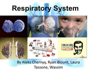
- 1. Respiratory System By Aleks Chernyy, Ryan Blount, Laura Tassone, Wassim
- 2. Respiratory System 1. Conducting 2. Respiratory
- 3. Conducting Portion 1. Nasal cavities 2. Pharynx 3. Larynx 4. Trachea 5. Bronchi
- 4. Conducting-Respiratory Junction Respiratory Bronchioles is the Conducting- Respiratory Junction. a. Alveoli out-pocketings b. Clara cells begin to predominate
- 5. Histology of the trachea Ciliated epithelium Cartilage
- 6. Within the Trachea we find? Trachea a. Pseudostratified ciliated columnar epithelium b. Goblet cells c. Basal cells d. Brush cells What types of Glands are present in RS? What do they excrete? Mucous - Mucin Serous - Glycoproteins, polysacharides & bacteriosidic proteins C-Shaped Cartilage What is its function? maintains patency, especially during forced expiration
- 7. Trachea Basal Bodies are associated with cilia Mucous gland vs. serous gland and are highly eosinophilic
- 8. Mucus Glands vs Serous Glands
- 9. Respiratory Portion Structures: 1. Respiratory bronchioles 2. Alveolar ducts 3. Alveoli
- 10. The Respiratory Segment • Respiratory Bronchioles – Give off Alveoli – Give off alveolar ducts • Alveolar ducts – Give off Alveoli only • Alveolar sacs – Spaces surrounded by clusters of alveoli
- 11. Upper Respiratory The larynx first appears as an outgrowth from the foregut. The outgrowth of tissue is called the respiratory diverticulum or the lung bud. The formation of the lung bud occurs when two lateral folds of splanchnic mesoderm and endoderm meet in the midline and separate the larynx and trachea from the esophagus. The lung bud is a ventral diverticulum of endoderm that arises from the floor of the foregut caudal to the pharynx. The diverticulum forms a groove in the floor of the pharynx called the laryngotracheal groove.
- 12. The Lower Respiratory System Develops during week 4 (26-27 days) Starts as a median laryngotracheal groove in the caudoventral wall of the primitive pharynx. The endoderm lining the groove gives rise to the epithelium and glands of the larynx, trachea, bronchi and the pulmonary epithelium. Connective tissue, cartilage and smooth muscle of these structures develop from the splanchnic mesenchyme surrounding the foregut.
- 13. Development The laryngotracheal groove deepens into a diverticulum ventrally which enlarges distally into a lung bud. The diverticulum becomes separated from the primitive pharynx by longitudinal trachoesophageal folds The folds fuse to form the trachoesophageal septum, dividing the foregut into the ventral laryngotracheal tube and the dorsal esophagus.
- 14. Fistula A fistula may exist connecting trachea and esophagus and resulting in abnormal communication between the two. This is usually associated with superior esophageal atresia. In a newborn infant, this is associated with coughing and choking upon swallowing. Gastric contents may reflux into the trachea and lungs resulting in pneumonia or pneumonitis (inflammation of the lungs). An excess of amniotic fluid (polyhydramnios) is associated with esophageal atresia and trachoesophageal fistula because amniotic fluid may not pass to the stomach and intestines for absorption and transfer via the placenta for disposal
- 15. STAGES OF THE DEVELOPMENT OF LUNGS Pseudoglandular 5 – 16 weeks Branching has Period continued to form terminal bronchioles Canalucular Period 16 – 26 weeks Each terminal bronchiole divides into 2 or more respiratory bronchioles which in turn divide into 3- 6 alveolar ducts Terminal Sac 26 weeks to birth Terminal sacs, Primitive alveoli form and capillaries establish close contact
- 16. Histology What is the basic name for the Cells within the Respiratory System? Pneumocytes 1. Type I 2. Type II 3. Clara Cell 4. Goblet Cells 5. Cartilage 6. Alveolar Macrophages
- 17. Type I Alveolar Epithelial Cell Features: cuboidal, lined bronchioles change into thin, flat cells Functions? 1. gas exchange between blood 2. air possible in primitive alveoli Clinical Correlations
- 18. Type II Alveolar Epithelial Cell Features: cuboidal, granular, alveolar septal junctions Function: 1. Produces surfactant (Lower Surface Tension Normally produced at end of 6th month maximum production 2 weeks before birth Clinical Correlation. Hyaline Membrane Disease (RDS)
- 19. TYPE I vs TYPE II They are roughly cuboidal Flattened squamous cells in shape. Line the alveolar surfaces and are Found interspresed among extremely attenuated. the type I alveolar cells with which they have occluding and desmosomal juntions. Make up 97% of the alveolar Make up 3% of the alveolar surface. surface. Have occluding junctions and Contains lamellar bodies desmosomes that stores pulmonary surfactants
- 20. Hyaline Membrane Disease SYMPTOMS: (RDS) 1. Respiratory difficulty at birth that gets progressively worse 2. Cyanosis 3. Flaring of the nostrils 4. Tachypnea (Rapid Breathing) 5. Grunting sounds with breathing 6. Chest retractions (pulling in at the ribs and sternum during breathing) What is the problem? 1. Not enough Surfactant. ( produced in the fetus 24 to 28 weeks) 2. Surfactant lowers surface tension in the airways keeping alveoli open. 3. Without Surfactant, the alveoli collapse. 4. Damaged cells collect in the airways and affect breathing ability. 5. These cells are called hyaline membranes. The baby works harder at breathing, trying to re-inflate the collapsed airways. 6. As the baby's lung function decreases, carbon dioxide builds up in the blood. This can lead to increased acid in the blood called acidosis, a condition that can affect other body organs.
- 21. RDS Treatment 1. Placing an endotracheal (ET) tube into the baby's windpipe 2. Mechanical breathing machine 3. Supplemental oxygen 4. Continuous positive airway pressure (CPAP) 5. Surfactant replacement with artificial surfactant - most effective if started in the first six hours of birth. 6. Surfactant is given as prophylactic (Preventive) treatment for some babies at very high risk for RDS. Surfactant is usually given in several doses. 7. Medications (Pain)
- 22. Clara Cell 1. Secrete CCSP 2. Protect Bronchiolar epitheliem 3. Detoxify harmful substances 4. Act as stem cells and multiple 5. Unique to Bronchioles Historical Controversy Max Clara, product of unethical research.
- 23. The Clara Cell Controversy
- 24. What are Goblet cells? What is their function? Clinical Correlation? Chronic Pulmonary Emphysema
- 25. MD 3 QUIZ
- 26. Answers
- 28. The Larynx Cartilage With the exception of the epiglottis, all larynx cartilage is hyaline cartilage. The Adam's apple is really the laryngeal prominence, where the curved disc shaped thyroid cartilage bond.
- 29. Trachea Cartilage The trachea is made up of between 16 and 20 “C” shaped rings The trachea is flexible and twistable, Without cartilage rings, it would collapse under the partial vacuum formed when inhaling.
- 30. The Bronchi • The first few levels of bronchi are supported by rings of cartilage. • Branches after that are supported by irregularly shaped discs of cartilage, while the latest levels of the tree have no support whatsoever.
- 31. Facts about alveolar macrophages? • An alveolar macrophage (or dust cell) is a type of macrophage found in the pulmonary alveolus, near the pneumocytes, but separated from the wall. • Alveolar macrophages are one of the many types of white blood cells(leukocytes) present in body tissues. They are important in immune response and cell stability because they mobilize in cell tissue to attack large foreign particles such as bacteria, yeast, and dead cells.
- 32. Where do alveolar macrophages derive from? • Macrophages are derived from precursor cells called monocytes that first develop in bone marrow. • Macrophages are any of the large, mononuclear, highly phagocytic cells derived from monocytes that occur in the walls of blood vessels (adventitial cells) and in loose connective tissue (histiocytes, phagocytic reticular cells).
- 33. Alveolar macrophage function • Alveolar macrophages enter the blood and travel throughout the body in the circulatory system. When needed circulatory monocytes move into tissue where they become macrophages. Here a lung (alveolar )macrophage is seeking foreign bacteria (Escherichia coli) with specialized cell extensions called filopodia. • Macrophages engulf and digest foreign materials in a process known as phagocytosis.
- 34. • Alveolar macrophages are frequently seen to contain granules of exogenous material such as particulate carbon that they have picked up from respiratory surfaces. Such black granules may be especially common in smoker's lungs or long-term city dwellers
- 35. • Inhaled air may contain particles or organisms which would be pathogenic. The respiratory pathway is a prime site for exposure to pathogens and toxic substances. • The respiratory tree, comprising the larynx, trachea, and bronchioles, is lined by ciliated epithelia cells that are continually exposed to harmful matter [1
- 36. • When these offensive agents infiltrate the superficial barriers, the body's immune system responds in an orchestrated defense involving a litany of specialized cells which target the threat, neutralize it, and clean up the remnants of the battle.
- 38. Epithelium of the Respiratory System Upper 1/3 of trachea has squamous cells Mid 1/3 of trachea is a combination Main respiratory epithelium is tall columnar ciliated epithelium The more you smoke, the longer the zone of squamous cells.
- 39. Conducting Epithelium What is the predominant type of epithelium that is found in the conducting portion of the respiratory system? Pseudostratified columnar epithelium ``````````````````````````````` ``````````````````````````````` Clinical Correlation? Metaplasia Metaplasia
- 40. Metaplasia
- 41. QUIZ A 65-year-old man with an 80- pack-year history of smoking presents with a cough and increasing dyspnea over the past 6 weeks. A 2-cm diameter mass is seen in the left lower lobe on x-ray of the chest. A sample of nonneoplastic tissue from the lung biopsy is shown in the image. Which of the following types of epithelium not normally present in the lung lines the bronchus shown in this image? (A) Pseudostratified columnar (B) Simple squamous (C) Stratified columnar (D) Stratified squamous (E) Transitional
- 42. Asthma What is Asthma? Inflammatory process of the airways characterized by reversible bronchospasm, and increased airway secretions. Clinical picture: Shortness of breath, wheezing and chronic cough. Treatment: 1. Steroids 2. Exercise Asthma/Albuterol 3. Beta 2 agonists (Short Acting) 4. Anticholinergic inhalers
- 43. Cilia Why do we have cilia in the conducting portion of the RS? a. Line the entire airway b. Beat in one direction c. Has the 9 + 2 configuration d. 9 microtubules surrounding 2 actin proteins e. Need a Dynein arm to have flexibility Clinical Correlation: Kartagener Syndrome
- 44. Kartagener Syndrome Symptoms: 1. Chronic sinus infection 2. Frequent lung infections, such as pneumonia and bronchitis 3. Bronchiectasis - lung damage from frequent infections 4. Frequent ear infections Whats the Problem? Dynein arm is defective Results in ? a. Obstructive lung disease b. Bronchiectasis c. Infertility d. Situs Inversus Treatment?
- 46. Lymphoid Tissue BALT (bronchus- associated lymphoid tissue) Similarity to Peyer’s Patches High percentage of IgA Lymph Drainage
- 47. Important Respiratory Functions 1. Hypoxia causes ______________everywhere in the body except the ______________, where it causes vasoconstriction. 2. CO2 is transported in the blood mainly in the form of _________ and to a lesser extent bound to___________. 3. Hering Bruer reflex: Inhibition of _____________due to stretch of lung tissue.
- 48. 4. Central: In medulla, and is stimulated by high CO2 and high H. 5. Peripheral: In carotid and aortic bodies. Stimulated by low oxygen, high CO2 and high H. So when they ask you in the USMLE about how breathing is stimulated in a patient with low oxygen but normal CO2, the answer is obviously peripheral receptors.
- 49. Case In August 1963, First Lady Jacqueline Bouvier Kennedy was hospitalized in her 34th week of pregnancy at the Otis Air Force Base Hospital. Her fetus was in distress, but labor did not progress. On August 7, she underwent a cesarean section to deliver Patrick Bouvier Kennedy, who weighed 4 pounds, 10.5 ounces (2,112 grams). After delivery, the baby developed difficulty breathing, The Physician ordered oxygen therapy, what was his diagnosis? What prenatal test, if available at the time would serve to predict the Child’s condition? What other steps would you take to improve the child probability of survival.
Hinweis der Redaktion
- Conducting Portion- warms, humidifies, and cleans the air as it passes down to The respiratory portion is where the actual gas exchange occursRespiratory portion
- The respiratory portion begins with the first branches of the respiratory bronchioles; essentially where hyaline cartilage ends and there is abundant smooth muscle, elastic fibers, reticular fibers, and epithelium transition from cuboidal to simple squamous.
- The epithelium of the larynx develops from the endoderm of the foregut. However, the muscles and cartilage arise from the 4th and the 6th arches. The development of these structures will be discussed in a later lecture.Cephalic to the laryngotracheal groove is the epiglottal swelling. On either side of this groove are the developing arytenoid swellings. TracheaThe trachea develops caudal to the larynx. The epithelium develops from the endoderm and the tracheal cartilage and muscles develop from splanchnic mesoderm. Early in development the trachea bifurcates into the left and right bronchi. Bronchi and BronchiolesAs the bronchi develop they continue to branch. The right bronchus gives off three diverticula and the left bronchus gives off two diverticula. These diverticula become the lobar bronchi and indicate that the right lung will have three lobes and the left lung will have two lobes. Each of the bronchi at this stage will divide into smaller bronchi. The branching of the bronchi continues until the bronchioles begin to form. In all there are 17 divisions of the bronchi until the sixth fetal month is reached. However, by early childhood there will be a total of 24 generations of branching that occurs.
- Type I cells. Also called type I alveolar cells, type I pneumocytes, and squamous alveolar cells, these are squamous epithelial cells that make up 97% of the alveolar surfaces. They are specialized to serve as very thin (often only 25 nm in width) gas-permeable components of the blood-air barrier. Their organelles leg, Golgi complex, endoplasmic reticulum, mitochondria) cluster around the nucleus.Much of the cytoplasm is thus unobstructed by organelles, except for the abundant small pinocytotic vesicles that are involved in the turn over of pulmonary surfactant and the removal of small particles from the alveolar surfaces. They attach to neighboring epithelial cells by desmosomes and occluding junctions.The Occluding junctions reduce pleural effusion--leakage of tissue fluid into the alveolar lumen. Type I cells can be distinguished from the nearby capillary endothelial cells by their position bordering the alveolar lumen and by their slightly more rounded nuclei.
- Type II cells. These cells, which are also called type II alveolar cells, type II pneumocytes, great alveolar cells, and alveolar septal cells, cover the remaining 3% of the alveolar surface. They are interspersed among the type I cells, to which they attach by desmosomes and occluding junctions.Type II cells are roughly cuboidal with round nuclei; they occur most often in small groups at the angles where alveolar septal walls converge. At the electron microscope level, they contain many mitochondria and a well-developed Golgi complex, but they are mainly characterized by the presence of large (0.2-um), membrane-limited lamellar (mutlilamellar) bodies. These structures, which exhibit many closely apposed concentric or parallel membranes (lamellae), contain phospholipids, glycosaminoglycans, and proteins.Type II cells are secretory cells. Their secretory product, pulmonary surfactant, is assembled and stored in the lamellar bodies, which also carry it to the apical cytoplasm. There, the bodies fuse with the apical plasma membrane and release surfactant onto the alveolar surface. 3. Alveolar marcrophages. Known also as dust cells, these large monocytc-derived representatives of the mononuclear phagocyte system are found both on the surface of alveolar septa and in the interstitium. Macrophages are important in removing any debris that escapes the mucus and cilia in the conducting portion of the system.They also phagocytose blood cells that enter the alveoli as a result of heart failure. These alveolar macrophagcs, which stain positively for iron pigment (hemosiderin), are thus designated heart failure cells.
- What causes RDS?RDS occurs when there is not enough of a substance in the lungs called surfactant. Surfactant is made by the cells in the airways and consists of phospholipids and protein. It begins to be produced in the fetus at about 24 to 28 weeks of pregnancy. Surfactant is found in amniotic fluid between 28 and 32 weeks. By about 35 weeks gestation, most babies have developed adequate amounts of surfactant.Surfactant is normally released into the lung tissues where it helps lower surface tension in the airways. This helps keep the lung alveoli (air sacs) open. When there is not enough surfactant, the tiny alveoli collapse with each breath. As the alveoli collapse, damaged cells collect in the airways and further affect breathing ability. These cells are called hyaline membranes. The baby works harder and harder at breathing, trying to re-inflate the collapsed airways.As the baby's lung function decreases, less oxygen is taken in and more carbon dioxide builds up in the blood. This can lead to increased acid in the blood called acidosis, a condition that can affect other body organs. Without treatment, the baby becomes exhausted trying to breathe and eventually gives up. A mechanical ventilator (breathing machine) must do the work of breathing instead.Who is affected by RDS?RDS occurs in over half of babies born before 28 weeks gestation, but only in less than one-third of those born between 32 and 36 weeks. Some premature babies develop RDS severe enough to need a mechanical ventilator (breathing machine). The more premature the baby, the higher the risk and the more severe the RDS.Although most babies with RDS are premature, other factors can influence the chances of developing the disease. These include the following:Caucasian or male babiesprevious birth of baby with RDScesarean deliveryperinatal asphyxiacold stress (a condition that suppresses surfactant production)perinatal infectionmultiple births (multiple birth babies are often premature)infants of diabetic mothers (too much insulin in a baby's system due to maternal diabetes can delay surfactant production)babies with patent ductusarteriosus
- Clara cells contain Tryptase clara, which is believed to be responsible for cleaving the hemagglutinin surface protein of influenza A virus, thereby activating it and causing the symptoms of flu.One of the main functions of Clara cells is to protect the bronchiolar epithelium. They do this by secreting a small variety of products, including Clara cell secretory protein (CCSP) and a solution similar to the component of the lung surfactant. They are also responsible for detoxifying harmful substances inhaled into the lungs. Clara cells accomplish this with cytochrome P450 enzymes found in their smooth endoplasmic reticulum. Clara cells also act as a stem cell and multiply and differentiate into ciliated cells to regenerate the bronchiolar epithelium.[5]"Clara cells" were originally described by their namesake, Max Clara in 1937. Clara was born in South Tyrol in 1899 and died in 1966. He was a Nazi doctor who used tissue from executed victims of the Third Reich for his research at Leipzig, including the work that led to his discovery of Clara cells.[3] Some scholars believe that the eponymous name of these cells should be changed because of the ethical controversy surrounding the discovery of the cells but other scholars disagree and think that the name should remain because it is a testament to a time when medicine crossed an ethical line.[4][edit]
- gland tissue and hyaline cartilage
- Also known as the voice box, the pharynx is what allows you to speak. The larynx has an inlet at the top that allows substances to pass through it or not. When food is being swallowed, the inlet is closed, forcing food into the stomach. When air is being breathed, the inlet is wide open so that air can enter your lungs.
- The trachea, or windpipe connects the larynx to the bronchi. This organ differs from others in the neck in that it is flexible, stretching to be between four and five inches long, and about one inch in diameter. The trachea is lined with mucous called the mucociliary escalator, which represents the mucous and cilia and carry the foreign substances up to be swallowed.
- The trachea branches off into two main bronchi, your left and right primary bronchi, which lead to the left and right lung respectively. Your right lung is slightly wider, shorter, and taller that the left, which makes it more vulnerable to foreign invasion. At this point in breathing, the air has been moistened, purified and warmed.Each bronchi enters its lung and begins on a series of branches, called the bronchial or respiratory tree. The first of these branches is the lobar (secondary) branch. On the left, there are two lobar branches, while on the right, there are three. Each lobar branches into one lobe. The next branch is called the segmental (tertiary) branch. Each branch continues to branch into smaller and smaller bronchioles. The final branch is called the terminal bronchioles. These bronchioles are smaller than 0.5 mm in diameter.
- he alveolar epithelium is comprised of two predominant cell types: the type I and II alveolar epithelial cells, or pneumocytes (Figure 1).[4] Type I cells are very flat cells, accounting for 80% of the alveolar surface area but only 20% of the total epithelial cells, and their thin morphology allows for rapid diffusion and exchange of gases. Type II cells are cuboidal and more numerous than type I cells, making up 80% of the total alveolar epithelium but only 20% of the surface area. Both cell types have roles in host defense and immunity. In contrast to the type II cells, type I cells are susceptible to injury and cell death. The more resistant type II cells proliferate and differentiate into type I cells, important in re-epithelialization of the epithelial barrier. These cells also produce surfactant and regulate fluid balance across the epithelium.The distal airway epithelium contains alveolar type I and type II cells and Clara cells, which possess various pumps and channels that achieve clearance of edema fluid. Sodium is transported through channels on the apical membrane and extruded from the cell by the Na+/K+-ATPase located on the basolateral membrane. This transport generates a sodium gradient that drives the transport of water, which is accomplished in part through water channels. AQP, aquaporin; CFTR, cystic fibrosis transmembrane conductance regulator; CNG, cyclic nucleotide-gated; ENaC, epithelial Na+channel. From Matthay and coworkers [3], with permission from the American Physiological Society.
- The respiratory epithelium lining the upper (cranial) airways is classified as ciliated pseudostratified columnar epithelium.[1] This designation is due to the arrangement of the multiple cell types composing the respiratory epithelium. While all cells make contact with the basement membrane and are, therefore, a single layer of cells, the nuclei are not aligned in the same plane. Hence, it appears as though several layers of cells are present and the epithelium is called pseudostratified.The majority of cells composing the ciliated pseudostratified columnar epithelium are of three types: a) ciliated cells, b) goblet cells, and c) basal cells. The ciliated cells are columnar epithelial cells with specialized ciliary modifications. Goblet cells, so named because they are shaped like a wine goblet, are columnar epithelial cells that contain membrane-bound mucous granules and secrete mucus, which helps maintain epithelial moisture and traps particulate material and pathogens moving through the airway. The basal cells are small, nearly cuboidal cells thought to have some ability to differentiate into other cells types found within the epithelium. For example, these basal cells respond to injury of the airway epithelium, migrating to cover a site denuded of differentiated epithelial cells, and subsequently differentiating to restore a healthy epithelial cell layer.
- The term pseudostratified is derived from the appearance of this epithelium in section which conveys the erroneous (pseudo means false) impression that there is more than one layer of cells, when in fact this is a true simple epithelium since all the cells rest on the basal lamina. The nuclei of these cells, however, are disposed at different levels, thus creating the illusion of cellular stratification. Not all ciliated cells extend to the luminal surface; such cells are capable of cell division providing replacements for cells lost or damaged.The medical significance of metaplasia is that in some sites where pathological irritation is present cells may progress from metaplasia, to develop dysplasia, and then malignant neoplasia (cancer). Thus, at sites where abnormal metaplasia is detected, efforts are made to remove the causative irritant, thereby decreasing the risk of progression to malignancy. The metaplastic area must be carefully monitored to ensure that dysplastic change does not begin to occur. A progression to significant dysplasia indicates that the area could need removal to prevent the development of cancer.Pseudostratified epithelia function in secretion or absorption. If a specimen looks stratified but has cilia, then it is a pseudostratified ciliated epithelium, since stratified epithelia do not have cilia.
- Answer is DThe correct answer is D. The letter E in theimage points to pseudostratified ciliated columnarepithelium, LP refers to the laminapropria, and C refers to hyaline cartilage. Insmokers, pseudostratified ciliated columnar epitheliumlining the bronchi can undergo metaplasiaand transformintostratifiedsquamousepithelium. Stratifiedsquamousepitheliumisclassified by the flattened shape of the cells inthe surface layer. Examples of tissues with stratified squamous epithelium include the skin,mouth, anus, vagina, and esophagus.Answer A isincorrect. Pseudostratifi ed columnarepithelium is the normal respiratory epitheliumon the right that is undergoing metaplasia.This type of epithelium only appearsstratified; however, all cells are in contact withbasal lamina and only some cells reach the surfaceof epithelium.Answer B is incorrect. Simple squamous epitheliumlines alveoli, loops of Henle, and endothelial linings of blood vessels. Simple epitheliumindicates that the epithelial membraneis composed of a single layer of cells. Underthe microscope, simple squamous epitheliumis characterized by a single sheet of flattenedcells lying on a basal lamina. It does not play arole in this case.Answer C isincorrect. Stratifi ed columnarepitheliumis found in only a few places in thebody, namely, the conjunctivae of the eye andregions of the male urethra. It is composed of alow polyhedral to cuboidal deeper layer in contactwith the basal lamina along with a superficial layer of columnar cells.Answer E is incorrect. The bladder is lined bytransitional epithelium, not the lung. Transitionalepithelium is characterized by severallayers of cuboidal cells, with the surface layerbeing large and dome-shaped.
- 1. Inhaled steroids is the mainstay of treatment: Steroids suppress the hyperreactivity of airways to various stimuli. Side effects: Oral thrush,which is prevented by washing the mouth after inhalation.2. Exercise induced asthma: Albuterol before exercise.3. Allergy induced asthma: Mast cell stabilizer inhaler, e.g., Nedocromil.4. Others: l Beta2 agonistbronchodilators: Shortacting,e.g., Albuterol, or long acting, e.g.,Anticholinergicinhalerscause bronchodilation and suppressionof the respiratory airways’ secretions. Side effects: Arrhythmia and seizures.
- cilia, which resemble microscopic "hairs” are complex organelles that beat synchronously in the respiratory tract, moving mucus toward the throat. Normally, cilia beat 7 to 22 times per second, and any impairment can result in poor mucociliary clearance, with subsequent upper and lower respiratory infection. Cilia also are involved in other biological processes (such as nitric oxide production), which are currently the subject of dozens of research efforts.
- BALT is populated by lymphocytes such as T cells and B cells, as well as plasma cells and macrophages, each of which is well situated to encounter antigens passing through the mucosal epithelium. In the case of BALT, Respiratory tissue identifies pathogens and deliver antigens to the lymphoid tissue.similarities to Peyer's patches found in the gut. Both possess a lymphoepithelium with selective antigen sampling properties, both appear in the apparent absence of direct antigen stimulation, both contain a high percentage of cells bearing IgA sdurface immunoglobulin and both can repopulate the bronchial and gut lamina propria with IgA containing cells. Good evidence now exists (and will be reviewed) in support of the concept of a common mucosal immunologic system. Cells potentially sensitized at or in a mucosal tissue such as the gut or lung would then migrate to the draining lymph node, thence into the circulation and localize in a variety of mucosal tissues. Factors involved but not essential for such localization include antigen.
- 1. Vasodilatation / lungs2. Bicarbonate / hemoglobin3. inspiration