

Empfohlen
Weitere ähnliche Inhalte
Was ist angesagt?
Was ist angesagt? (20)
Andere mochten auch
Andere mochten auch (19)
Ähnlich wie 4 u1.0-b978-1-4160-4224-2..50042-9..docpdf
Ähnlich wie 4 u1.0-b978-1-4160-4224-2..50042-9..docpdf (20)
Mehr von Loveis1able Khumpuangdee
Mehr von Loveis1able Khumpuangdee (20)
Kürzlich hochgeladen
Kürzlich hochgeladen (20)
4 u1.0-b978-1-4160-4224-2..50042-9..docpdf
- 1. Chapter 39 Cardiac Diseases Daniel G. Blanchard, MD, and Ralph Shabetai, MD pregnancy. The most important of these are increases in blood volume, cardiac output, and heart rate. These adaptations exacerbate the symp- Diagnosis of Heart Disease toms and clinical signs of heart disease and may necessitate significant in Pregnancy escalation in treatment. Cardiac risk varies among the specific forms of heart disease and Pregnant women with heart disease are at higher risk for cardiovascu- also with severity. During prepregnancy counseling, the physician lar complications during pregnancy and also have a higher incidence should describe the nature of the heart disease in terms comprehensi- of neonatal complications.1 However, the significant hemodynamic ble to the prospective parents. The risk to the woman, which can vary changes that accompany pregnancy make the diagnosis of certain from negligible to prohibitive, should be spelled out as clearly as pos- forms of cardiovascular disease difficult. During normal pregnancies, sible.7 On this basis, the patient may be advised either that the con- women frequently experience dyspnea, orthopnea, easy fatigability, templated pregnancy is safe, will be uncomfortable and will necessitate dizzy spells, and, occasionally, even syncope. On physical examination, treatment, carries a significantly increased risk, or would be extremely dependent edema, rales in the lower lung fields, visible neck veins, and dangerous and should not be undertaken. cardiomegaly are commonly found. Systolic murmurs occur in more In the case of certain cardiac conditions, the patient should be than 95% of pregnant women, and internal mammary flow murmurs strongly advised to undergo the necessary treatment before pregnancy and venous hums are common. A third heart sound (S3 gallop) is often and to allow several months to elapse before becoming pregnant. present.2 Nevertheless, certain findings indicate heart disease in preg- Examples in this category include the following: nancy and should suggest the presence of a significant cardiovascular abnormality. These symptoms include severe dyspnea, syncope with Large intracardiac shunt (atrial or ventricular septal defect) with exertion, hemoptysis, paroxysmal nocturnal dyspnea, and chest pain mild to moderate pulmonary hypertension related to exertion. Physical signs of organic heart disease include a Patent ductus arteriosus (PDA) with mild to moderate pulmonary fourth heart sound (S4 gallop), cyanosis, clubbing, diastolic murmurs, hypertension sustained cardiac arrhythmias, and loud, harsh systolic murmurs.3 Severe coarctation of the aorta If there is a strong suspicion of heart disease during pregnancy, Severe mitral stenosis or regurgitation confirmatory diagnostic tests should be initiated. The changes of Severe aortic stenosis or regurgitation normal pregnancy must be recognized so that the findings are not Tetralogy of Fallot misinterpreted. For example, nonspecific ST segment and T-wave Various congenital malformations and acquired heart diseases abnormalities and shifts in the electrical axis can occur.4 Pregnancy also produces changes in the echocardiogram, including alterations in Again, it is imperative that the palliative procedure be carried out cardiac dimensions and performance. The internal dimensions of all before pregnancy is undertaken and that a year or so elapse before the cardiac chambers are increased, and slight regurgitation through pregnancy occurs. Flexibility in clinical judgment is necessary, however. the four valves is frequently observed. The ejection fraction (EF) and A woman with moderately severe valvular disease may require a pros- stroke volume are concomitantly larger, and the cardiac output is thetic valve in the future. In such a case, the patient should be advised increased.5 A small pericardial effusion can be a normal finding in to have her family before valve replacement—with its associated anti- pregnant women.6 Both radiographic and radionuclide diagnostic pro- coagulant risk—is required8 (see Pregnancy in Patients with Artificial cedures should be avoided during pregnancy unless the procedure is Heart Valves, later). The valvular heart lesions associated with high and deemed essential for the health and safety of the mother. low maternal and fetal risk during pregnancy are listed in Tables 39-1 and 39-2. As previously noted, some cardiac disorders are so serious in nature Pre-Conception Counseling that the physiologic changes of a superimposed pregnancy pose pro- hibitive risks to the mother; they carry such a high maternal mortality If a woman plans to become pregnant but knows that she has heart risk that pregnancy is contraindicated. In such circumstances, patients disease, she and her physicians must be fully aware of several funda- must be strongly cautioned against becoming pregnant. If such a mental principles. The cardiovascular system undergoes specific adap- patient is seen for the first time when she is already pregnant, termina- tations to meet the increased demands of the mother and fetus during tion of the pregnancy is recommended. The most serious of the cardiac
- 2. 798 CHAPTER 39 Cardiac Diseases TABLE 39-1 VALVULAR HEART LESIONS TABLE 39-3 HIGH-RISK MATERNAL ASSOCIATED WITH HIGH CARDIOVASCULAR DISORDERS MATERNAL AND/OR FETAL RISK Estimated Maternal DURING PREGNANCY Disorder Mortality Rate (%) Severe AS with or without symptoms Aortic valve stenosis 10-20 AR with NYHA functional class III-IV symptoms Coarctation of the aorta 5 MS with NYHA functional class II-IV symptoms Marfan syndrome 10-20 MR with NYHA functional class III-IV symptoms Peripartum cardiomyopathy 15-60 Aortic and/or mitral valve disease resulting in severe pulmonary Severe pulmonary hypertension 50 hypertension (pulmonary pressure greater than 75% of Tetralogy of Fallot 10 systemic pressures) Aortic and/or mitral valve disease with significant LV dysfunction (EF < 40%) Mechanical prosthetic valve requiring anticoagulation Marfan syndrome with or without AR If a patient with one of these disorders presents when she is already pregnant, she should be strongly urged to consider early termina- AR, aortic regurgitation; AS, aortic stenosis; EF, ejection fraction; LV, tion. A carefully planned suction curettage before 13 weeks’ gestation left ventricular; MR, mitral regurgitation; MS, mitral stenosis; NYHA, would place such a patient at minimal risk. Termination of pregnancy New York Heart Association. Reproduced with permission from Bonow RO, Carabello B, DeLeon AC, beyond 13 weeks increases the risk to the mother, because many of et al: ACC/AHA 2006 guidelines for the management of patients with the cardiovascular alterations that occur in pregnancy have taken valvular heart disease. Circulation 114:84, 2006. place. Infective endocarditis often causes rapid and serious deterioration of the cardiac status, posing a major threat to the life and health of the TABLE 39-2 VALVULAR HEART LESIONS mother and, therefore, of the fetus as well. Scrupulous attention to ASSOCIATED WITH LOW prophylaxis against endocarditis is critical during pregnancy. Pregnant MATERNAL AND FETAL RISK women must pay meticulous attention to their dental health; if they have cardiac lesions susceptible to infective endocarditis, neglect of DURING PREGNANCY antibacterial prophylaxis could have dire consequences. In general, Asymptomatic AS with low mean gradient (<25 mm Hg and aortic women with valvular heart disease should have antibiotic prophylaxis valve area >1.5 cm2) in the presence of normal LV systolic at the time of delivery.10,11 function (EF > 50%) The prospective parents will want to know not only about the risk NYHA functional class I or II AR with normal LV systolic function to the health and life of the future mother but also about the fetal risks. NYHA functional class I or II MR with normal LV systolic function One of the most important questions is whether the mother’s heart MVP with no MR or mild to moderate MR with normal LV systolic disease is hereditary and, if so, what is the risk that the infant will be function Mild MS (mitral valve area >1.5 cm2, gradient <5 mm Hg) without born with the same defect. A detailed family cardiac history must be severe pulmonary hypertension obtained before pregnancy, especially if the prospective mother has Mild to moderate pulmonary valve stenosis heart disease. Some of the cardiomyopathies, especially hypertrophic forms, may AR, aortic regurgitation; AS, aortic stenosis; EF, ejection fraction; LV, be inherited in a mendelian manner.12 Familial dilated cardiomyopathy left ventricular; MR, mitral regurgitation; MS, mitral stenosis; MVP, has also been described. Approximately 20% of idiopathic dilated car- mitral valve prolapse; NYHA, New York Heart Association. Reproduced with permission from Bonow RO, Carabello B, DeLeon AC, diomyopathy is inherited.13 There is a strong familial tendency in et al: ACC/AHA 2006 guidelines for the management of patients with certain congenital malformations, such as PDA and arterial septal valvular heart disease. Circulation 114:84, 2006. defect (ASD). Additionally, mothers with congenital heart disease may have children with unrelated congenital malformations: this risk appears to be approximately 5%.3,14 Also, pregnant women with disorders are those involving pulmonary hypertension, particularly advanced heart disease, especially those with low cardiac output or those associated with a right-to-left shunt in cardiac blood flow severe hypoxia, experience a greatly increased incidence of spontane- (Eisenmenger syndrome). Low cardiac output states and entities ous abortion, stillbirths, and small or deformed children.9 For most in which there is an increased risk of aortic dissection (Marfan pregnant women with heart disease, vaginal delivery (with a low syndrome) also represent an extraordinarily high risk of maternal threshold for forceps or vacuum assistance) is recommended. Elective mortality. These high-risk maternal cardiovascular disorders are cesarean section is recommended in cases of Marfan syndrome or listed in Table 39-3. aortic aneurysm of any cause.3 In some women with specific dangerous cardiovascular diseases, Today’s prospective mother wants to know about the risks to her pregnancy is contraindicated because of the substantial risk of mater- fetus of drugs, other therapies, and diagnostic tests that are used to nal death.9 Examples include the following: treat heart disease. Echocardiography poses no threat to the fetus, but radiation incurred with radionuclide angiography, cardiac catheteriza- Dilated cardiomyopathy or left ventricular dysfunction tion with contrast angiography, or computed tomography pose a (EF < 40%) of any cause potential hazard to the fetus. If these studies are required, they should Severe pulmonary hypertension of any cause be performed before pregnancy occurs; they should be repeated there- Marfan syndrome, especially with aortic root dilation (diameter after only if mandated for the safety of the mother, and pelvic shielding > 4 cm) should be used.
- 3. CHAPTER 39 Cardiac Diseases 799 Maternal infection with the virus that causes German measles vena cava, accounting for lower cardiac output. This is one reason why (rubella) is associated with a high risk of congenital malformation of some obstetricians prefer to manage labor with the patient in the left the fetal heart as well as PDA. If the patient has not had German decubitus position. measles as a child and has never been inoculated against it and her antibody titer confirms the absence of immunity, she should be vacci- nated some months before becoming pregnant. Cardiac Performance Every pregnant woman who is known or thought to have heart Echocardiographic studies have shown increases in the left ventricular disease should, at a minimum, be evaluated once by a cardiologist who fiber shortening velocity and in EF. These changes do not necessarily understands the cardiovascular adaptations to pregnancy. The cardi- indicate increased myocardial contractility but may simply be the ologist will prescribe necessary diagnostic studies and treatments and, result of decreased peripheral vascular resistance and increased preload. of equal importance, will not allow unnecessary ones. The effects of In any case, stroke volume is increased, and cardiac output is further heart disease can often be ameliorated by correcting coexisting medical augmented by the 10% to 15% increase in heart rate that characterizes problems, such as anemia, chronic infection, anxiety, thyroid dysfunc- normal pregnancy.18 tion, hypertension, and arrhythmia. Demands on the cardiovascular system increase significantly during labor and delivery. Pain increases sympathetic tone, and uterine contractions induce wide swings in the systemic venous return. With placental separation, autotransfusion of at least 500 mL takes place, Cardiovascular Adaptations placing an acute load on the diseased heart, unless offset by blood loss. These large shifts in blood volume can precipitate, on the one hand, to Pregnancy shock, and on the other, pulmonary edema in women with severe heart disease. Increased Blood Volume If a chest radiograph is obtained in a pregnant woman, the cardiac The cardiovascular alterations observed in pregnancy are discussed in silhouette often appears slightly enlarged owing to the combined detail in Chapter 7 but are reviewed here briefly. It is worthwhile to effects of volume overload and elevation of the diaphragm. Routine reconsider and emphasize some of the most important cardiovascular echocardiographic studies have demonstrated that a small, silent peri- changes that occur during pregnancy, because they may significantly cardial effusion is quite common.6 alter the course of cardiac disease or may themselves be influenced by a specific disorder. Blood volume and cardiac output increase during pregnancy.15 The Electrocardiographic Changes uterus hypertrophies, endometrial vascularization is greatly increased, The mean QRS axis may shift to the left19 as a result of the elevated and the placenta becomes a highly vascular structure that functions to diaphragm. In later pregnancy, the axis may shift to the right when the some extent as an arteriovenous shunt. In addition, generalized arte- fetus descends into the pelvis. Minor ST segment and T-wave changes riolar dilation develops, mediated most probably by estrogen. These may be observed, usually in lead III but sometimes aVF as well. Less mechanisms combine to lower systemic vascular resistance and increase often, T inversions may appear transiently in the left precordial leads. the pulse pressure. The total blood volume rises steadily during the Occasionally, small Q waves may accompany T-wave inversion in leads first trimester and is increased by almost 50% by the 30th week, III and aVF. These changes are seldom of sufficient magnitude to raise remaining more or less constant thereafter.16 Several mechanisms are the question of ischemic heart disease, which in any case is relatively responsible for increasing blood volume in pregnancy, including uncommon in pregnancy, especially if the mother is young and free steroid hormones of pregnancy, elevated plasma renin activity, and from symptoms. Extrasystoles and supraventricular tachycardia are elevated plasma aldosterone levels. Human placental lactogen, atrial more common during pregnancy. Symptoms of palpitations are natriuretic factor, and other peptides may also play significant roles in common during pregnancy but only rarely signify the presence of governing changes of blood volume in pregnancy. Hypervolemia also organic heart disease. occurs with trophoblastic disease, indicating that a fetus is not essential for its development. Heart rate increases by 10 to 20 beats/min. In a normal pregnancy, blood pressure does not increase, because the increased intravascular volume is balanced by decreased peripheral General Guidelines vascular resistance mediated by the placenta. Plasma volume tends to increase more than the red blood cell mass, accounting for a “physio- for Management logic anemia” that is common in pregnancy. Treatment with iron cor- During treatment of all pregnant patients with heart disease, priority rects the anemia which, if left untreated, may become significant must be given to maternal health, but all possible therapeutic measures (hematocrit as low as 33% and hemoglobin 11 g/dL). should also be taken to protect the developing fetus. The aspects of management are outlined in Table 39-4. Because pregnancy increases the demands on the heart, physical Cardiac Output exertion frequently must be restricted, especially if it causes symptoms. Cardiac output rises during the first few weeks of pregnancy and is Some women with certain forms of cardiac disease, such as significant 30% to 45% above the nonpregnant level by the 20th week, remaining mitral stenosis and cardiomyopathy, tolerate pregnancy poorly and there until term.15 The increase in cardiac output in the first trimester cannot endure physical exertion. They may require strict bed rest for begins rapidly and peaks between the 20th and 26th week. Early in the duration of the pregnancy, particularly during the last trimester. pregnancy, the dominant factor is elevated stroke volume; later, Women with heart disease have a limited ability to increase cardiac increased heart rate predominates.17 In late pregnancy, the enlarged output to meet increased metabolic demands and should minimize the uterus partially impedes venous return by compressing the inferior demands placed on the heart from physical activity.
- 4. 800 CHAPTER 39 Cardiac Diseases TABLE 39-4 CARDIAC DISEASE IN inhibitors during pregnancy.25,26 These complications suggest a pro- PREGNANCY: ASPECTS found and deleterious effect on fetal renal function, leading to decreased renal function and oligohydramnios, as well as neonatal renal failure. OF MANAGEMENT ACE inhibitors are absolutely contraindicated during pregnancy. Activity restriction Another class of antihypertensive drugs, the angiotensin receptor Diet modification blockers, also may affect fetal renal function and are likewise absolutely Team approach for medical care contraindicated in pregnancy. Infection control The indications and possible adverse effects of commonly pre- Immunizations scribed cardioactive drugs during pregnancy are summarized in Prophylaxis against bacterial endocarditis Table 39-5. Prophylaxis against rheumatic fever Interruption of pregnancy Counseling Contraception or sterilization Team Approach to Medical Care Cardiovascular surgery Medical care for pregnant women with heart disease is best provided Cardiovascular drugs through the cooperative efforts of a cardiologist who is familiar with the hemodynamic changes of pregnancy and an obstetrician. Frequent visits to both specialists, along with open consultations, can provide the patient with consistent advice and reassurance and can circumvent Cardiovascular Drugs the worry and anxiety created by confusing and conflicting informa- Some of the drugs commonly used in the management of cardiovas- tion. In addition, the anesthesiologist needs to be consulted during cular disease have potentially harmful effects on the developing embryo the antepartum period to outline the anticipated approach to intra- and fetus. For example, there is no question that oral anticoagulants partum management, a time of maximum risk for most of these are potential teratogens when administered in the first trimester (see women. The role of the anesthesiologist and the approach to women Chapter 20 and later discussion in this chapter). The “warfarin embry- with pregnancies complicated by cardiac disease are summarized in opathy syndrome,” consisting of nasal hypoplasia, optic atrophy, digital Chapter 56. abnormalities, and mental impairment, occurs in a minority of cases. The actual risk of warfarin embryopathy is difficult to estimate and has ranged from 4% to 67% in various reports.20,21 A risk of 4% to 10% seems more reasonable.22 There is some evidence that embryopathy is Congenital Heart Disease less likely if the warfarin dose is 5 mg/day or less.23 The fetal risks A number of simple congenital malformations are compatible with a continue beyond the first trimester, because warfarin increases the normal or nearly normal pregnancy. Congenital malformations previ- possibility of both fetal and intrauterine bleeding. ously associated with high maternal morbidity and mortality and fetal Anticoagulation presents a significant practical problem in the wastage now frequently end with a satisfactory outcome because of management of atrial fibrillation, systemic or pulmonary embolism, palliative or corrective surgery. Despite recent advances, however, thrombophlebitis, and pulmonary hypertension in pregnancy. The most women with congenital heart disease who become pregnant still have vexing problem arises in the setting of prosthetic heart valves10,11 (dis- a significant risk of miscarriage, cardiac complications, and premature cussed later). In the case of mechanical valve prostheses, warfarin delivery.27 appears to be superior to heparin in preventing valvular thrombosis. The care of adults with congenital heart malformation is an impor- Although heparin is safer for the fetus, there is probably an increased tant and growing branch of cardiology7,28,29 that requires the coopera- risk for the mother.20,24 This is a complex medical issue, and no random- tive efforts of medical and pediatric cardiologists, cardiac surgeons, ized trial to determine the optimal anticoagulant therapy for women and, in the case of pregnancy, obstetricians and anesthesiologists.30,31 with a prosthetic valve has been conducted. Therefore, recommenda- tions are based on smaller studies and on clinical judgment.10,11 No single regimen is likely to be applicable to all such cases, because the Left-to-Right Shunt issue is complicated by the type and generation of the prosthetic valve, the cardiac rhythm, and the size and contractility of the cardiac Atrial Septal Defect chambers. ASD may be undiscovered before pregnancy, because symptoms are β-Adrenergic blocking agents, used for the treatment of hyper- often absent and the physical findings are not blatant. Other causes of tension and tachyarrhythmia, have been associated with neonatal left-to-right shunt, such as PDA and ventricular septal defect (VSD), respiratory depression, sustained bradycardia, and hypoglycemia when are more likely to be discovered and treated in infancy or childhood. administered late in pregnancy or just before delivery. However, if they Physicians should be alert to the higher possibility of uncorrected are used judiciously in selected cases (e.g., in women with cardiomy- defects in women who have immigrated from an undeveloped opathy and heart failure), β-blockers are usually well tolerated. country. The thiazide diuretics are another class of drugs that can produce Closure of uncomplicated large ostium secundum ASD is straight- harmful effects on the fetus—especially if they are used in the third forward and safe and usually is curative. Therefore, the procedure trimester or for extended periods—and may impair normal expansion should be done before pregnancy. Many ASDs can now be closed per- of plasma volume. Rarely, severe neonatal electrolyte imbalance, cutaneously, using a “clamshell” or “umbrella” device inserted via a jaundice, thrombocytopenia, liver damage, and even death have been transvenous catheter.32 If the patient is unwilling to undergo ASD reported. closure, she can be advised that the lesion is unlikely to complicate There have been numerous reports of fetal and neonatal renal pregnancy and labor, provided that pulmonary hypertension is not complications after the use of angiotensin-converting enzyme (ACE) present.3
- 5. CHAPTER 39 Cardiac Diseases 801 TABLE 39-5 INDICATIONS FOR AND POSSIBLE ADVERSE EFFECTS OF COMMONLY PRESCRIBED CARDIOACTIVE DRUGS ON MOTHER AND FETUS Drug Use in Pregnancy Potential Side Effects Breastfeeding Risk Category* Adenosine Maternal and fetal arrhythmias No side effects reported; data on use during Data NA C first trimester are limited Amiodarone Maternal arrhythmias IUGR, prematurity, congenital goiter, Not C hypothyroidism and hyperthyroidism, recommended transient bradycardia, prolonged QT in the newborn ACEIs and Hypertension Oligohydramnios, IUGR, prematurity, neonatal Compatible X angiotensin hypotension, renal failure, anemia, death, receptor skull ossification defect, limb contractures, blockers patent ductus arteriosus β-Blockers Hypertension, maternal arrhythmias, Fetal bradycardia, low placental weight, possible Compatible; Acebutolol: B myocardial ischemia, mitral IUGR, hypoglycemia; no information on monitoring Labetalol: C stenosis, hypertrophic carvedilol of infant’s Metoprolol: C cardiomyopathy, hyperthyroidism, heart rate Propranolol: C Marfan syndrome recommended Atenolol: D Digoxin Maternal and fetal arrhythmias, No evidence for unfavorable side effects on the Compatible C heart failure fetus Diltiazem Myocardial ischemia, tocolysis Limited data; increased incidence of major birth Compatible C defects Disopyramide Maternal arrhythmias Limited data; may induce uterine contraction Compatible C and premature delivery Diuretics Hypertension, congestive heart Hypovolemia leads to reduced uteroplacental Compatible C failure perfusion, fetal hypoglycemia, thrombocytopenia, hyponatremia, hypokalemia; thiazide diuretics can inhibit labor and suppress lactation Flecainide Maternal and fetal arrhythmias Limited data; 2 cases of fetal death after Compatible C successful treatment of fetal SVT reported, but relation to flecainide uncertain Heparin Anticoagulation None reported Compatible C Hydralazine Hypertension None reported Compatible C Lidocaine Local anesthesia, maternal No evidence for unfavorable fetal effects; high Compatible C arrhythmias serum levels may cause CNS depression at birth Nifedipine Hypertension, tocolysis Fetal distress related to maternal hypotension Compatible C reported Nitrates Myocardial infarction and ischemia, Limited data; use is generally safe; few cases Data NA C hypertension, pulmonary edema, of fetal heart rate deceleration and tocolysis bradycardia have been reported Procainamide Maternal and fetal arrhythmias Limited data; no fetal side effects reported Compatible C Propafenone Fetal arrhythmias Limited data; fetal death reported after direct Data NA C intrauterine administration in fetuses with fetal hydrops Quinidine Maternal and fetal arrhythmias Minimal oxytocic effect, high doses may cause Compatible C premature labor or abortion; transient neonatal thrombocytopenia and damage to eighth nerve reported Sodium Hypertension, aortic dissection Limited data; potential thiocyanate fetal toxicity, Data NA C nitroprusside fetal mortality reported in animals Sotalol Maternal arrhythmias, hypertension, Limited data; 2 cases of fetal death and 2 cases Compatible; B fetal tachycardia of significant neurologic morbidity in monitoring newborns reported, as well as bradycardia in of infant’s newborns heart rate recommended Verapamil Maternal and fetal arrhythmias, Limited data; other than 1 case of fetal death of Compatible C hypertension, tocolysis uncertain cause, no adverse fetal or newborn effects reported Warfarin Anticoagulation Crosses placental barrier; fetal hemorrhage in Compatible X utero, embryopathy, CNS abnormalities ACE, angiotensin-converting enzyme inhibitor; CNS, central nervous system; IUGR, intrauterine growth retardation; NA, not available; SVT, supraventricular tachycardia. *U.S. Food and Drug Administration classification of drug risk. B: Either animal reproduction studies have not demonstrated a fetal risk but there are no controlled studies in pregnant women, or animal reproduction studies have shown an adverse effect that was not confirmed in controlled studies in women. C: Either studies in animals have revealed adverse effects on the fetus and there are no controlled studies in women, or studies in women and animals are not available. Drug should be given only if the potential benefits justify the potential risks to the fetus. D: There is positive evidence of human fetal risk, but the benefits from use in pregnant women may be acceptable despite the risk. X: Studies in animals or human beings have demonstrated fetal abnormalities. The risk of the use of the drug in pregnant women clearly outweighs any possible benefit. The drug is contraindicated in women who are or may become pregnant. Source: Drug Information for the Health Care Professional (USDPI Vol 1). Micromedex, 23rd ed, January 1, 2003. (Adapted and modified from Elkayam U. Pregnancy and cardiovascular disease. In Zipes DP, Libby P, Bonow RO, Braunwald E (eds): Braunwald’s Heart Disease: A Textbook of Cardiovascular Medicine, 7th ed. Philadelphia: Elsevier, 2005, p 1965.)
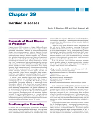
- 6. 802 CHAPTER 39 Cardiac Diseases Metcalfe and colleagues33 reported one maternal death among 219 pregnancies in 113 women with ASD. Peripheral vasodilation, if any- thing, reduces the left-to-right shunt.17 Because ASD in young women is not associated with heart failure, diuretics and extreme limitation of LA intravenous infusion are not warranted. A small percentage of patients with ASD have atrial flutter or fibrillation, which usually is paroxysmal. This arrhythmia can be managed along conventional lines, often with digoxin if necessary. The prospective mother should be informed that closure of the defect does not prevent atrial fibrillation once the arrhythmia has occurred. ASD can be difficult to diagnose during pregnancy. The murmur RA associated with ASD may be inconspicuous, being a pulmonary ejec- RV tion systolic murmur and therefore not unlike the physiologic murmur of pregnancy. However, the second heart sound is widely split and may be fixed throughout the respiratory cycle, a distinctly abnormal finding. The electrocardiogram (ECG) shows incomplete right bundle branch block and, in the case of the much more common ostium secundum defect, right axis deviation. In the less common ostium primum defect, marked left axis deviation accompanies incomplete right bundle branch block. The chest radiograph shows right atrial and right ven- tricular enlargement, prominent pulmonary arteries, and plethoric lung fields. Echocardiography establishes or confirms the diagnosis LA (Fig. 39-1), obviating the need for cardiac catheterization in many cases. Complicated Atrial Septal Defect If atrial arrhythmias recur frequently—and especially if the heart rate is difficult to control—catheter ablation is successful in restoring RA normal sinus rhythm without the need for antiarrhythmic drugs. In most cases, this procedure should not be done until after delivery RV because of the extensive radiation exposure that is needed. In rare instances, pregnancy and labor may be associated with a paradoxical systemic embolus resulting from a thrombus migrating from the infe- rior vena cava across the ASD into the left atrium. In the uncommon event that the patient is more than 35 years old and has an uncorrected large ASD, the likelihood of chronic atrial fibrillation, right ventricular dysfunction, and pulmonary hyperten- FIGURE 39-1 Transesophageal echocardiographic image of atrial sion rises significantly. Pregnancy is not advised if any of these sequelae septal defect (ostium secundum). In the upper panel, a large defect is present. If the patient insists on going through with the pregnancy, in the interatrial septum is present. In the lower panel, the blue/ prolonged bed rest will be required, and vigorous treatment of heart yellow color represents blood flow from the left atrium (LA) into the failure may be needed. The maternal risk is increased, and there is sig- right atrium (RA). RV, right ventricle. nificant risk of fetal loss. Although warfarin is generally recommended for chronic atrial fibrillation, its use is best avoided (especially in the first trimester); aspirin would be a reasonable compromise. Severe These findings constitute the maladie de Roger. Prophylaxis against pulmonary hypertension is an uncommon feature of an ostium secun- infective endocarditis is indicated, but otherwise this lesion has no dum ASD but is a contraindication to pregnancy. The ostium primum effect on pregnancy or labor. ASD, which is associated with Down syndrome and poses a risk of When the defect is in the membranous septum, spontaneous endocarditis, is more often associated with severe pulmonary hyper- closure is rare. In the absence of significant pulmonary vascular disease, tension. Infective endocarditis rarely, if ever, complicates a simple the same pansystolic murmur and thrill are found. If the shunt is large, ostium secundum ASD; therefore, prophylaxis during labor is not however, the lung fields are plethoric on chest radiography, and the warranted. heart and pulmonary arteries are enlarged. The classic ECG shows a pattern of biventricular hypertrophy. In such cases, flow through the Ventricular Septal Defect pulmonary vascular bed is usually at least twice the systemic cardiac The clinical spectrum and risk of VSD may range from so mild that it output. Patients with a relatively large, uncomplicated left-to-right has little or no effect on pregnancy to so high that maternal or fetal shunt through a VSD tolerate pregnancy well and, in this respect, are death can occur. Small defects in the muscular ventricular septum comparable to patients with ASD. However, prophylaxis against endo- frequently close spontaneously during childhood. However, these carditis is essential in cases of VSD. defects occasionally persist, allowing a small left-to-right shunt, mani- Here it is appropriate to detour from clinical description to fested by a loud pansystolic murmur along the left sternal border pathophysiology. Pulmonary vascular resistance is calculated as the accompanied by a coarse thrill. The chest radiograph is often normal, pressure drop across the pulmonary vascular bed divided by the flow as is the ECG. The echocardiogram is usually diagnostic (Fig. 39-2). through it:
- 7. CHAPTER 39 Cardiac Diseases 803 where P is the pressure drop and Q is the flow across the pulmonary vascular bed. A patient at one extreme may have a large shunt with pulmonary flow of 20 L/min and an R of 3 units, yielding an MPAP of 55 mm Hg (assuming a normal MPCWP of 5 mm Hg). At the other extreme, a patient with pulmonary vascular disease may have a pulmonary blood flow of 7 L/min and an R of 7 units, yielding an MPAP of 44 mm Hg. The higher the pulmonary vascular resistance (R), the greater the maternal risk. The risk is prohibitive when R reaches the systemic level (approximately 15 Wood units). In borderline cases (e.g., patients with R between 5 and 8 units), a pulmonary arteriolar vasodilating agent is rv lv sometimes administered to determine whether the increased resistance is partially reversible or completely irreversible. An increase in pulmo- nary vascular resistance of 3 to 4 units is considered mild, 5 to 7 units is moderate, and more than 8 units is severe. VSD may be associated with considerable increase in pulmonary vascular resistance, reflecting occlusive disease of the small pulmonary arteries and arterioles. This development, if it is to occur, usually does so in childhood; unless corrected, it leads to the Eisenmenger syn- drome (discussed later). However, a small number of adults may survive with VSD and pulmonary vascular resistance that is signifi- la cantly elevated but falls short of the Eisenmenger syndrome. Such patients are at high risk for death during pregnancy or labor, and there is a high risk of fetal impairment or loss. The patient should be told ra that early therapeutic abortion would be the safest option, and that later pregnancy would be hazardous and would require intensive care, with physical exercise strictly curtailed and prolonged bed rest enforced. The combination of decreased physical activity, pulmonary hyperten- lv sion, and pulmonary vascular disease would constitute a sound ratio- rv nale for instituting anticoagulation, which is another reason why pregnancy is better avoided or terminated. Some authorities strongly advise that delivery be effected prema- turely by means of cesarean section and urge sterilization at the same operation. The dangers must be thoroughly understood by women in this category who insist on continuing pregnancy. FIGURE 39-2 Transesophageal echocardiographic image of a Patent Ductus Arteriosus small muscular ventricular septal defect. In the upper panel, a The loud, continuous or machinery murmur of typical PDA with small communication is seen (arrow) between the right ventricle (rv) a large left-to-right shunt and no pulmonary vascular disease is so and left ventricle (lv). In the lower panel, color imaging confirms blood striking that the lesion is almost invariably detected and corrected in flow between the two chambers. la, left atrium; ra, right atrium. infancy or childhood. Occasionally, however, women of childbearing age or pregnant women from underdeveloped countries may present R = (MPAP − MPCWP)/Qpulm with a PDA. If the left-to-right shunt is large, the circulation is hyper- dynamic, with a wide arterial pulse pressure, low arterial diastolic where R is pulmonary vascular resistance in Wood units; MPAP and pressure, and hyperactive precordium. The heart may be somewhat MPCWP (in mm Hg) are the mean pulmonary arterial and capillary enlarged to clinical and radiologic examination, and the ECG may wedge pressures, respectively; and Qpulm is total flow through the right show left ventricular hypertrophy. The echocardiogram is useful for heart and pulmonary circulation (i.e., cardiac output plus left-to-right demonstrating a shunt between the two great vessels (Fig. 39-3). The shunt) in liters per minute. signs of hyperdynamic circulation resulting from the PDA are Resistance can be described in Wood units or in dyne·s/cm5. The exaggerated by pregnancy. Wood unit has the merit of simplicity and is derived from clinical units Because the murmur of PDA is systolic-diastolic, it is commonly of pressure and flow. The more fundamental but less friendly dyne·s/ referred to as a “continuous” murmur, although it usually peaks late in cm5 can be obtained by multiplying Wood units by 80. Normal pul- systole. Because of its characteristics, the murmur is also referred to as monary vascular resistance is 0.5 to 1.5 units. When a clinician is faced a “machinery” murmur. It is maximal in the left infraclavicular region. with a pregnant woman with pulmonary hypertension, the key to her It must be distinguished from a venous hum, which is loudest in the risk during pregnancy lies in the pulmonary vascular resistance. High neck rather than the infraclavicular area. Venous hum is common in flow, by itself, can be the mechanism for pulmonary hypertension pregnant women, and it changes dramatically with changes in the without dangerous elevation in the resistance. This mechanism can be position of the head. appreciated by rewriting the resistance equation to read Division or occlusion of the PDA should be accomplished before pregnancy is undertaken. Currently, most PDAs can be closed by the P=Q·R insertion of an occluder device delivered via a percutaneous intravas-
- 8. 804 CHAPTER 39 Cardiac Diseases Head and upper extremities AO SVC Aorta PA PA Right PA lung PV Left lung LA PV PA RA LV FIGURE 39-3 Transesophageal echocardiographic image of a Descending patent ductus arteriosus. A communication is present between the aorta RV proximal portion of the descending aorta (AO) and the pulmonary artery (PA). Color imaging (arrow) confirms blood flow from the aorta into the PA. IVC cular catheter.34 If a patient does become pregnant before PDA occlu- sion, an uncomplicated left-to-right shunt can be managed safely. Endocarditis is a risk in patients with PDA, and antibiotic prophylaxis is required. Embolic complications of infective endocarditis and end- arteritis secondary to PDA may take the form of infected pulmonary emboli. The patient becomes febrile with respiratory symptoms, and the chest radiograph shows multiple opacities and infiltrates. Trunk and lower extremities The leading cause of Eisenmenger syndrome is a large VSD, fol- lowed in prevalence by a large PDA. As with VSD, individuals with PDA Arterial blood Venous and arterial may sustain severe increases in pulmonary vascular resistance with the (fully saturated) blood corresponding pulmonary hypertension and right ventricular hyper- Cyanosis visible trophy, yet fall short of Eisenmenger physiology. The maternal risk Small admixture of Venous blood during pregnancy is high in this situation, similar to that encountered venous blood in VSD with equivalent pathology. Treatment is the same as in VSD No visible cyanosis with Eisenmenger syndrome. When the pulmonary pressure rises, the aortopulmonary shunt decreases, and the murmur becomes progres- FIGURE 39-4 Eisenmenger complex. Here the cause of right-to-left sively quieter and shorter, until it finally disappears. shunt across the ventricular septal defect is increased pulmonary In general, the woman with uncomplicated PDA tolerates preg- vascular resistance arising in the small pulmonary arteries and nancy well. If pulmonary hypertension supervenes, the risk to the arterioles. IVC, inferior vena cava; LA, left atrium; LV, left ventricle; PA, pulmonary artery; PV, pulmonary vein; RA, right atrium; RV, right mother becomes significant. Therefore, if pulmonary hypertension is ventricle; SVC, superior vena cava. (Reprinted by permission of the suspected and documented, termination of pregnancy is strongly publisher. From Taussig HB: Congenital Malformations of the Heart. recommended. Cambridge, Mass, Harvard University Press, 1960. Copyright © 1960 by the Commonwealth Fund by the President and Fellows of Harvard College.) Eisenmenger Syndrome Eisenmenger syndrome is characterized by a congenital communica- tion between the systemic and pulmonary circulations and increased ventricles. It is more common in girls and develops at a young age. pulmonary vascular resistance, either to systemic level (so that there is Therefore, when increased pulmonary vascular resistance is detected no shunt across the defect) or exceeding systemic (allowing right-to- in a child with a large VSD, operative closure must be done as soon as left shunting). As mentioned, the most common underlying defect is possible to prevent the development of Eisenmenger pathophysiology. a large VSD, followed in prevalence by a large PDA. Eisenmenger Once this has appeared, pulmonary hypertension is irreversible, and pathophysiology is less common in ASD. Occasionally, this type of the VSD is consequently inoperable (unless lung transplantation is pathophysiology develops in other, less common defects. By the time performed as well). the syndrome is fully developed, it is often difficult clinically to diag- The major clues that pulmonary vascular resistance is increasing nose the underlying defect. For this discussion, the VSD serves as a are (1) diminution of evidence of a left-to-right shunt and (2) the good model (Fig. 39-4). appearance of progressively severe pulmonary hypertension. The pan- Eisenmenger pathophysiology develops only if the defect is large systolic murmur of VSD or the continuous murmur of PDA is replaced and is not restrictive, resulting in equal systolic pressure in the two by a short ejection systolic murmur. The lungs are no longer plethoric
- 9. CHAPTER 39 Cardiac Diseases 805 but show large central pulmonary arteries and small peripheral arteries until term, treated with oxygen and heparin, and delivered by cesarean characteristic of severe pulmonary hypertension. Because the shunt section. One patient died a month after delivery. Most of the infants has disappeared, the radiographic cardiothoracic ratio returns to were small, and one died. Despite this better-than-average outcome, normal but the main pulmonary segment is prominent. There is pregnancy should not be encouraged in women with Eisenmenger usually a striking right ventricular heave, a loud and palpable pulmo- syndrome or in those with a systemic level of pulmonary hypertension nary valve closure sound, and an ejection sound in early systole. When of any cause. concentric ventricular hypertrophy gives way to dilatation and right- sided heart failure, evidence of tricuspid regurgitation appears. Until then, the mean venous pressure is normal but the amplitude of the a Primary Pulmonary Hypertension wave may be increased, reflecting decreased right ventricular diastolic Severe idiopathic (“primary”) pulmonary hypertension, like the compliance. Eisenmenger syndrome, carries a high risk in pregnancy, and the If pulmonary vascular resistance is significantly higher than sys- same principles apply to its management. Pregnancy is not advised in temic levels, right-to-left shunting of blood occurs and causes cyanosis women with this condition, because the mortality rate approaches and clubbing of the fingers and toes. This shunting of deoxygenated 50%.40 blood into the systemic circulation leads to hypoxemia and triggers a Severe pulmonary hypertension can result from taking appetite- reactive erythrocytosis as the system attempts to increase peripheral suppressing drugs. The fenfluramine-phentermine regimen (“fen- oxygen delivery. This increases blood viscosity and can cause sludging phen”) was a notorious culprit41 and was withdrawn from the market. and decreased flow of blood, especially in small vessels. A high hemat- Treatment strategies include vasodilators, sometimes by chronic intra- ocrit value, however, is not an automatic indication for serial phle- venous infusion, and nitric oxide inhalation. In some 25% of cases, botomy, because this approach can lead to iron deficiency and pulmonary arterial pressure is lowered by prostacyclin infusion.42 This microcytosis. Tissue hypoxia may then actually worsen, a particularly response predicts a favorable response to chronic oral nifedipine undesirable result in pregnancy. Phlebotomy is reserved for patients administration and a good prognosis. Balloon atrial septostomy,43,44 without evidence of iron deficiency on laboratory testing who have through the foramen ovale or via transseptal puncture, can be used, in symptoms of hyperviscosity, including headache, dizziness, visual dis- extreme cases of pulmonary hypertension, to relieve right heart pres- turbance, myalgia, and bleeding diathesis. Quantitative volume replace- sure, usually as a bridge to transplantation. ment is necessary during phlebotomy. Attempted surgical correction of a congenital cardiac shunt after Eisenmenger syndrome is present usually results in the death of the patient.35 Many patients ultimately die of right-sided heart failure, Congenital Obstructive pulmonary hypertension, or pulmonary hemorrhage.36 Lesions The woman with Eisenmenger syndrome must be informed that pregnancy carries a mortality risk of about 50%.37 Even if the mother Some congenital cardiac malformations are characterized by obstruc- survives, the outcome for the fetus is likely to be poor, because the fetal tion to left or right ventricular outflow. The more common examples mortality rate exceeds 50% in cyanotic women with Eisenmenger syn- include pulmonary stenosis, aortic stenosis, and coarctation of the drome.36 Sudden death may occur at any time, but labor, delivery, and aorta. The hypoplastic left heart syndrome seldom allows survival to particularly the early puerperium seem to be the most dangerous childbearing age, and those who do survive usually have undergone a periods.38 Any significant fall in venous return, regardless of cause, major palliative procedure, such as construction of a ventriculoaortic impairs the ability of the right heart to pump blood through the high, conduit with a prosthetic valve, that would constitute a strong contra- fixed pulmonary vascular resistance. Hypotension and shock can occur indication to pregnancy. quickly and are often unresponsive to medical therapy. The major physiologic difficulty in pulmonary hypertension is maintenance of adequate pulmonary blood flow. Any event or condi- Mitral Stenosis tion that decreases venous return, such as vasodilation on the systemic Congenital mitral stenosis is a rare malformation. When it is associated side of the circulation from epidural anesthesia or pooling of blood in with an ASD, it constitutes the Lutembacher syndrome. Survival to the lower extremities from vena caval compression, decreases preload childbearing age is usual. Both lesions tend to promote atrial fibrilla- to the right ventricle and pulmonary blood flow. Therefore, manage- tion. Ideally, the mitral valve and the atrial defect should be repaired ment during pregnancy centers on the maintenance of pulmonary before pregnancy. blood flow. If the patient insists on continuing her pregnancy, limita- tion of physical activity is essential, as is the use of pressure-graded elastic support hose, low-flow home oxygen therapy, and monthly Aortic Stenosis monitoring of blood and platelet counts. Because of the precarious (see also the later section on aortic stenosis under physiologic balance, a planned delivery should be performed with Aortic Valve Disease) intensive care monitoring, including a Swan-Ganz catheter and provi- sions for skilled obstetric anesthesia care. Anesthetic considerations for Bicuspid aortic valve is one of the more common congenital malfor- this entity are discussed in Chapter 56. mations that may lead to aortic stenosis, regurgitation, or both (Fig. On a more optimistic note, a report published in 1995 described 39-5). Often, aortic stenosis is not present during early life but pro- 13 pregnancies in 12 women with Eisenmenger syndrome who elected gresses over time because of valve calcification and gradual restriction not to accept advice to terminate pregnancy.39 Mean systolic pulmo- in leaflet motion. The bicuspid valve may occur as an isolated defect nary arterial pressure was 113 mm Hg. Three spontaneous abortions, or in combination with other congenital anomalies, most commonly one premature labor, and two maternal deaths occurred. The seven aortic coarctation. Congenital aortic stenosis, on the other hand, can patients who reached the end of the second trimester were hospitalized be severe at birth and may cause severe left ventricular hypertrophy
- 10. 806 CHAPTER 39 Cardiac Diseases LA AV PA RVOT RV FIGURE 39-5 Transesophageal echocardiographic image of a bicuspid aortic valve. During systole, only two aortic valve (AV) leaflets are seen. LA, left atrium; RVOT, right ventricular outflow tract. FIGURE 39-6 Transesophageal echocardiographic image of pulmonic stenosis. The pulmonic valve leaflets exhibit characteristic that limits the ability of the heart to respond to demands for increased doming (arrow). PA, pulmonary artery; RV, right ventricle. cardiac output. In the syndrome of severe congenital aortic stenosis, the pulses are of slow upstroke and diminished amplitude. Unlike adults with acquired aortic stenosis, children and young adults with congenital Vasodilators, helpful in patients with heart failure of other etiology, aortic stenosis have an abnormally loud aortic valve closure sound. Left are dangerous in patients with aortic stenosis, because the impeded left ventricular ejection is prolonged, so that the aortic valve closure sound ventricle may not be able to fill the dilated peripheral vascular bed. It may occur after the pulmonary valve closure sound. Therefore, split- should be remembered that the lowered systemic vascular resistance ting of the second heart sound is paradoxical and is heard in expiration of pregnancy adversely affects aortic stenosis. The obstructed left ven- instead of inspiration. Often a loud ejection sound is heard in early tricle is limited in its ability to fill the dilated peripheral bed, a situation systole. The duration of the ejection murmur and the time to its peak that can lead to syncope or more serious manifestations of limited, intensity increase with worsening severity of aortic stenosis. relatively fixed cardiac output. The ECG shows severe left ventricular hypertrophy. The chest radiograph is characterized by poststenotic dilatation of the aorta. Although some patients complain of dyspnea, chest pain, and syncope, Pulmonic Stenosis others remain asymptomatic. The lesion can be recognized and its The murmur of pulmonic stenosis is loud and is often accompanied severity assessed by Doppler echocardiography. by a thrill. The lesion is usually detected in early childhood and is likely Critical calcific aortic stenosis is usually treated by aortic valve to have been corrected before the childbearing age. Expectant mothers replacement in older patients, but aortic valve repair is often possible who have not had adequate health supervision in childhood may have in younger women of childbearing age with congenital aortic stenosis. unrecognized pulmonary stenosis. If aortic stenosis is severe—and especially if it is symptomatic—the The diagnosis is suggested by a long, harsh systolic murmur over woman should be advised against becoming pregnant. She should be the upper left sternal border that is usually preceded by an ejection advised that, if the aortic valve must be replaced, pregnancy and labor sound. The venous pressure is normal, but there are striking a waves would be difficult and dangerous because of the need for anticoagulant in the jugular venous pulse. The pulmonary valve closure sound is treatment after a mechanical prosthesis is implanted. Maternal mortal- usually too soft to hear when pulmonary stenosis is severe. Severe ity rates as high as 17% have been reported,45 although more recent pulmonary stenosis causes massive concentric right ventricular data have suggested a somewhat lower risk. However, these studies also hypertrophy; this is manifested by a left parasternal heave and by tall emphasize the adverse effects of severe maternal aortic stenosis on fetal R waves and deeply inverted T waves in the right precordial leads of outcomes, including increased rates of preterm delivery and intrauter- the ECG. Tall, pointed P waves are also present, denoting right atrial ine growth restriction.46 If aortic stenosis is moderate in severity, the enlargement. patient should be advised to complete her pregnancies before the aortic Right ventricular enlargement and poststenotic dilatation of the valve is replaced. Labor can be managed in such cases without a high main and left pulmonary arteries are seen on the chest radiograph, maternal or fetal risk, but assisted shortening of the second stage of which also may show slightly diminished peripheral pulmonary vas- labor is recommended.47 culature. Echocardiography demonstrates limited opening of the pul- Strict limits on physical exertion and prolonged periods of bed rest monic valve leaflets (Fig. 39-6), right ventricular hypertrophy, and may be required. Left ventricular failure may appear and may necessi- abnormally high velocity of blood flow in the pulmonary artery. tate the use of diuretic agents and digitalis. Rarely, even in the presence Doppler echocardiography also allows calculation of the right ven- of severe aortic stenosis, heart failure may be due to another cause (e.g., tricular pressure and the systolic pressure gradient across the valve. peripartum cardiomyopathy).48 Prophylaxis against bacterial endocar- These pressures can also be measured directly in the hemodynamics ditis at delivery is recommended. laboratory (Fig. 39-7).
- 11. CHAPTER 39 Cardiac Diseases 807 Aorta LV LV Septal RV defect RV Pulmonary artery Tetralogy of Fallot Normal FIGURE 39-8 Tetralogy of Fallot. The anatomic pathology (left) compared with normal (right). Note the ventricular septal defect, the aorta (which overrides the defect), the pulmonary stenosis, and the right ventricular hypertrophy. LV, left ventricle; RV, right ventricle. (Reprinted by permission of the publisher. From Taussig HB: Congenital Malformations of the Heart. Cambridge, Mass, Harvard University Press, 1960. Copyright © 1960 by the Commonwealth Fund of the President and Fellows of Harvard College.) FIGURE 39-7 Pressure tracings in severe pulmonary stenosis. Pulmonary pressure is extremely low and appears damped. Right ventricular pressure is suprasystemic. (From Shabetai R, Adolph RJ: Principles of cardiac catheterization. In Fowler NO [ed]: Cardiac Diagnosis and Treatment. Hagerstown, MD: Harper & Row, 1980, p 106.) RV Pulmonic stenosis is generally well tolerated so that neither preg- nancy nor labor poses a significant threat.49 Prophylaxis against infec- tive endocarditis is necessary. More severe pulmonary stenosis requires LV AO treatment. Unlike aortic stenosis, however, critical pulmonary stenosis does not require valve replacement or open repair. Most cases are treated successfully with transvenous balloon valvuloplasty.50 Ideally, MV this should be carried out before pregnancy is undertaken; if a woman does become pregnant and develops intractable right-sided heart failure, the procedure can still be safely performed (but at some risk to the fetus). Extreme pulmonary stenosis (right ventricular systolic pressure > systemic systolic pressure) is a contraindication to preg- FIGURE 39-9 Transthoracic echocardiographic image of tetralogy nancy until the lesion is adequately treated. of Fallot. A large ventricular septal defect is present, and the aorta (AO) overrides the interventricular septum. LV, left ventricle; MV, mitral valve; RV, right ventricle. Right-to-Left Shunt without Pulmonary Hypertension (Tetralogy of Fallot) outflow obstruction that diverts blood flow through the VSD. In the The congenital cyanotic heart diseases discussed so far have been asso- typical case, right and left ventricular systolic pressures are equal but ciated with a communication between the pulmonary and systemic the pulmonary artery pressure is exceedingly low. A loud, long systolic circulations and pulmonary vascular resistance sufficiently high to murmur is audible along the left sternal border. The murmur is caused cause a right-to-left shunt. However, cyanosis occurs in other congeni- by an abnormal flow pattern through the obstructed right ventricular tal malformations, in which there is a defect between the right and left outflow tract. The pulmonary valve closure sound is usually inaudible. sides of the heart but also right ventricular outflow obstruction (Figs. Patients are usually cyanotic and often have significant clubbing of the 39-8 and 39-9). Examples include the tetralogy of Fallot and tricuspid fingers and toes. The hematocrit value is greatly elevated because of atresia. the severe erythrocytosis. Phlebotomy is not indicated to treat the Tetralogy of Fallot is used to illustrate this class of congenital mal- hematocrit level per se but is indicated if symptoms of hyperviscosity formation of the heart, because it is by far the most common form of occur. Ignoring this important therapeutic principle leads to a micro- cyanotic congenital heart disease encountered in pregnancy. Moreover, cytic anemia that further complicates pregnancy. The ECG shows the offspring of a mother with tetralogy of Fallot has a 2% to 13% severe right ventricular hypertrophy. The chest radiograph is charac- chance of inheriting the condition.51 The syndrome includes (1) a large terized by a normal-sized heart and a concavity in the region where defect high in the ventricular septum; (2) pulmonary stenosis, which the pulmonary artery should be (Fig. 39-11). As in all malformations may be at the valve itself but more commonly is in the infundibulum of this general type, the lung fields are oligemic, showing small vessels of the right ventricle; (3) dextroposition of the aorta so that the aortic throughout. orifice sits astride the VSD and overrides, at least in part, the right Most adults born with the tetralogy of Fallot and lesions with ventricle; and (4) right ventricular hypertrophy (Fig. 39-10). similar pathophysiology have undergone surgical treatment before A wide spectrum of clinical presentations may be present, depend- reaching young adulthood. Children raised in undeveloped countries ing on the relative size of the VSD and the degree of right ventricular are an important exception. Many patients have had surgery to close
- 12. 808 CHAPTER 39 Cardiac Diseases The cyanotic patient with tetralogy of Fallot has special problems Head and upper extremities during pregnancy. The reduced systemic vascular resistance of preg- nancy causes more blood to shunt from right to left, leaving less to flow to the pulmonary circulation. This intensifies hypoxemia and can lead to syncope or death. Maintenance of venous return is crucial. The SVC most dangerous times for these women are late pregnancy and the Aorta early puerperium, because venous return is impeded by the large PA PA gravid uterus near term and by peripheral venous pooling after deliv- ery. Pressure-graded elastic support hose are recommended. Blood loss Right lung PV Left during labor may compromise venous return, and blood volume must PA lung be promptly and adequately restored. Anesthetic considerations during delivery are discussed in detail in Chapter 56. Antibiotic prophylaxis LA PV should be used in these susceptible patients at delivery. Because of the combined high maternal risk and high incidence of fetal loss, pregnancy is discouraged in women with uncorrected tetral- RA ogy of Fallot. The prognosis is particularly bleak in those women with LV a history of repeated syncopal episodes, a hematocrit level greater than Descending 60%, or a right ventricular systolic pressure greater than 120 mm Hg. RV aorta If a young woman with untreated tetralogy of Fallot requests prepreg- nancy counseling, she should be advised to undergo surgical correction before pregnancy. Pregnancy does not represent a significantly IVC increased risk for patients in whom the VSD has been patched and the pulmonary stenosis corrected. Coarctation of the Aorta Coarctation of the aorta is a congenital defect in the area of the aorta where the ligamentum arteriosum and the left subclavian artery insert (the distal portion of the aortic arch). The malformation may be Trunk and simple or complex, and it is either isolated or associated with PDA and lower extremities other malformations, notably aortic stenosis and aortic regurgitation Arterial blood Venous and arterial secondary to a bicuspid aortic valve. It may also occur in women with (fully saturated) blood Turner syndrome. The lesion should be detected and treated surgically Cyanosis visible or by balloon dilation in infancy or childhood, but it may be present in women who are, or want to become, pregnant. Small admixture of Venous blood venous blood Typical features include the following: No visible cyanosis Upper extremity hypertension but lower extremity hypotension FIGURE 39-10 Tetralogy of Fallot. Blood shunts from left to right Visible and palpable collateral arteries in the scapular area through the ventricular septal defect because its flow to the lungs is A late systolic murmur, usually loudest over the interscapular impeded by pulmonary stenosis; this results in cyanosis. IVC, inferior region vena cava; LA, left atrium; LV, left ventricle; PA, pulmonary artery; PV, Femoral pulses that lag behind the carotid pulses and are of pulmonary vein; RA, right atrium; RV, right ventricle; SVC, superior diminished amplitude vena cava. (Reprinted by permission of the publisher. From Taussig Notching of the inferior rib borders seen on the chest radiograph HB: Congenital Malformations of the Heart. Cambridge, Mass, Harvard University Press, 1960. Copyright © 1960 by the and resulting from erosion by arterial collaterals that bridge the Commonwealth Fund by the President and Fellows of Harvard coarctation College.) Electrocardiographic evidence of severe left ventricular hypertro- phy strongly suggests associated aortic stenosis. Surgical grafting or the VSD and relieve the pulmonary stenosis, constituting virtual “total percutaneous intravascular balloon dilation reduces the upper extrem- repair” and rendering them potentially safe candidates for pregnancy ity hypertension, but blood pressure does not always return to normal, and delivery. However, the operation is not curative. Significant and hypertension may recur in later life. arrhythmia and conduction defects that may eventually lead to the Whenever possible, the operation should be performed before need for electronic cardiac pacing or an implantable defibrillator may pregnancy; otherwise, the maternal mortality rate is approximately occur years after an apparently successful operation. Other sequelae 3%. Coarctation is associated with congenital berry aneurysm of the and residua include only partial relief of the right ventricular outflow circle of Willis and hemorrhagic stroke. The risk of stroke may increase obstruction and pulmonic regurgitation. This latter problem is usually during labor because of transient elevations in blood pressure. Patients well tolerated early but may lead to right-sided heart failure, necessitat- are at risk for aortic dissection and infective endocarditis involving ing reoperation. In addition, women with repaired tetralogy of Fallot an abnormal aortic valve; these risks increase during pregnancy.53 and significant pulmonic regurgitation have a higher risk of decom- Hypertension often worsens as well.54 Coarctation is also associated pensation during pregnancy.52 with an increased frequency of preeclampsia.31 The operation does not
- 13. CHAPTER 39 Cardiac Diseases 809 A B C D FIGURE 39-11 Tetralogy of Fallot. A, Chest radiograph. Note concavity in the area of the pulmonary artery, oligemic lungs, and right aortic arch. B, Right ventriculogram. Note the narrow right ventricular outflow tract. C, Further clarification of the pulmonary arteries. The left ventricle is slightly opacified via the ventricular septal defect. D, The associated right-sided aortic arch is now visible. (From Shabetai R, Adolph RJ: Principles of cardiac catheterization. In Fowler NO [ed]: Cardiac Diagnosis and Treatment. Hagerstown, MD: Harper & Row, 1980, p 106.) require cardiopulmonary bypass and can be carried out with safety for dissection and rupture. A number of centers are now performing the mother and with less fetal risk than accompanies open heart balloon dilation with stent implantation for adults with unoperated surgery with cardiopulmonary bypass. Although transvascular balloon aortic coarctation, but large, multicenter studies are currently not dilation of aortic coarctation is a viable option for children and infants available.56 with coarctation, its use in adults is controversial.55 The procedure If delivery must be undertaken in cases of unoperated coarctation, is well accepted for treatment of postsurgical renarrowing of the blood pressure can be titrated with β-adrenergic–blocking agents coarctation, but de novo balloon angioplasty carries a risk of aortic delivered by intravenous drip.
- 14. 810 CHAPTER 39 Cardiac Diseases more rapid than in acquired complete atrioventricular block. Although these patients appear to do well during childhood and young adult- Other Congenital Cardiac hood, the lesion is associated with an unexpectedly high mortality rate. Therefore, treatment with a pacemaker is indicated in many Malformations of the cases.58 The pacemaker used should be dual-chamber and rate- responsive, so that normal cardiovascular dynamics at rest and exercise Ebstein Anomaly will be preserved. Patients who are untreated or who have received a Ebstein anomaly is a malformation of the tricuspid valve in which the pacemaker are at slight to no increased risk during pregnancy. septal leaflet is displaced apically and the anterior leaflet is abnormally large in size. The deformed tricuspid valve apparatus may be signifi- cantly incompetent or stenotic, depending on the location of the Additional Malformations anomalously placed cusps of the valve. In some cases, the malforma- A number of other malformations may be present in women of child- tion causes impediment to right ventricular outflow. bearing age, including The clinical features are easily recognized by a cardiologist, and the echocardiogram is characteristic and reliable (Fig. 39-12). This syn- Other left-to-right or right-to-left shunts drome is frequently associated with anomalous atrioventricular con- Transposition of the great vessels duction pathways and with the Wolff-Parkinson-White syndrome. Truncus arteriosus Patients may also have an ASD with right-to-left shunting and cyano- Single-ventricle double-outlet right ventricle sis. Supraventricular arrhythmias are also common. Various obstructive lesions The most favored treatment is reconstruction of the tricuspid valve, for which satisfactory techniques have now been developed. The opera- The malformations may be multiple and complex. Survival to tion should be performed before pregnancy is undertaken. Interrup- adulthood depends on at least partial correction, which may have been tion of anomalous conduction pathways also can be performed during furnished by surgical operation or may be part of the malformation. surgery. For example, in D-type transposition of the great vessels, the aorta The Mayo Clinic group57 reported on 111 pregnancies in 44 women arises from the right ventricle and the pulmonary artery from the left. with Ebstein anomaly resulting in 95 live births, although most of the Survival requires a shunt at some level (ASD, VSD, or PDA) so that infants had low birth weight. Vaginal delivery was performed in 89% oxygenated blood can enter the systemic circulation. and cesarean section in 9%; 23 deliveries were premature. Nineteen Some of these women with untreated and delicately balanced pregnancies ended with spontaneous abortion, and seven ended with lesions bear children, but usually this is not wise to attempt. Transposi- therapeutic abortion. Congenital heart disease occurred in 6% of the tion of the great vessels is now treated by anastomosis of the aorta to children of mothers with Ebstein anomaly. the morphologic left ventricle and of the pulmonary artery to the morphologic right ventricle. Lesions such as single ventricle may be palliated by the Fontan procedure, in which venous return is connected Congenital Atrioventricular Block directly to the pulmonary circulation, bypassing the right side of the Congenital atrioventricular block differs somewhat from heart block heart. Neither procedure constitutes a cure, but successful pregnancy in adults. The pacemaker is usually junctional, and therefore the QRS can occur. complex is normal or only slightly widened and the ventricular rate is In summary, patients should be evaluated and tracked by a cardi- ologist who is experienced in congenital heart disease and by a mater- nal-fetal medicine specialist with knowledge and experience in managing pregnancy in women with congenital cardiac lesions.7,31 RV Rheumatic Heart Disease Rheumatic Fever Rheumatic fever is now distinctly uncommon in the United States, Canada, Western Europe, and Great Britain, but it is still prevalent in less economically developed countries. Young female immigrants to the Western world constitute a large proportion of the patients with a RA history of rheumatic fever. These women are at risk of developing rheumatic valvular heart disease 10 to 20 years after the initial episode of rheumatic fever. Chronic Rheumatic Heart Disease In the United States, acute rheumatic fever with carditis has been uncommon for many years, and chronic rheumatic heart disease, FIGURE 39-12 Ebstein anomaly. The right atrium (RA) and right which manifests years to decades after the episode of acute rheumatic ventricle (RV) are markedly dilated, and the tricuspid valve is fever, is becoming uncommon among the native childbearing popula- displaced toward the cardiac apex. tion. Control of rheumatic fever has largely shifted the burden of