orthopedics.Flat foot and halux valgus.(dr.omer barawe)
•Als PPTX, PDF herunterladen•
0 gefällt mir•2,276 views


Empfohlen
Weitere ähnliche Inhalte
Andere mochten auch
Andere mochten auch (16)
Mehr von student
Mehr von student (20)
Kürzlich hochgeladen
Kürzlich hochgeladen (20)
orthopedics.Flat foot and halux valgus.(dr.omer barawe)
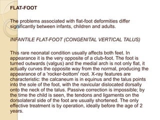
- 1. FLAT-FOOT The problems associated with flat-foot deformities differ significantly between infants, children and adults. INFANTILE FLAT-FOOT (CONGENITAL VERTICAL TALUS) This rare neonatal condition usually affects both feet. In appearance it is the very opposite of a club-foot. The foot is turned outwards (valgus) and the medial arch is not only flat, it actually curves the opposite way from the normal, producing the appearance of a 'rocker-bottom' root. X-ray features are characteristic: the calcaneum is in equinus and the talus points into the sole of the foot, with the navicular dislocated dorsally onto the neck of the talus. Passive correction is impossible; by the time the child is seen, the tendons and ligaments on the dorsolateral side of the foot are usually shortened. The only effective treatment is by operation, ideally before the age of 2 years.
- 2. FLAT-FOOT IN CHILDREN AND ADOLESCENTS Flat-foot is a common complaint among children and teenagers, or rather their parents - the children themselves usually don't seem to mind! When weight-bearing, the foot is turned outwards and the medial border of the foot is in contact (or nearly in contact) with the ground. The heel becomes valgus (hence the medical terms pes planus and pes valgus). Two forms of the condition are recognized: flexible and rigid.
- 3. Flexible flat-foot appears in toddlers as a normal stage in development, and it usually disappears after a few years when medial arch development is complete; sometimes, though, it persists into adult life. The arch can often be restored by simply dorsiflexing the great toe (the jack, or great-toe extension, test) and during this manoeuvre the tibia rotates externally. Many of these children have ligamentous laxity and there may be a family history of both flat-feet and joint hypermobility. Stiff (or 'rigid') flat-foot which cannot be corrected passively should alert the examiner to an underlying abnormality. In older children and adolescents, conditions to be considered are tarsal coalition (often a bar of bone connecting the calcaneum to the talus or the navicular), an inflammatory joint condition or a neurological disorder.
- 4. Clinical assessment In the common flexible flat-foot there are no symptoms, but the parents notice that the feet are flat or that the shoes wear badly. The deformity becomes noticeable when the youngster stands. The first test is to ask the patient to go up on tiptoes: if the heels invert, it is a flexible (or mobile) deformity. Next, examine the foot with the child sitting or lying. Feel for localized tenderness and test the range of movement in the ankle, the subtalar and midtarsal joints. A tight Achilles tendon may induce a compensatory flat-foot deformity.
- 5. Teenagers and young adults sometimes present with a painful, rigid flat-foot. On examination, the peroneal and extensor tendons appear to be in spasm (the condition is sometimes called spasmodic flat-foot). In some cases a definite cause may be found (e.g. a tarsal coalition or inflammatory arthritis), but in many no specific cause is identified. The spine, hips and knees should always be examined. The clinical assessment is completed by a swift general examination for joint hypermobility and signs of neuromuscular abnormalities.
- 6. Imaging X-rays are unnecessary for asymptomatic, flexible flat-feet. For pathological flat-feet (which are usually painful or stiff), standing anteroposterior, lateral and oblique views may help to identify underlying disorders. CT scanning is the most reliable way of demonstrating tarsal coalitions. Treatment Young children with flexible flat-feet require no treatment. Parents need to be reassured and told that the 'deformity' will probably correct itself in time; even if it does not fully correct, function is unlikely to be impaired. Where the condition is obviously due to an underlying disorder such as poliomyelitis, splintage or operative correction and muscle rebalancing may be needed. Spasmodic flat-foot is relieved by rest in a cast or a splint. If there is an abnormal tarsal bar or other bony irregularity, this may have to be removed; in late cases, if pain is intolerable, a triple arthrodesis may be necessary
- 7. FLAT-FOOT IN ADULTS When adults present with symptomatic flat- feet, the first thing to ask whether they always had flat-feet or whether it is of recent onset. Constitutional flat-feet which have been more or less asymptomatic for many years may start causing nagging pain after a change in daily activities (e.g. taking on work which requires a lot of standing and walking). More recent deformities may be due to an underlying disorder such as rheumatoid arthritis or generalized muscular weakness; and unilateral flat-foot should make one think of tibialis posterior synovitis or rupture.
- 8. Where there e is no underlying deformity, little can be done apart from giving advice about sensible footwear and arch supports. Patients with painful rigid flat-foot may require more robust splintage (and of course, treatment for any generalized conditions such as rheumatoid arthritis). Those with tibialis posterior rupture can be helped by operative repair or replacement of defective tendon.
- 11. HALLUX VALGUS Hallux valgus is the commonest of the foot deformities (and probably of all musculoskeletal deformities). In people who have never worn shoes the big toe is in line with the first metatarsal, retaining the slightly fan-shaped appearance of the forefoot In people who habitually wear shoes the hallux assumes a valgus position; but only if the angulation is excessive is it referred to as 'hallux valgus'. Splaying of the forefoot, with varus angulation of the first metatarsal, predisposes to lateral angulation of the big toe in people who wear shoes. This metatarsus primus varus may be congenital, or it may result from loss of muscle tone in the forefoot in elderly people. Hallux valgus is also common in rheumatoid arthritis. The elements of the deformity are lateral deviation and rotation of the hallux, together with hypertrophy ('exostosis') of the medial part of the metatarsal head and an overlying bursa which together form a prominent bump (or bunion) on the medial side. Lateral deviation of the hallux may lead to overcrowding of the lateral toes and sometimes overriding.
- 13. Clinical features Hallux valgus is most common in women between 50 and 70 years, and is usually bilateral. An important sub-group, with strong familial tendency, appears during late adolescence. Often there are no symptoms apart from the deformity. Pain, if present, may be due to (1) shoe pressure on a large or an inflamed bunion, (2) splaying of the forefoot and muscle strain (metatarsalgia), (3) associated deformities of the lesser toes, or (4) secondary osteoarthritis of the first metatarsophalangeal joint. X-rays X-rays should be taken with the patient standing, to show the degree of metatarsal and hallux angulation. The first metatarsophalangeal joint may be subluxated, or it may look osteoarthritic.
- 14. Treatment Adolescents Deformity is usually the only 'symptom', but the mother is anxious to prevent it becoming as severe as her own. Conservative treatment is justified as a first measure (operative correction carries a 20--40 per cent recurrence rate in this age group). The patient is encouraged to wear shoes with deep toe-boxes, soft uppers and low heels. If the deformity progresses, a corrective osteotomy of the first metatarsal and soft tissue rebalancing around the metatarsophalangeal joint may produce a satisfactory correction. Adults Surgical treatment is more readily offered to older patients. This usually takes the form of excision of the bunion, metatarsal osteotomy and soft-tissue rebalancing. However, if the metatarsophalangeal joint is frankly osteoarthritic, arthrodesis of the joint may be a better option.
- 15. HALLUX RIGIDUS 'Rigidity' of the first metatarsophalangeal J01nt may be due to local trauma or osteochondritis dissecans of the first metatarsal head, but in older people it is usually caused by long-standing joint disorders such as gout, pseudogout or osteoarthritis. In contrast to hallux valgus, men and women are affected with equal frequency. Clinical problems are due to the fact that, because the big toe cannot extend (dorsiflex), push-off at the end of the stance phase of gait becomes painful and clumsy.
- 16. Clinical features Pain on walking, especially on .slopes or rough ground, is the predominant symptom. The hallux is straight and the metatarsophalangeal joint feels knobbly; a tender dorsal 'bunion' (actually a large osteophyte) is characteristic. Dorsiflexion is restricted and painful; plantarflexion is also limited, but less so. X-ray The changes· are those of osteoarthritis: the joint space is narrowed, there is bone sclerosis and. often, large osteophytes at the joint margins.
- 17. Treatment A rocker-soled shoe may abolish pain by allowing the foot to 'roll' without the necessity for dorsiflexion at the metatarsophalangeal joint. If walking is painful despite this type of shoe adjustment, an operation is advised. For young patients, the best procedure is a simple extension osteotomy of the proximal phalanx, to mimic dorsiflexion at the interphalangeal joint. In older patients, cheilectomy is the procedure of choice: the dorsal osteophytes and the dorsal edge of the metatarsal head are removed in an attempt to restore extension (dorsiflexion) at the metatarsophalangeal joint. Arthrodesis of the metatarsophalangeal joint is a good solution for the badly arthritic joint.