
Empfohlen
Weitere ähnliche Inhalte
Was ist angesagt?
Was ist angesagt? (20)
Andere mochten auch
Andere mochten auch (20)
Ähnlich wie Introduction to the Digestive System
Ähnlich wie Introduction to the Digestive System (20)
Mehr von Rajesh Goit
Mehr von Rajesh Goit (16)
Kürzlich hochgeladen
Kürzlich hochgeladen (20)
Introduction to the Digestive System
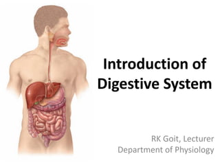
- 1. Introduction of Digestive System RK Goit, Lecturer Department of Physiology
- 2. • Digestive system – performs the mechanical & chemical processes of digestion, absorption of nutrients, & elimination of wastes – consists of the mouth, esophagus, stomach, intestine, & accessory organs • medical specialty that deals with the structure, function, diagnosis, & treatment of diseases of the stomach & intestines is called gastroenterology • medical specialty that deals with the diagnosis & treatment of disorders of the rectum & anus is called proctology
- 3. • basic chemical unit of our food & our tissue are the same • our food looks so different from the tissue because the units are arranged very differently • in order to achieve rearrangement of the building blocks of our food, it is first necessary to break the food molecules into their basic constituents
- 4. • 2 groups of organs compose the digestive system: 1. gastrointestinal (GI) tract, or alimentary canal – a continuous tube that extends from the mouth to the anus through the thoracic & abdominopelvic cavities – organs include the mouth, most of the pharynx, esophagus, stomach, small intestine, & large intestine – length of the GI tract is about 5–7 meters in a living person (longer in a cadaver- about 7–9 meters) 2. accessory digestive organs – include the teeth, tongue, salivary glands, liver, gallbladder, & pancreas – teeth aid in the physical breakdown of food – tongue assists in chewing & swallowing – salivary glands, liver, gallbladder, & pancreas produce or store secretions that flow into the GI tract through ducts
- 7. Functions of the digestive system 1. Ingestion – taking food into mouth 2. Secretion – release of water, acid, buffers, & enzymes into lumen of GIT 3. Mixing & propulsion – churning & propulsion of food through GI tract 4. Digestion – mechanical & chemical breakdown of food 5. Absorption – passage of digested products from GIT into blood & lymph 6. Defecation – elimination of feces from GI tract
- 9. • Mucosa 1. epithelium – in the mouth, pharynx, esophagus, & anal canal is mainly nonkeratinized stratified squamous epithelium – in stomach & intestine is simple columnar epithelium – several types of endocrine cells (enteroendocrine cells) secrete hormones 2. lamina propria – is areolar connective tissue containing many blood & lymphatic vessels – also contains mucosa associated lymphatic tissue (MALT) 3. muscularis mucosae – throws the mucous membrane of the stomach & small intestine into many small folds, which increase the surface area for digestion & absorption
- 10. • Submucosa – consists of areolar connective tissue that binds the mucosa to the muscularis – contains many blood & lymphatic vessels that receive absorbed food molecules – located in the submucosa is an extensive network of neurons known as the submucosal plexus – also contain glands & lymphatic tissue
- 11. • Muscularis – mouth, pharynx, & superior & middle parts of the esophagus contains skeletal muscle that produces voluntary swallowing – skeletal muscle also forms the external anal sphincter, which permits voluntary control of defecation – throughout the rest of the tract, the muscularis consists of smooth muscle that is generally found in two sheets: an inner sheet of circular fibers & an outer sheet of longitudinal fibers – between the layers of the muscularis is a second plexus of neurons—the myenteric plexus
- 12. • Serosa – composed of areolar connective tissue & simple squamous epithelium – esophagus lacks a serosa; instead only a single layer of areolar connective tissue called the adventitia forms the superficial layer of this organ
- 14. Summary of organs of the digestive system & their functions ORGAN FUNCTION(S) Tongue Maneuvers food for mastication, shapes food into a bolus, maneuvers food for deglutition, detects sensations for taste, & initiates digestion of triglycerides. Salivary glands Saliva produced by these glands softens, moistens, & dissolves foods; cleanses mouth & teeth; initiates the digestion of starch. Teeth Cut, tear, & pulverize food to reduce solids to smaller particles for swallowing. Pancreas Pancreatic juice buffers acidic gastric juice in chyme, stops the action of pepsin from the stomach, creates the proper pH for digestion in the small intestine, & participates in the digestion of carbohydrates, proteins, triglycerides, & nucleic acids. Liver Produces bile, which is required for the emulsification & absorption of lipids in the small intestine. Gallbladder Stores & concentrates bile & releases it into the small intestine. Mouth (functions of the tongue, salivary glands, & teeth). Additionally, the lips & cheeks keep food between the teeth during mastication, & buccal glands lining the mouth produce saliva.
- 15. ORGAN FUNCTION(S) Pharynx Receives a bolus from the oral cavity & passes it into the esophagus. Esophagus Receives a bolus from the pharynx & moves it into the stomach; this requires relaxation of the upper esophageal sphincter & secretion of mucus. Stomach Mixing waves combine saliva, food, & gastric juice, which activates pepsin, initiates protein digestion, kills microbes in food, helps absorb vitamin B12, contracts the lower esophageal sphincter, increases stomach motility, relaxes the pyloric sphincter, & moves chyme into the small intestine. Small intestine Segmentation mixes chyme with digestive juices; peristalsis propels chyme toward the ileocecal sphincter; digestive secretions from the small intestine, pancreas, & liver complete the digestion of carbohydrates, proteins, lipids, & nucleic acids; circular folds, villi, & microvilli help absorb about 90 percent of digested nutrients. Large intestine Haustral churning, peristalsis, and mass peristalsis drive the colonic contents into the rectum; bacteria produce some B vitamins and vitamin K; absorption of some water, ions, and vitamins occurs; defecation.
- 16. Regulation of GI functions regulated mainly by neural & hormonal influences: Neural regulation: • Intrinsic neural regulation – Enteric nervous system • Extrinsic neural regulation – Autonomic nervous system • Reflex control (gastrointestinal reflexes): 1. Reflexes that are integrated entirely within the gut wall enteric nervous system – gastrointestinal secretion, peristalsis
- 17. 2. Reflexes from the gut to the prevertebral sympathetic ganglia & then back to the gastrointestinal tract – gastrocolic reflex- signals from the stomach to cause evacuation of the colon – enterogastric reflexes- signals from the colon & small intestine to inhibit stomach motility & stomach secretion – colonoileal reflex- reflexes from the colon to inhibit emptying of ileal contents into the colon
- 18. 3. Reflexes from the gut to the spinal cord or brain stem & then back to the gastrointestinal tract – reflexes from the stomach & duodenum to the brain stem & back to the stomach by way of the vagus nerves to control gastric motor & secretory activity – pain reflexes that cause general inhibition of the entire gastrointestinal tract – defecation reflexes that travel from the colon & rectum to the spinal cord & back again to produce the powerful colonic, rectal, & abdominal contractions required for defecation
- 19. Hormonal regulation: • Intrinsic hormones – many hormones are secreted from endocrine cells of GI tract. • Extrinsic hormones – GI functions are also influenced by hormones secreted from other endocrine glands like thyroxine & cortisol.
- 20. Thank You